4586
Cardiac-incoherent sampling of transverse signal decay mitigates cardiac-induced noise in brain maps of R2*1Laboratory for Research in Neuroimaging, Department for Clinical Neuroscience, Lausanne University Hospital and University of Lausanne, Lausanne, Switzerland, 2Department of Diagnostic and Interventional Radiology, Lausanne University Hospital and University of Lausanne, Lausanne, Switzerland, 3Center for Biomedical Imaging (CIBM), Lausanne, Switzerland
Synopsis
Keywords: Pulse Sequence Design, Pulse Sequence Design
Motivation: Robust measures of the relaxation rate R2* are essential for use in neuroscience studies. With standard acquisition techniques, data are sampled consecutively at multiple echo times at the same phase of the cardiac cycle. This coherent sampling leads to a high level of cardiac-induced noise in brain maps of R2*.
Goal(s): We design a new data acquisition strategy that mitigates the impact of cardiac-induced noise in brain quantitative R2* maps.
Approach: We incoherently sample cardiac-induced noise by shifting k-space location during the multi-echo acquisition.
Results: Compared to standard techniques, this strategy reduces the variability of R2* estimates across repetitions by 30-40%.
Impact: The proposed k-space shifting acquisition strategy reduces the level of cardiac-induced noise in brain maps of R2*. This will increase the sensitivity of brain change analyses in future neuroscience studies that include R2* mapping.
Introduction
The transverse relaxation rate R2* (=1/T2*) is a marker of iron and myelin concentration within brain tissue and is frequently used in neuroscience research for the study of brain change.1–3 In inferior brain regions, cardiac-induced noise brings an additional source of variability of R2* estimates and reduces the sensitivity of the data to tissue microstructure.R2* maps are computed from data acquired at different echo times. With standard acquisition techniques, the multi-echo data are sampled consecutively at the same phase of the cardiac cycle. This cardiac phase varies across k-space locations. This coherent sampling within the cardiac cycle leads to a high level of cardiac-induced noise in brain maps of R2*.4 Successful mitigation strategies have focused on the design of tailored k-space sampling strategies, but have not considered the sampling of cardiac-induced noise within the echo train.5
In this work, we introduce a novel strategy to mitigate the impact of cardiac-induced noise on R2* maps of the brain. This strategy is based on the incoherent sampling of cardiac-induced noise across the multi-echo images used for the computation of R2*.
Methods
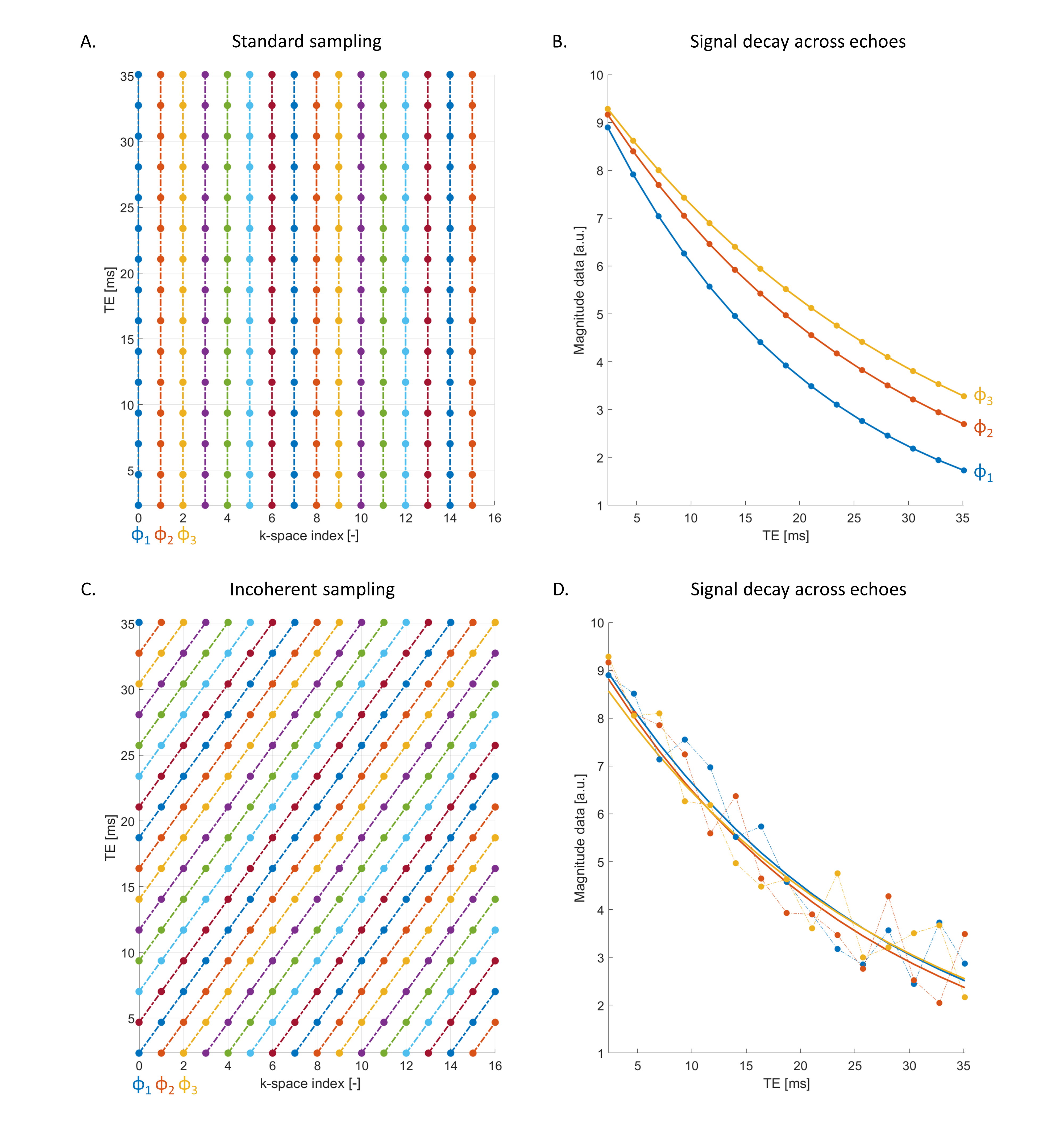
With standard acquisition techniques, multi-echo data are acquired consecutively at a given phase of the cardiac cycle, for each k-space location (Figure 1A). As a result, cardiac pulsation leads to an apparent change in R2* across the cardiac cycle (Figure 1B).4 With the proposed mitigation strategy, k-space location is shifted between the consecutive echoes of a multi-echo train (Figure 1C). With this acquisition strategy, the decay of the MRI signal with echo time is sampled incoherently with respect to the cardiac cycle (Figure 1D).The properties of cardiac-induced noise in multi-echo MRI data were assessed from data acquired in 5 healthy participants using a 3T scanner (MAGNETOM Prisma, Siemens Healthineers, Erlangen, Germany) and a custom-written 3D Cartesian FLASH sequence.4 Multi-echo data were acquired continuously for one hour and retrospectively binned according to the phase of the cardiac cycle at which they were acquired. This resulted in five-dimensional datasets including three spatial dimensions, one dimension for the echo time (2.34ms to 35.10ms with 2.34ms spacing), and one dimension for the phase of the cardiac cycle (12 bins).
From these five-dimensional datasets, we conducted numerical simulations of MRI data acquired using standard techniques and using the proposed mitigation strategy. The simulated four-dimensional data (3 spatial dimensions and 1 echo time dimension) was created by selecting points along the cardiac cycle dimension of the five-dimensional dataset according to the k-space trajectory and a time course of cardiac pulsation recorded with a pulse-oximeter. This simulation was repeated 30 times with a variable start of the k-space sampling relative to the pulse-oximeter recording. R2* maps were computed by fitting the echo trains of the simulated data with an exponential decay function. The ability of each sampling strategy to mitigate cardiac-induced noise was assessed from the variability of the R2* estimates across repetitions and from the goodness of fit (root-mean-square error, RMSE). For comparison, these simulations were also conducted with uncorrelated Gaussian-distributed noise, with variance equal to that of cardiac-induced noise at each echo time.
Results
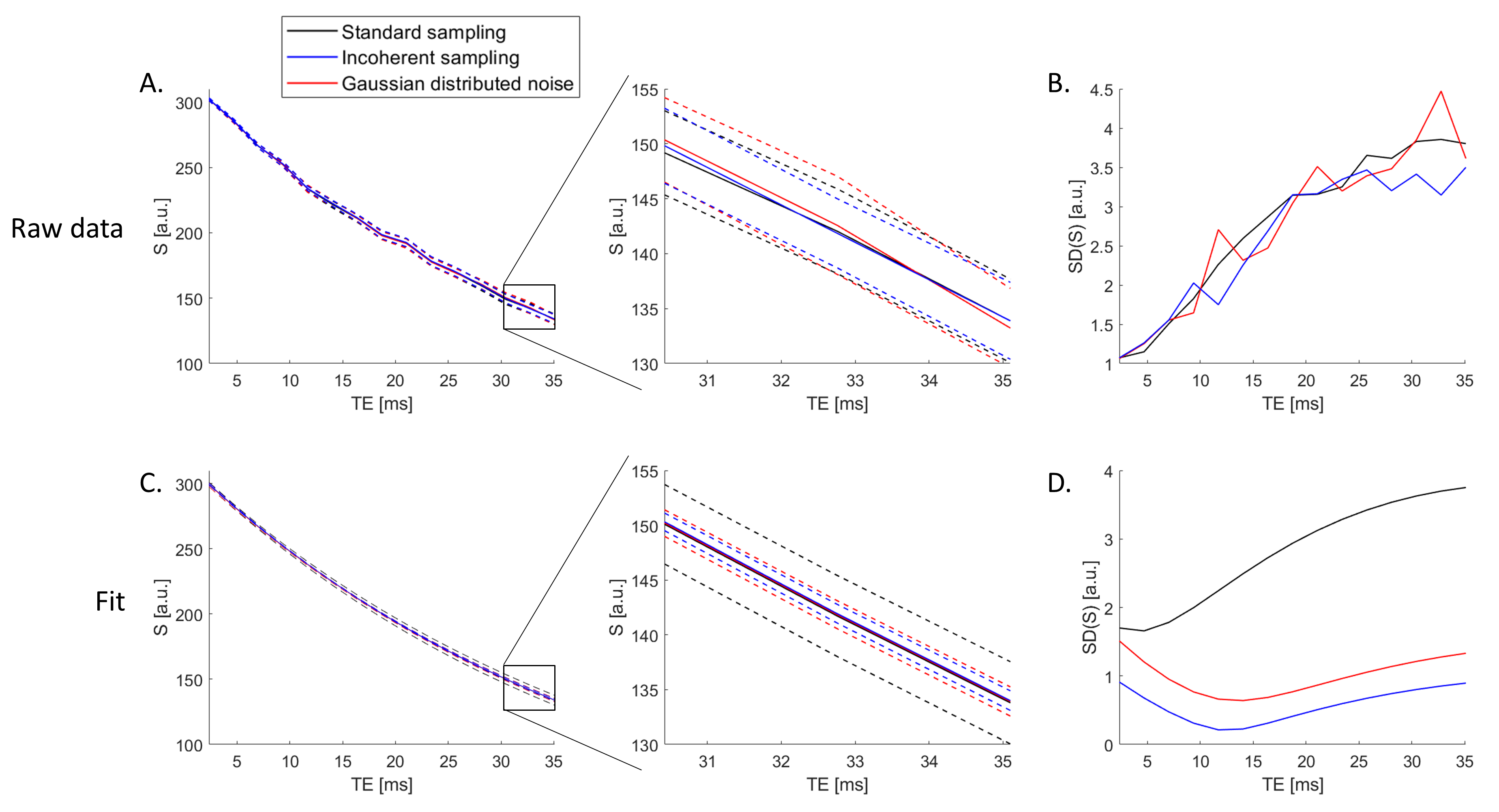
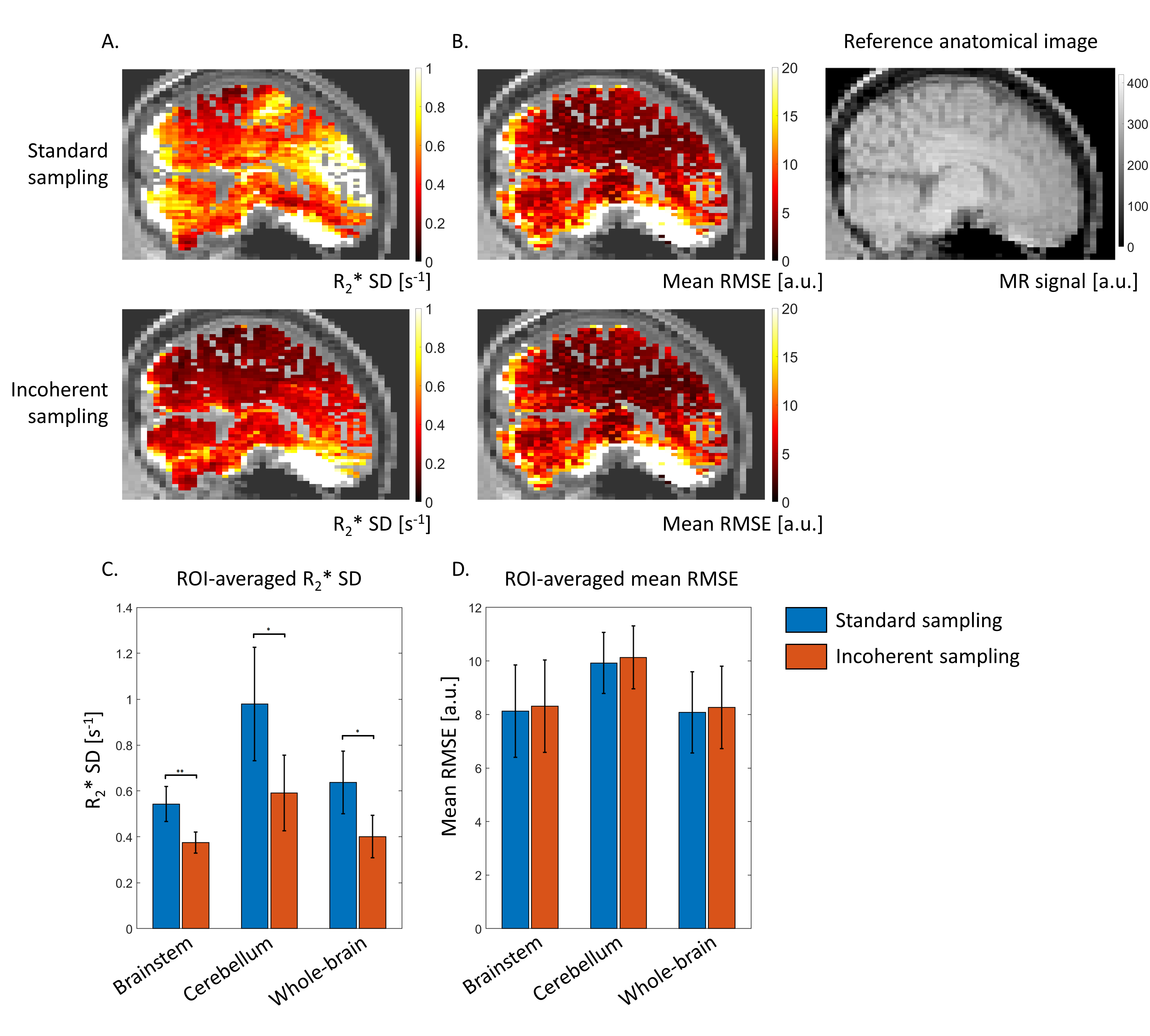
The raw magnitude signal shows the same level of variability across repetitions with the standard sampling strategy (black), incoherent sampling (blue), and uncorrelated noise (red) (Figure 2A). The standard deviation (SD) of the signal magnitude between repetitions increases fourfold from short echo times to long echo times (Figure 2B). However, the variability of the exponential fit of the data is much reduced with the incoherent sampling strategy and the uncorrelated noise data compared to the standard strategy (Figure 2C). With cardiac-incoherent sampling, the standard deviation of the fits across repetitions is low for all echo times, consistent with uncorrelated noise data (Figure 2D).Compared to the standard acquisition strategy, incoherent sampling reduces the variability of R2* across repetitions by 31%, 40% and 37% in the brainstem, cerebellum, and whole brain respectively (p<0.05, Figure 3A). In these regions, the mean RMSE across repetitions remains similar between strategies (Figure 3B).
Discussion & conclusion
We propose a novel acquisition strategy to mitigate the impact of cardiac-induced noise on R2* maps of the brain. This strategy allows the incoherent sampling of cardiac-induced noise across the multi-echo images used for the computation of the R2* maps. This cardiac-incoherent sampling led to a 30-40% reduction of the variability of R2* maps across repetitions compared to a standard acquisition scheme. The proposed strategy also ensures incoherent noise across the echo train, which might provide additional benefits for model-based image reconstruction techniques6 and sparse-domain denoising methods.7Acknowledgements
No acknowledgement found.References
- Barbosa JHO, Santos AC, Tumas V, et al. Quantifying brain iron deposition in patients with Parkinson’s disease using quantitative susceptibility mapping, R2 and R2*. Magn Reson Imaging. 2015;33(5):559-565. doi:10.1016/j.mri.2015.02.021
- Ulla M, Bonny JM, Ouchchane L, Rieu I, Claise B, Durif F. Is R2* a New MRI Biomarker for the Progression of Parkinson’s Disease? A Longitudinal Follow-Up. PLoS One. 2013;8(3):1-8. doi:10.1371/journal.pone.0057904
- Khalil M, Enzinger C, Langkammer C, et al. Quantitative assessment of brain iron by R2* relaxometry in patients with clinically isolated syndrome and relapsing-remitting multiple sclerosis. Mult Scler. 2009;15(9):1048-1054. doi:10.1177/1352458509106609
- Raynaud Q, Di Domenicantonio G, Yerly J, Dardano T, van Heeswijk RB, Lutti A. A characterization of cardiac-induced noise in R2* maps of the brain. Magn Reson Med. 2023. doi:10.1002/mrm.29853
- Raynaud Q, Dardano T, Roy CW, et al. Data acquisition strategies to mitigate cardiac-induced noise in quantitative R2* maps of the brain. In: International Society for Magnetic Resonance in Medicine. Toronto, Canada; 2023:Abstract #1151
- Tamir JI, Uecker M, Chen W, et al. T2 shuffling: Sharp, multicontrast, volumetric fast spin-echo imaging. Magn Reson Med. 2017;77(1):180-195. doi:10.1002/mrm.26102
- Does MD, Olesen JL, Harkins KD, et al. Evaluation of principal component analysis image denoising on multi-exponential MRI relaxometry. Magn Reson Med. 2019;81(6):3503-3514. doi:10.1002/mrm.27658
Figures