4585
Rapid multi-contrast multi-parametric whole brain imaging based on AI-accelerated multi-shot echo-planar imaging (msEPI)1MR Reseach Collaboration Team, Siemens Healthineers, Beijing, China, 2Radiology Department, Peking Union Medical College Hospital, Beijing, China, 3Siemens Healthcare GmbH, Erlangen, Germany, 4Siemens Medical Solutions USA, Boston, MA, United States
Synopsis
Keywords: Quantitative Imaging, Multi-Contrast, Artificial Intelligence; Quantitative Mapping; Ultra-fast Acquisition
Motivation: To enhance MRI efficiency for brain imaging, improve image quality, and enable multi-parametric mapping for diagnosing neurological diseases.
Goal(s): Develop a rapid imaging protocol using AI-accelerated multi-shot echo-planar imaging (msEPI) to simultaneously acquire multi-contrast images and quantitatively map R2, R2', R2*,R1, M0, and MTR across the entire brain.
Approach: Utilize msEPI with AI-enhanced reconstruction, scan five healthy volunteers, adjust parameters for different contrasts, and conduct whole-brain quantification using MATLAB.
Results: The acquisition technique collects FLAIR and FGATIR-like multi-contrast images with high SNR and enable multi-parameter quantification in just 5 minutes. This approach holds the potential to streamline diagnostics and enhance the patient’s experience.
Impact: Our multi-contrast fast quantification MRI protocol, founded on an AI-accelerated multi-shot echo-planar imaging sequence, substantially shortens scanning time while delivering high-quality multi-parametric brain images, offering a promising advancement in efficient and effective diagnostic processes for neurological diseases.
Introduction
Quantitative MRI probes brain tissue properties through parametric mappings that are directly linked to the biological properties of the tissue and enables better comparability across sites. While a wide variety of parameters can be quantified, commonly utilized indicators for depicting biological/pathological changes include transverse relaxation time T2, T2* and T2’, longitudinal relaxation time T1, magnetization transfer ratio (MTR) and T2’. These parameters offer insights into alterations in iron fraction, water fraction and macromolecules in the brain1-5. Previous studies have investigated multi-parametric imaging, but most of these protocols are long in duration6,7. While recent techniques are efficient at data collection, they may fall short of providing sufficient quantitative indicators. For instance, multi-dynamic multi-echo (MDME)8 cannot quantify T2* related to magnetic susceptibility as it collects spin echo signals. Similarly, Strategic Acquisition of Gradient Echo (STAGE)9 collects gradient echo signals, so it cannot quantify T2 relaxation time. Echo Planar Time-Resolved Imaging (EPTI)10 does not take the longitudinal relaxation time T1 into quantitative consideration. MR fingerprinting (MRF)11 represents an advanced quantitative concept with the potential to encompass all relevant quantitative parameters. However, as the number of simultaneously quantitative parameters increases, the size of the dictionary grows substantially, leading to an unacceptably long reconstruction time. In this work, we adopted a multi-shot echo-planar imaging (msEPI) research sequence that offers rapid protocols with tunable machine learning reconstruction, which was validated through radiological review12, to simultaneously collect multi-contrast images for the simultaneous quantification of R2, R2’, R2*, R1, M0 and MTR maps.Method
Five healthy volunteers were scanned on a 3T scanner (MAGNETOM Vida, Siemens Healthcare, Erlangen, Germany) with the following acquisition parameters: msEPI was repeated with four different TI’s (220, 400, 800 and 1200 ms, respectively); six echoes (10.9, 29.5, 48, 82, 100.5 and 119 ms) were collected in each TI. TR = 4600 ms, resolution of 0.9 × 0.9 × 4.0 mm3, receiver bandwidth of 1028 Hz/pixel, echo spacing = 1.2ms, in-plane acceleration of 3 and partial Fourier of 7/8, MTC preparation enabled, and an AI-Enhanced reconstruction. At TI = 1200ms, volunteers were scanned with magnetic transfer contrast (MTC) first, then without MTC, to calculate MTR. Total acquisition time for one volunteer was 5min 40s.R2 and R2’ was calculated by the following equations: $$$S = \left\{ \begin{array}{lr} S0*e^{-t*(R2+R2')}, & \text{if } i \lt max(TE)/2\\ S0*e^{-t*(R2-R2')}, & \text{if } i \ge max(TE)/2 \end{array}\right\}$$$, R2*=R2 + R2'.
T1 was calculated by $$$S(TI_n )=e^{-iθ}*(r_a+r_b*e^{-TI_n/T_1})$$$, with θ, ra, and rb real parameters, and R1 was calculated by 1/T1.
MTR was calculated by MTR = (S0 - SMTR) / S0.
Whole brain analysis was performed for those six parameters with MATLAB (Mathworks, Natick, MA, USA).
Results
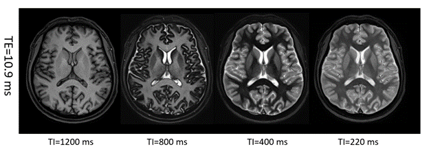
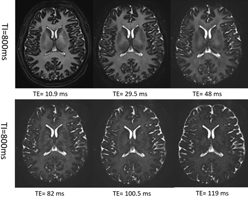
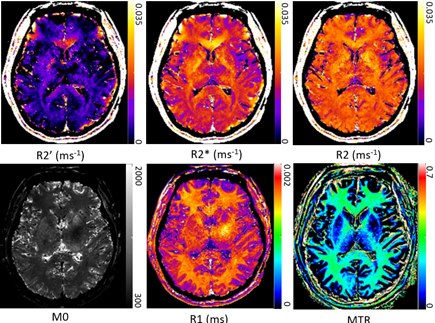
Figure 1. demonstrated brain images from one volunteer at four different TIs. We could notice that the brain image showed a FLAIR-like contrast at TI=1200 ms and a FGATIR-like contrast at TI = 400 ms. Typically, acquiring FLAIR images requires approximately a 5-min scan, while FGATIR necessitates a 10-min scan, both of which already exceeding the total scanning time allotted for msEPI. Figure 2. displays six images at TE= 10.9, 29.5, 48, 82, 100.5 and 119 ms with TI=800 ms, respectively. From this figure, we could find that brain images still maintained high SNR at TE= 119 ms, which may be attributed to the AI-enhanced reconstruction. Other contrast could be easily obtained by adjusting TI and TE. In Figure 3, we showed voxel-wise parametric mapping from one volunteer after analyzing images. White matter (WM) fiber bundles were clearly shown in these mappings, especially in the MTR map.Discussion
Through our experiments, we discovered that using a fast-imaging sequence such as msEPI will substantially reduce the scanning duration, alleviating the burden on patients. There are a number of neurological diseases in white matter and gray matter that require multi-modality imaging protocols to become diagnostic and prognostic. To address this problem, the msEPI provides multi-contrast images containing structural and qualitative information, while multi-parametric mapping provides quantitative tissue characteristics. Compared to a specific MRI modality, the msEPI will collect all information that radiologists need in a short time and provide uniform signal across the entire brain. Routine usage of msEPI in clinics will advance the whole diagnostic process.Conclusion
This rapid msEPI protocol with machine learning reconstruction enables multi-contrast images with a high SNR. A 5-minute scan could collect FLAIR-like and FGATIR-like contrast, and high quality multi-parametric mapping could be obtained through processing, which will advance the diagnostic efficiency of radiologists. Future application of msEPI is a promising approach to shorten scan times considerably while maintaining access to tissue characteristics.Acknowledgements
Yanglei Wu and Yuan Li contributed equally to this work.References
1. Deoni SC. Magnetic resonance relaxation and quantitative measurement in the brain. Method s Mol Biol (2011) 711:65–108.
2. Schenker C, Meier D, Wichmann W, Boesiger P, Valavanis A. Age distribu-tion and iron dependency of the T2 relaxation time in the globus pallidus and putamen. Neuroradiology (1993) 35:119–24.
3. Langkammer C, Krebs N, Goessler W, Scheurer E, Ebner F, Yen K, et al. Quantitative MR imaging of brain iron: a postmortem validation study. Radiology (2010) 257:455–62.
4. Deloire-Grassin MS, Brochet B, Quesson B, Delalande C, Dousset V, Canioni P, et al. In vivo evaluation of remyelination in rat brain by magne-tization transfer imaging. J Neurol Sci (2000) 178:10–6.
5. Mottershead JP, Schmierer K, Clemence M, Thornton JS, Scaravilli F, Barker GJ, et al. High field MRI correlates of myelin content and axonal density in multiple sclerosis - a post-mortem study of the spinal cord. J Neurol (2003) 250:1293–301.
6. Jack, C.R., Jr., Bernstein, M.A., Fox, N.C., Thompson, P., Alexander, G., Harvey, D., Borowski, B., Britson, P.J., L. Whitwell, J., Ward, C., Dale, A.M., Felmlee, J.P., Gunter, J.L., Hill, D.L.G., Killiany, R., Schuff, N., Fox-Bosetti, S., Lin, C., Studholme, C., DeCarli, C.S., Gunnar Krueger, , Ward, H.A., Metzger, G.J., Scott, K.T., Mallozzi, R., Blezek, D., Levy, J., Debbins, J.P., Fleisher, A.S., Albert, M., Green, R., Bartzokis, G., Glover, G., Mugler, J. and Weiner, M.W. (2008), The Alzheimer's disease neuroimaging initiative (ADNI): MRI methods. J. Magn. Reson. Imaging, 27: 685-691.
7. Rovira, À., Wattjes, M., Tintoré, M. et al. MAGNIMS consensus guidelines on the use of MRI in multiple sclerosis—clinical implementation in the diagnostic process. Nat Rev Neurol 11, 471–482 (2015). 8. Konar AS, Paudyal R, Shah AD, Fung M, Banerjee S, Dave A, Lee N, Hatzoglou V, Shukla-Dave A. Qualitative and Quantitative Performance of Magnetic Resonance Image Compilation (MAGiC) Method: An Exploratory Analysis for Head and Neck Imaging. Cancers (Basel). 2022 Jul 26;14(15):3624.
9. Haacke EM, Chen Y, Utriainen D, Wu B, Wang Y, Xia S, He N, Zhang C, Wang X, Lagana MM, Luo Y, Fatemi A, Liu S, Gharabaghi S, Wu D, Sethi SK, Huang F, Sun T, Qu F, Yadav BK, Ma X, Bai Y, Wang M, Cheng J, Yan F. STrategically Acquired Gradient Echo (STAGE) imaging, part III: Technical advances and clinical applications of a rapid multi-contrast multi-parametric brain imaging method. Magn Reson Imaging. 2020 Jan;65:15-26.
10. Wang F, Dong Z, Reese T G, et al. Echo planar time‐resolved imaging (EPTI)[J]. Magnetic resonance in medicine, 2019, 81(6): 3599-3615.
11. Ma, Dan, et al. "Magnetic resonance fingerprinting." Nature 495.7440 (2013): 187-192.
12. Clifford B, Conklin J, Huang SY, Feiweier T, Hosseini Z, Goncalves Filho ALM, Tabari A, Demir S, Lo WC, Longo MGF, Lev M, Schaefer P, Rapalino O, Setsompop K, Bilgic B, Cauley S. An artificial intelligence-accelerated 2-minute multi-shot echo planar imaging protocol for comprehensive high-quality clinical brain imaging. Magn Reson Med. 2022 May;87(5):2453-2463.
Figures