4578
Revolutionizing 3T Animal Imaging: Metamaterials' Advantages in Enhancing Contrast1Radiology, Huashan hospital, Fudan university, Shanghai, China, 2MR Research Collaboration Team, Siemens Healthineers Ltd., Shanghai, China, 3Application Developments, Siemens Shenzhen Magnetic Resonance Ltd., Shenzhen, China
Synopsis
Keywords: Data Acquisition, Data Acquisition, Metamaterials
Motivation: To achieve high-quality animal imaging, metamaterials can offer an array of electromagnetic properties that can enhance 3T MRI by improving signal-to-noise ratio (SNR), image quality, and reducd scan times.
Goal(s): Achieve superior imaging quality in 3T MRI by implementing metamaterial coils, thereby improving animal imaging capabilities.
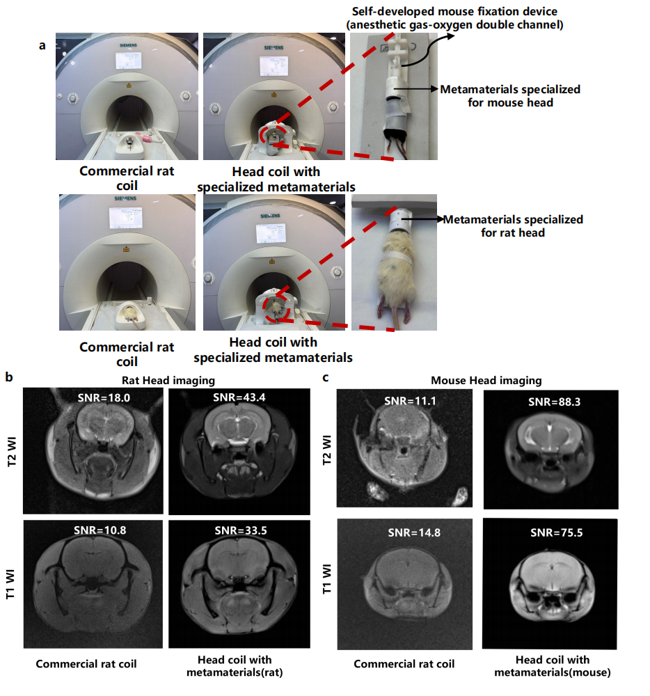
Approach: Utilized metamaterials on a 3T MRI to scan rats and mice, comparing image resolution, SNR, and CNR against traditional coils. Images were assessed by radiologists for quality.
Results: Metamaterial coils provided markedly better resolution and contrast, with significantly higher SNR and shorter scan times, enabling clearer anatomical detail.
Impact: The study's findings pave the way for enhanced diagnostic precision in veterinary and preclinical studies, raise questions about broader applications in small animal research.
Introduction
Leveraging the bespoke electromagnetic properties of metamaterials, this research investigates their capacity to augment the performance of 3T MRI by elevating the signal-to-noise ratio and image fidelity, whilst concurrently accelerating acquisition times. The objective is to surpass conventional coil capabilities in achieving higher resolution and enhanced contrast, and to refine advanced neuroimaging modalities such as Diffusion Tensor Imaging (DTI) and Magnetization Transfer (MT) in both healthy and cerebrovascular-compromised rodent models. This may consequently minimize the necessity for multiple imaging sessions, streamlining the diagnostic process.Methods
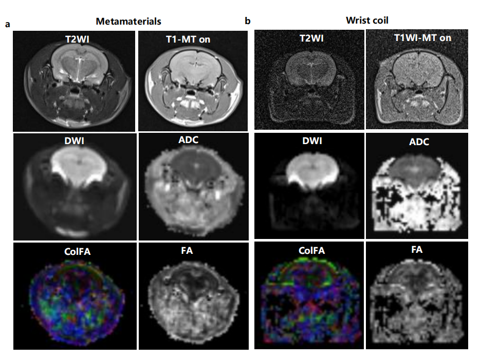
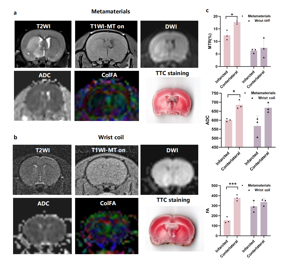
The study was conducted on MAGNETOM Prisma 3T MR scanner (Siemens Healthcare, Erlangen, Germany). Healthy SD rats and C57 mice were imaged using metamaterials(TsingMeta, Beijing, China) and a standard 64-channel head coil versus a 12-channel commercial animal-specific coil. T2WI and T1WI sequences were performed with identical settings for both coils to ensure comparability. Radiologists evaluated the quality and anatomical detail in images, employing SNR for quantitative analysis. Additionally, metamaterials were compared with a conventional wrist coil on three healthy rats and two ischemic stroke-induced rats using DTI, T1WI (with MT), and T2WI, maintaining consistent imaging parameters. Stroke was induced by MCAO. Experts assessed the delineation between normal and ischemic tissues in the images. For DTI, T2 MR images helped map the infarcted areas, with FA and ADC values measured at the striatum's ischemic boundary. ROI analyses were repeated thrice for reliability. MTR calculations further quantified tissue contrasts.Results
In a study comparing the imaging performance of metamaterials with commercial animal-specific and wrist coils in a 3T MRI setting, metamaterials significantly outperformed in terms of resolution and signal-to-noise ratio (SNR). For both mice and rats, the metamaterials achieved finer resolution in T2-weighted imaging (T2WI) and T1-weighted imaging (T1WI), with a substantial increase in SNR, particularly enhancing anatomical details in various brain regions. The scan times were also reasonable, taking slightly over three minutes for mice and under five minutes for rats on T1WI.The metamaterials' superiority extended to self-referenced comparisons on three rats, demonstrating improved image quality and sensitivity in detecting different tissues. This included better delineation of structures in regions prone to distortion and a significantly higher SNR in the cortex and striatum on T2WI.The SNR demonstrated significant improvements, showing a 3.6-fold increase for the cortex (8.45 vs. 30.31 for wrist coil and metamaterials, respectively) and 4.9-fold increase for striatum (5.31 vs 26.13) on T2WI. As for scan times, T2WI scans took 3minutes and 47seconds, while both DTI and T1WI with MT scans required 6 minutes and 5 second.In functional imaging, the metamaterials afforded clearer contrasts in infarcted versus healthy tissues, vital for understanding physiological and disease processes. Diffusion tensor imaging (DTI) and magnetization transfer (MT) sequences showed greater resolution and reduced artifact susceptibility. This precision was confirmed via TTC staining, emphasizing the metamaterials' reliability in accurately reflecting tissue conditions and microstructural changes.Quantitatively, within the infarcted areas, fractional anisotropy (FA) and apparent diffusion coefficient (ADC) values were significantly altered, indicating changes in tissue microstructure and diffusion patterns, as confirmed by high-contrast DTI images and MT ratio analyses. within the infarcted region, the fractional anisotropy (FA) values were significantly (P=0.0008, t=35.05) altered compared to the healthy region, indicating changes in tissue microstructure and anisotropy. ADC values in the infarcted region were considerably higher (P=0.02, t=7.92) relative to the healthy region, signifying greater overall water diffusion.The metamaterials exhibited higher precision in differentiating between infarcted and healthy regions compared to wrist coils, particularly in MT imaging where magnetization transfer contrast is crucial.The MT ratio was significantly (P=0.02, t=7.89) lower compared to the healthy region.
The advantages of volumetric metamaterials lie in responding efficiently to the circularly polarized magnetic fields of MRI, enhancing both transmitting and receiving fields without wasting RF power or increasing specific absorption rate (SAR). This enables not only improved SNR but also better uniformity of the magnetic field, crucial for accurate functional imaging like DTI and MT imaging.
Conclusions
Metamaterials offer superior resolution and contrast over traditional coils, enhancing functional imaging and lesion characterization, especially in low-field MRI systems. They improve diagnostic accuracy in small animal models, offering advantages over conventional coils in clinical and research settings.Acknowledgements
No acknowledgement found.References
1. E. Mark Haacke, Robert W. Brown, Michael R. Thompson RV. Magnetic Resonance Imaging: Physical Principles and Sequence Design. Wiley-Liss. 1999. doi: 10.1002/9781118633953. 2. Chi Z, Yi Y, Wang Y, et al. Adaptive Cylindrical Wireless Metasurfaces in Clinical Magnetic Resonance Imaging. Adv Mater. 2021;33(40). doi: 10.1002/adma.202102469.Figures