4577
Motion robust MR-ARFI using timeseries single-shot spiral acquisition1Radiology, Stanford University, Stanford, CA, United States, 2Stanford, Stanford, CA, United States
Synopsis
Keywords: Data Acquisition, Neuro, MR-ARFI
Motivation: 2DFT MR-ARFI methods used to map ultrasound beam profiles can fail with small amounts of head motion because the signal is based on phase shifts from um-sized Acoustic Radiation Force displacement of tissue.
Goal(s): To demonstrate that a single-shot timeseries method is more robust to bulk motion.
Approach: Employing an efficient spiral redout trajectory, we compared the two methods’ ARFI images in a tissue-mimicking phantom undergoing controlled random translations.
Results: The timeseries method demonstrated good quality tissue translation maps, while the 2DFT method failed with motions as small as ~50 microns RMS.
Impact: Our single-shot method may allow routine use of MR-ARFI in humans for guiding US neuromodulation and US-based ablative therapy.
INTRODUCTION
MR-based Acoustic Radiation Force Imaging (MR-ARFI) employs low-intensity focused ultrasound (FUS) to displace tissue during motion-encoding gradients to create maps of the US beam profile from the phase difference between FUS on vs. off conditions [1]. Typically, 2DFT spin echo pulse sequences are utilized to acquire two (FUS-on/FUS-off) complex images, from which the phase difference is extracted to create tissue displacement (ARFI) maps [2]. While the reconstructed magnitude images are essentially immune to small bulk motions in 2DFT, the corresponding phase images can be significantly distorted, as, therefore are the ARFI phase difference maps, especially because tissue displacement is only a few microns compared to bulk motion that can be substantially larger in human scans. Alternatively, single-shot spiral acquisitions have been employed to generate the two complex images [3] used to create ARFI maps. However, this leads to the same motion sensitivity problem when only one ON and one OFF image is acquired. Here we use a timeseries of singe-shot spiral images while triggering the FUS on and off in blocks, analogous to fMRI block task designs and compare ARFI results with those of the 2DFT design. Pseudo-random motion of the phantom allows accurate comparison of the motion sensitivity of the two methods.METHODS
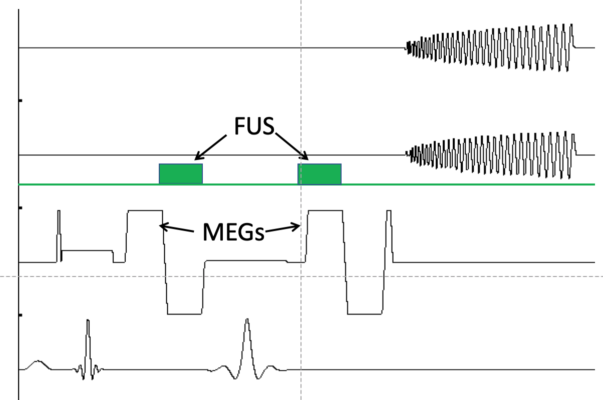
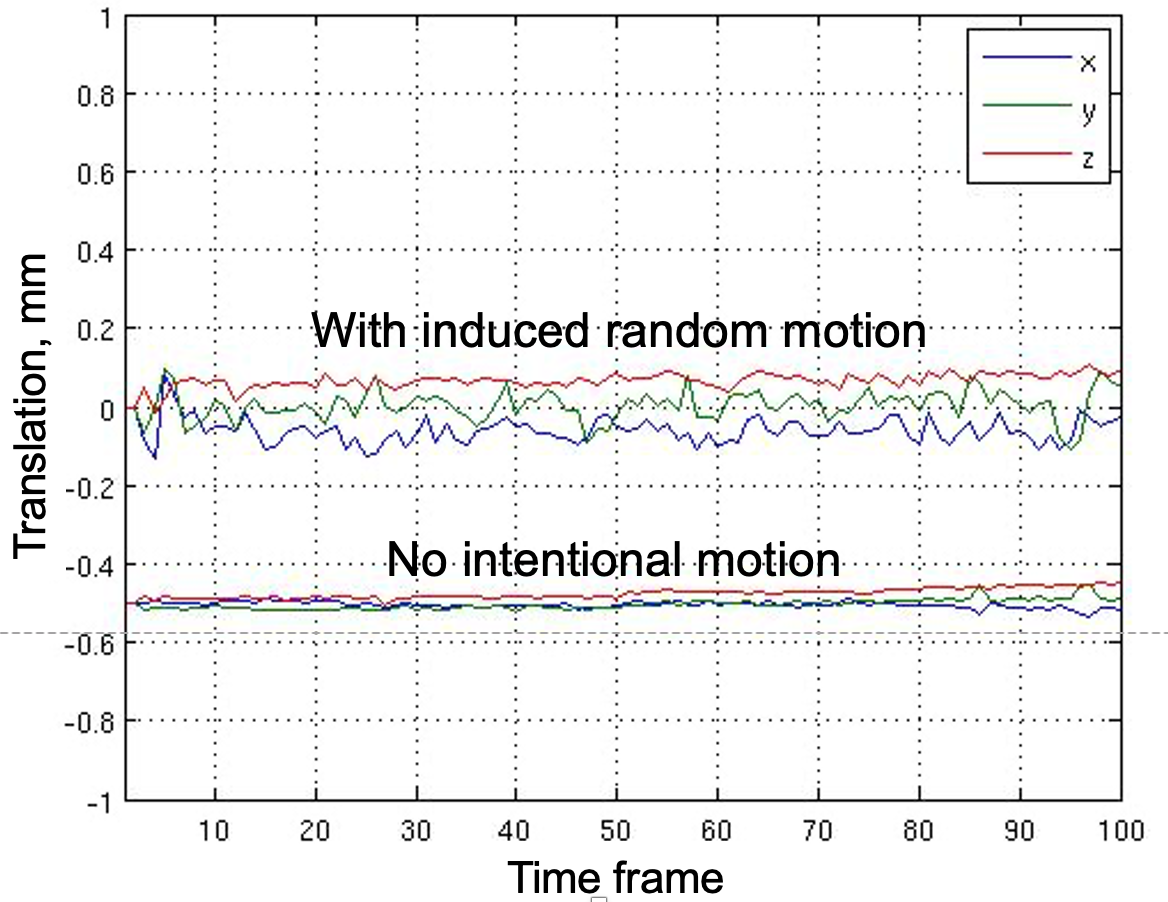
2DFT MR-ARFI maps were acquired on a 3T scanner (GE Healthcare, Milwaukee, WI) using an 8-channel head coil with a spin echo sequence having 800 ms repetition time, 40 ms echo time, 5 4mm slices. A 1.0 MHz 128-element phased array US transducer (Image Guided Therapy, Paris, France) was focused to a depth of 60 mm in a soft tissue-mimicking phantom (Computerized Imaging Referencing System, Virginia, USA). Scan times were 102.4s each for the two separate acquisitions. Repeated bipolar MEGs (motion encoding gradients) [4] had a total duration of 20ms. The spiral acquisition was identical except the 2DFT readout was replaced by a single-shot spiral-out trajectory, with identical FUS, TR and slice positioning (Fig. 1). 100 time frames of complex images were acquired in 80.0s total, half with FUS triggered on and half with FUS off. To generate controlled motion, a small balloon was attached to the end of the phantom and supplied by an electric compressed air valve with pseudo-random duration on times, not synchronized to TR. Plots of motion without and with motion applied (54 ms RMS total) during the spiral acquisitions are shown in Fig. 2. The complex timeseries were processed by separately averaging the two halves, complex division and extraction of the phase difference from the ratio with atan2().RESULTS
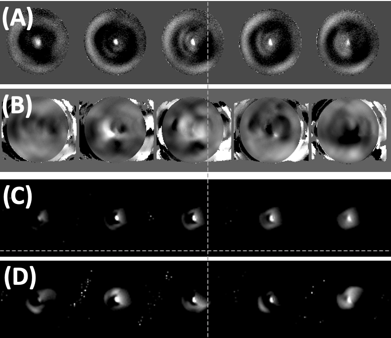
Figure 3 shows ARFI maps acquired with (A) 2DFT, no motion; (B) 2DFT with motion; (C) Timeseries spiral, no motion; (B) Timeseries spiral with motion. As may be seen, the two methods are roughly equivalent with no intentional motion, but the 2DFT ARFI maps are unusable with even the small motion applied, while the spiral maps are degraded slightly but remain usable. It is notable that the spiral scan time was only 40% that of the 2DFT acquisition, suggesting that even better ARFI could be obtained in the presence of motion if a larger number of time frames are acquired in the single-shot methodDISCUSSION
Bulk motion of the head can cause large artifacts in 2DFT ARFI methods because the motion occurs over acquisition of phase-encoding lines of k-space, which takes many seconds or minutes. After Fourier reconstruction, small motions such as demonstrated here have little effect on the reconstructed image magnitude, but major effect on its phase, which is discarded in most other MRI applications but is the signal of interest for ARFI. By contrast, the complex time series single-shot method introduced in this study employs temporal averaging to diminish the influence of motion in the combination of the timeseries. Furthermore, an additional correction step may be added to account for the phase shifts resulting from bulk motion of the entire phantom/head (not just the tissue displaced in the US beam) during the MEGs. This step uses magnitude images to generate S/I motion as in Fig. 2, from which phase values are then employed during the timeseries averaging.CONCLUSION
In preliminary results on a phantom, the timeseries single-shot MR-ARFI method introduced here has substantial advantages over 2DFT methods, by virtue of its reduced sensitivity to interference from head motion. This suggests that human studies may be more reliable with our method, because bulk head motion of the order of ARF-generated tissue displacement is unavoidable even when stabilization is employed.Acknowledgements
Funding for MM and KBP is supplied by NIH MH131684; Funding for GHG in part from NIH P41 EB015891.References
1. McDannold N, Maier SE. Magnetic resonance acoustic radiation force imaging.Med Phys 2008;35:3748–3758.
2. Radicke M, Engelbertz A, Habenstein B, Lewerenz M, Oehms O, Trautner P,Weber B, Wrede S, Maier K. New image contrast method in magnetic resonanceimaging via ultrasound. Hyperfine interactions, Vol. 181. The Netherlands. Springer; 2008. 21–26.
3. Asaf Ilovitsh, Brett Z Fite, Tali Ilovitsh and Katherine W Ferrara. Acoustic radiation force imaging using a singleshot spiral readout. 2019 Phys. Med. Biol. 64 1250044.
4. Chen J, Watkins R, Pauly KB. Optimization of Encoding Gradients for MR-ARFI. Magnetic Resonance in Medicine (2010) 63:1050–1058.Figures