4567
3D-TSE Dixon sequence with preserved short echo spacing for second and subsequent echoes1Siemens Healthineers AG, Erlangen, Germany
Synopsis
Keywords: Data Acquisition, MSK, Dixon
Motivation: Common to existing TSE Dixon sequences is that the chemical shift between water and fat is encoded by shifting the readout window away from the spin-echo time point. The shifting prolongs the echo spacing by twice the shift. A short echo spacing is an important component of modern 3D-TSE sequences to enable high resolution 3D imaging in clinically acceptable times.
Goal(s): Develop a 3D-TSE Dixon sequence with unchanged echo spacing.
Approach: Encode the chemical shift via the excitation. Acquire in-phase and opposed-phase data for 2pt-Dixon symmetric around the spin-echo time point.
Results: Homogeneous fat-water separation in knee, hand and ankle.
Impact: 3D-TSE 2pt-Dixon sequence with unchanged echo spacing between second and subsequent echoes compared to a conventional 3D-TSE sequence with same acquisition parameters.
Introduction
In reference 1, the authors describe a T2-preparation module that, in addition to T2 weighting of longitudinal magnetization, saturates the fat signal. The excitation pulse of the T2-preparation module is replaced by a novel pulse that generates a specific phase ∆φ between on-resonance spins (water) and off-resonance spins (fat) at the time of the flip-back pulse. The phase is chosen such that only on-resonance spins are restored. The idea of the present work is to use this pulse as excitation pulse of a 3D-TSE sequence to generate the phase shift between water and fat needed for Dixon reconstruction.Methods – Sequence design
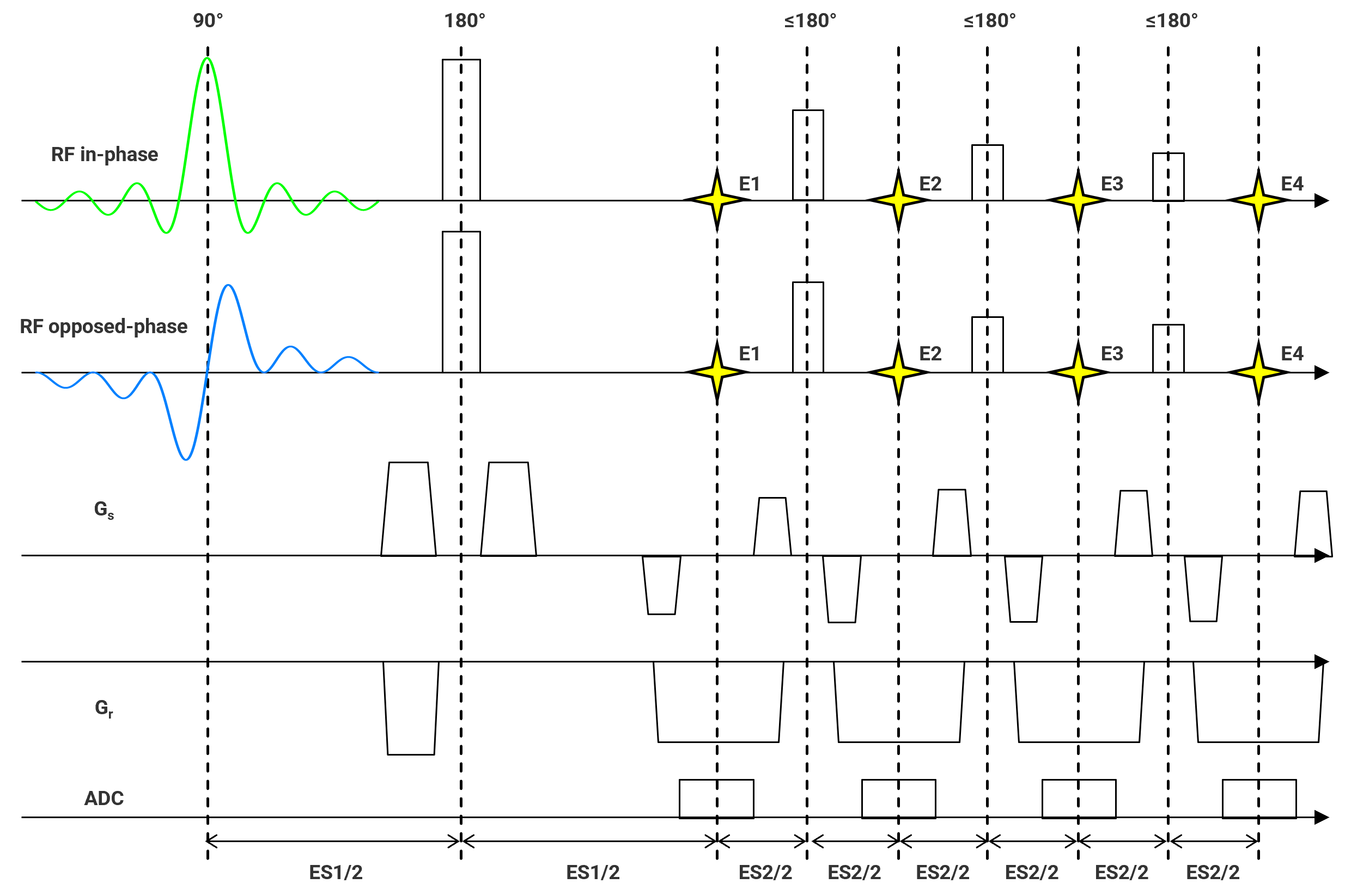
The excitation pulse of a research 3D-TSE sequence is replaced by the novel pulse of reference 1. Data with different phase shift are acquired in separate echo trains (Figure 1). The phase ∆φ of the respective excitation pulse is adapted to the desired phase shift. Specific design strategies include:- The CPMG-condition limits the possible phase shift to ∆φ=0° or ∆φ=180°. Fortunately, phase ∆φ=0° corresponds to the in-phase condition and ∆φ=180° corresponds to the opposed phase condition. This phase choice is optimal for a 2pt-Dixon technique.
- At 3T the duration of the excitation pulse with appropriate bandwidth and frequency profile for Dixon is around 8 ms. To create temporal space for the long excitation pulse, a strategy used for slab-selective 3D-TSE2 is adopted as follows: The 1st echo-spacing is prolonged compared to spacing between subsequent echoes. To avoid the formation of stimulated echoes involving the first refocusing RF pulse, the flip angle of the 1st refocusing RF is set to 180°. The stimulated echo signal formatted despite the nominal flip angle of 180°, due to the unavoidable variation of the B1-Field across the volume, is de-phased by crushers.
- FID artifacts can occur, if longitudinal magnetization regrows during the echo train and is re-excited by a refocusing pulse. Since the excitation pulse is not involved in the formation of FID artefacts and since the readout window is not shifted, the FID signal has the same phase in the in-phase and the opposed phase data sets and hence would be assigned to the water image by the Dixon reconstruction. By alternating the phase of the refocusing pulses by 180° between echo trains used to acquire corresponding data of the in-phase and opposed-phase image the FID artefacts can be moved to the fat image – regardless of whether the FID emerges from water or fat tissue.
Methods – Data acquisition
3D-TSE SPAIR and 3D-TSE Dixon knee, hand, and ankle images were acquired in healthy volunteers with a 3T scanner (MAGNETOM Vida, Siemens Healthineers, Erlangen, Germany). All image parameters of 3D-TSE SPAIR and 3D-TSE Dixon sequence were identical, except for the number of averages, which were doubled for the 3D-TSE SPAIR protocol to adjust the number of echo trains and hence the acquisition time.Results
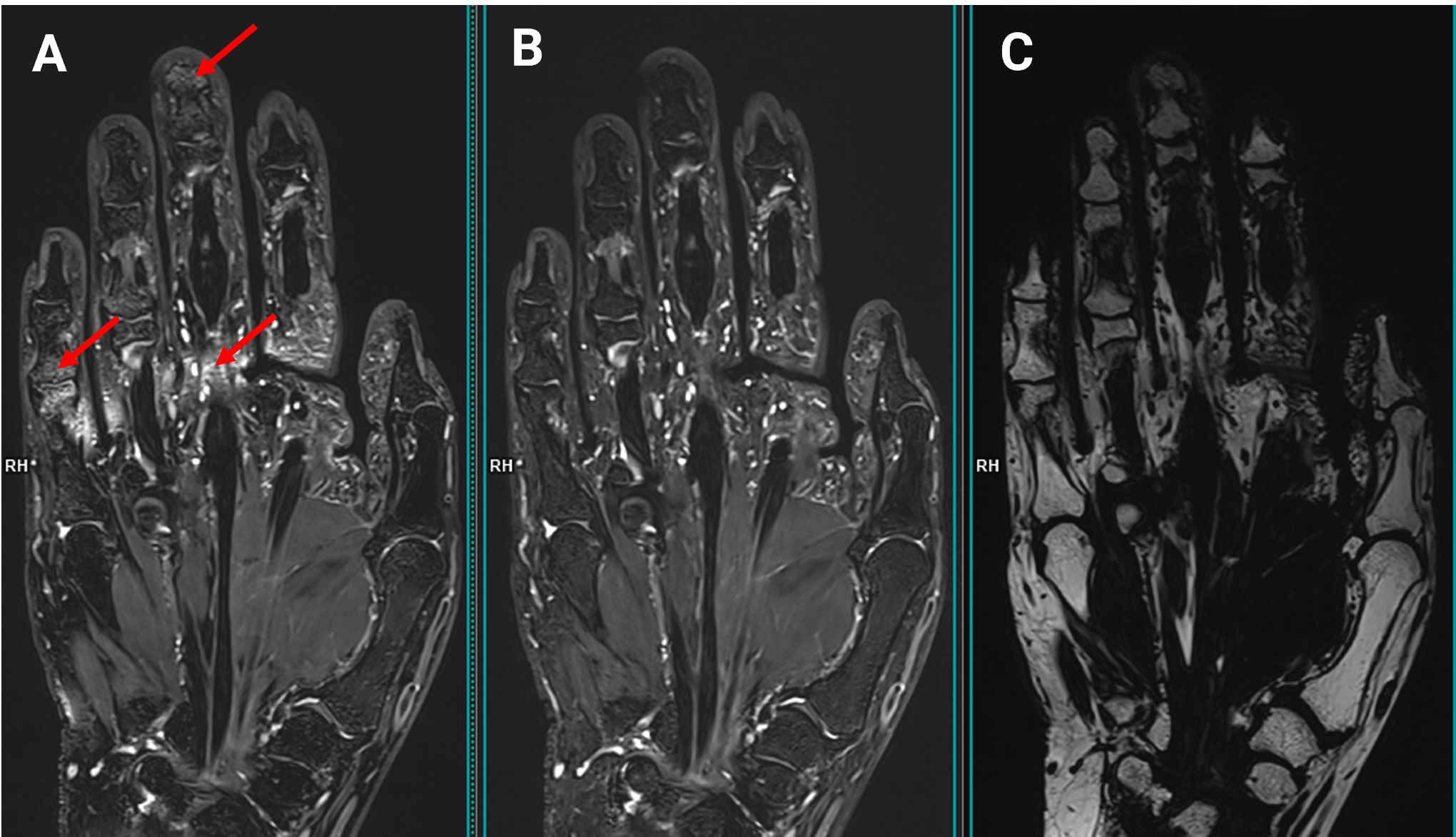
Figure 2 exemplary compares hand images acquired with 3D-TSE SPAIR and 3D-TSE Dixon. The homogeneity of the fat suppression is markedly improved in the Dixon water image.Discussion
The unchanged echo-spacing of the new Dixon method allows to acquire 3D-TSE 2pt-Dixon fat and water images using the same imaging parameters used for 3D-TSE with conventional fat saturation techniques. In conventional 3D-TSE two averages are often used to eliminate FID artefacts by alternating the phase of the refocusing pulses by 180° between averages. The phase-cycling scheme of the new Dixon method allows to shift FID artefacts into the Dixon fat image. Hence the new Dixon method allows to acquire FID artifact-free 2pt-Dixon water images in the same time and with the same SNR efficiency as with the conventional sequence. The doubling of the number of echo trains in the conventional sequence for eliminating the FID artifact corresponds to the doubling of echo trains needed to acquire in-phase and opposed-phase data sets for the Dixon method. Initial results indicate that the fat saturation homogeneity and completeness is improved compared to conventional fat saturation methods. Furthermore, the additional contrasts (Dixon fat image, in-phase, and opposed-phase image) may be of diagnostic use. However, since the new Dixon method creates the phase shift between water and fat via the excitation it cannot deal with B0-inhomogenities which are in the order of the fat-water shift. The usefulness of this approach at lower field strength (1.5T, 0.55T), where the echo spacing penalty of conventional Dixon methods is even higher, still has to be proven.Acknowledgements
No acknowledgement found.References
- Arn L, van Heeswijk RB, Stuber M, Bastiaansen JAM. A robust broadband fat-suppressing phaser T2 -preparation module for cardiac magnetic resonance imaging at 3T. Magn Reson Med. 2021 Sep;86(3):1434-1444.
- Mugler JP 3rd. Optimized three-dimensional fast-spin-echo MRI. J Magn Reson Imaging. 2014 Apr;39(4):745-67.
Figures