4560
k-space Based Signal Processing Approach for Automated Detection of Streaky Coils in Radial k-space Sampling MRI1Department of Radiology, Northwestern University Feinberg School of Medicine, Chicago, IL, United States, 2Division of Cardiology, Internal Medicine, Northwestern University Feinberg School of Medicine, Chicago, IL, United States
Synopsis
Keywords: Artifacts, Data Processing, Visualization
Motivation: Radial k-space sampling is preferred over cartesian k-space sampling due to its many advantages. One major drawback of radial k-space is the streaking artifacts that arise from the non-linear gradients from the peripheral of FOV.
Goal(s): The goal of this study is to reduce streaky artifacts.
Approach: We developed an algorithm that aims to detect the streaky coils based on post-processing of the coils’ images and then remove these streaky coils for a cleaner image.
Results: Our results show that the proposed method precisely predicts streaky coils and improves the appearance of streaky artifacts in the CS reconstruction after removing the selected coils.
Impact: Radial k-space sampling is improved using our new precise streaky coils detection algorithm that effectively removes them to produce a clean image with no streaky artifacts. An unmet need in the radial k-space sampling in the MRI field in general.
Introduction
Radial k-space sampling provides many advantages over Cartesian k-space sampling, including more benign aliasing artifacts, better motion properties, and potentially higher signal-to-noise ratio when under-sampled. One disadvantage of radial sampling is streaky artifacts resulting from a mismatch in spatial encoding due to non-linear magnetic field gradients near the periphery and chemical shift of fat. Xue et al. proposed an image-based post-processing approach to detect streaky coils[1], but its efficacy has not been evaluated for cardiac MRI. We proposed a k-space-based post-processing approach to detecting streaky coils and compared its efficacy with the method proposed by Xue et al. for first-pass cardiac perfusion MRI.Methods
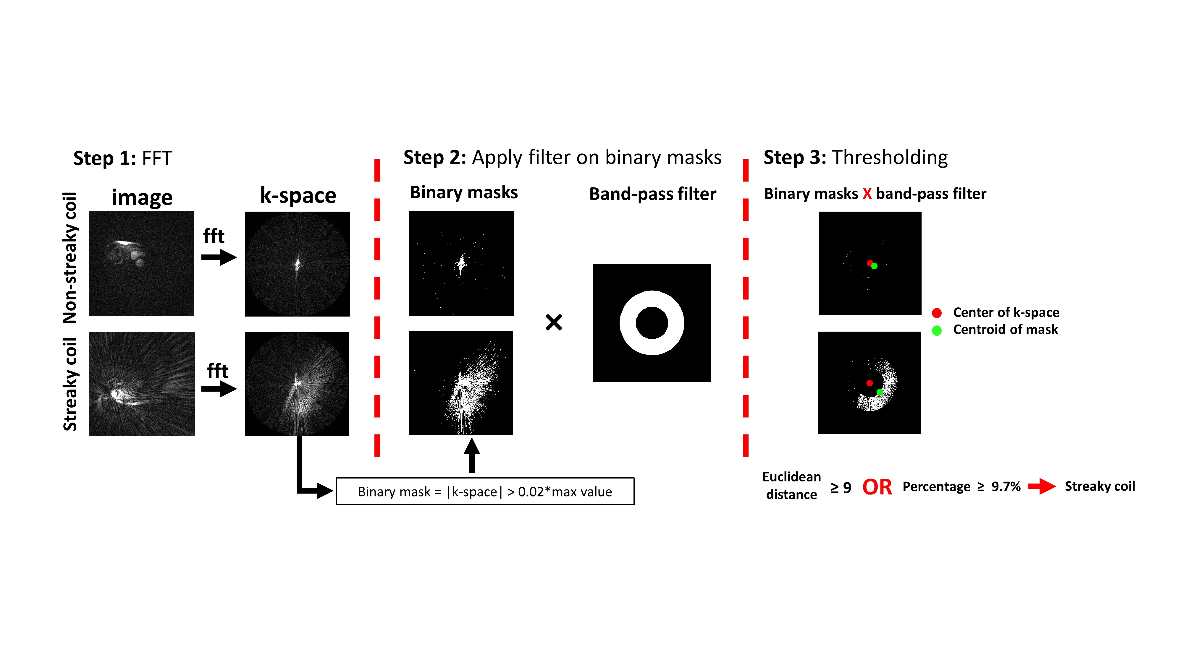
Human Subjects and Pulse Sequence: We retrospectively identified cardiac perfusion data from 11 patients (4 females; mean age = 58.3 ± 13.6 years). The relevant image parameters included: FOV = 384x384 mm2, slice thickness = 8 mm, acquisition matrix = 192x192, readout duration = 109, flip angle = 15°, receiver bandwidth = 744 Hz/pixel, 42 radial spokes per frame, 100 repetitions, number of slices ranged from 5 to 6, and acceleration factor = 4.6. In total, this study entailed 1683 coil-by-coil time series.Post-Processing: As the pre-processing step, we combined 7 timeframes (frames 21 to 27 representing post-gadolinium) to achieve 294, which is nearly 301 – the Nyquist condition for radial k-space sampling with 192 kx points, and performed coil-by-coil non-uniform fast Fourier transform (NUFFT). In step one, we performed FFT to derive coil-by-coil k-space representing the Nyquist condition. In step two, we performed thresholding to create a binary mask of k-space, in order to capture streaking artifacts distributed across k-space as shown. We performed a preliminary experiment on several training data to empirically derive 0.02 of the maximal k-space value as the optimal cut point. We created a bandpass filter to remove the central lobe of k-space (DC component) that is unrelated to streaks and to suppress the peripheral of k-space since the number of spokes (294) is slightly below Nyquist condition. To detect streaky coils, we computed the following: (a) we calculated the “mass” (i.e., area) of the streaking artifacts reflected in k-space as a fraction of total k-space dimension; we empirically defined percent area of 9.7% based on training data; (b) we calculated the centroid of the k-space mask to ensure that it is away from the center of k-space space and used the Euclidean distance of 9 (equal to the inner radius of the annulus filter). Streaky coil is defined as Euclidean distance > 9 or perfect area > 9.7%.
Comparison Method: We used the method developed by Xue et al. as reference. We used a 64-point Hanning window, computed $$$Rstreak = \frac{mean(abs(Iorig-Iref))}{mean(Iref)}$$$, where Iorig is unprocessed reconstruction and Iref is Hanning windowed reconstruction and adjusted the threshold for Rstreak to 4.5 based on training data.
Accuracy Calculation: We used one human expert as reference for detecting streaky coils. We calculated the sensitivity, specificity, and accuracy for both methods.
Image reconstruction: To further evaluate the benefit of removing streaking artifacts, we performed iterative Golden-angle Radial Sparse Parallel Imaging (GRASP)[2] implemented in MATLAB, for all coils and after removal of streaky coils by our method and the method by Xue et al.
Results
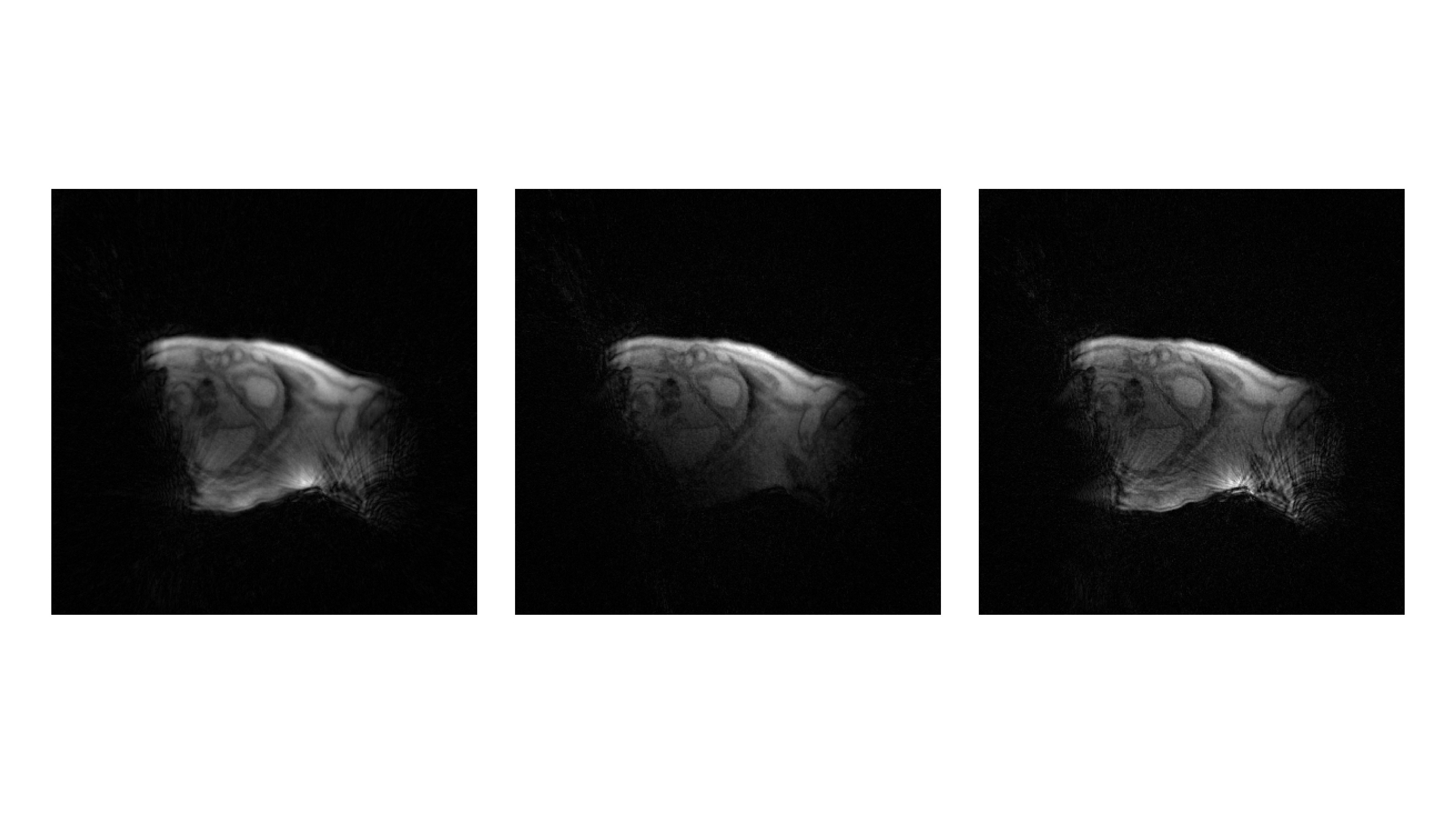
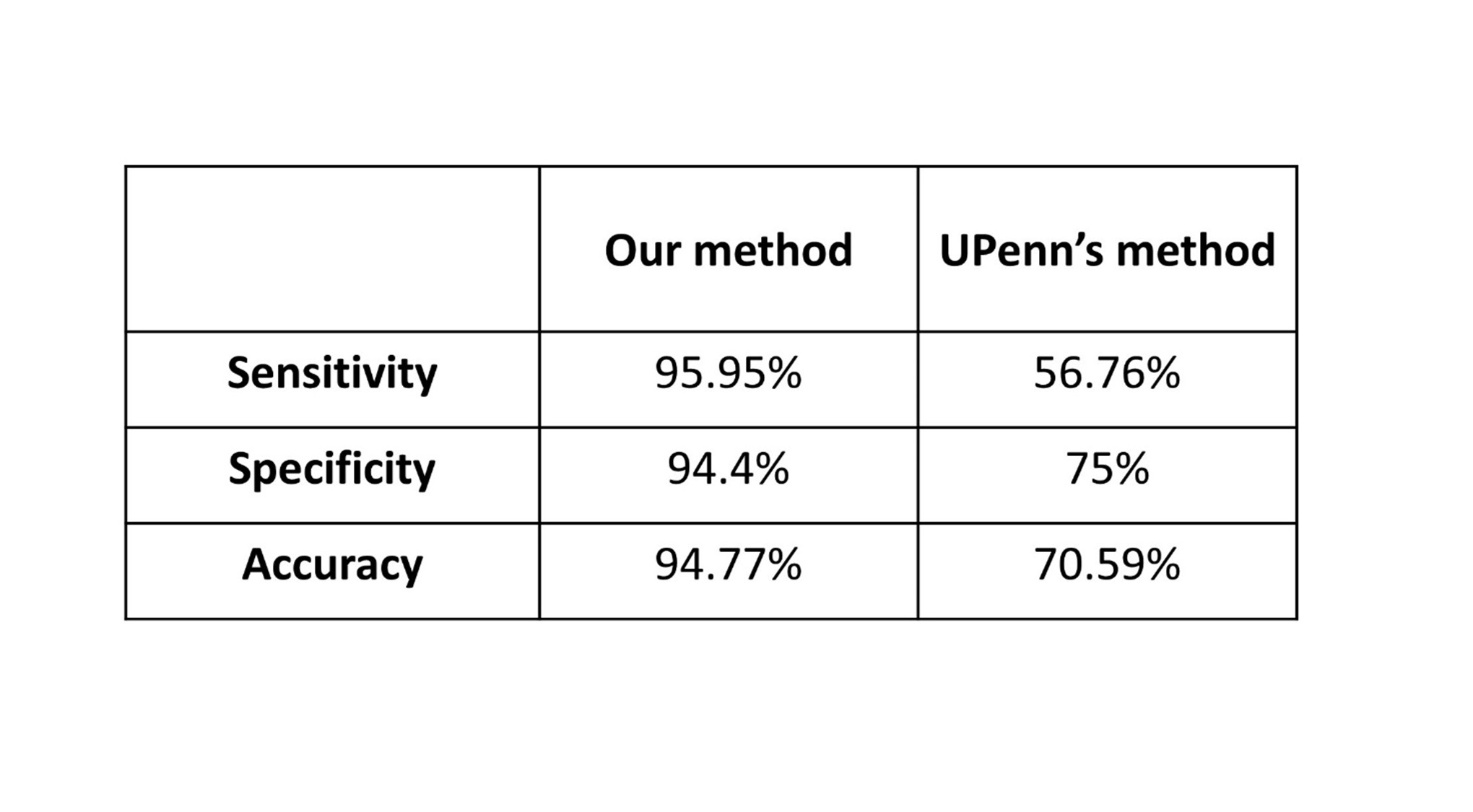
As summarized in Table 1, the sensitivity, specificity, and accuracy of our method were superior to that of Xue et al for predicting N=390 streaky coils out of 1683 coils. Figure 2 compares representative cardiac perfusion GRASP reconstructed images using all coils and after removing streaky coils by our method and by Xue’s method. As shown, our method produced less streaky artifacts compared with all coils and the method by Xue et al.Discussion and Conclusion
Our k-space based post-processing approach detects streaky coils more accurately than the method proposed by Xue et al. Future study including different types of radial k-space data is warranted to test the generalizability of our method.Acknowledgements
The authors would like to thank funding support from the National Institutes of Health (R01HL116895, R01HL151079, R21EB030806A1, 1R01HL167148‐01A1), the American Heart Association (19IPLOI34760317, 949899), and the Radiological Society of North America (EILTC2302).References
1. Xue Y, Yu J, Kang HS, Englander S, Rosen MA, Song HK. Automatic coil selection for streak artifact reduction in radial MRI. Magn Reson Med. 2012 Feb;67(2):470-6.
2. Feng L, Grimm R, Block KT, Chandarana H, Kim S, Xu J, Axel L, Sodickson DK, Otazo R. Golden-angle radial sparse parallel MRI: combination of compressed sensing, parallel imaging, and golden-angle radial sampling for fast and flexible dynamic volumetric MRI. Magn Reson Med. 2014 Sep;72(3):707-17.
Figures