4558
Ultrashort Echo Time Quantitative Susceptibility Mapping (UTE-QSM) of The Human Knee with Motion Registration1UCSD, San Diego, CA, United States, 2Dept of Medicine, UCSD, San Diego, CA, United States, 3Radiology Service, Veterans Affairs San Diego Healthcare System, San Diego, CA, United States, 4Research Service, Veterans Affairs San Diego Healthcare System, San Diego, CA, United States, 5Dept of Bioengineering, UCSD, San Diego, CA, United States
Synopsis
Keywords: Motion Correction, Quantitative Susceptibility mapping
Motivation: There is a need for improving UTE-QSM in the human knee joint, which is prone to motion artifacts due to multiple repeated scans required for a short echo spacing.
Goal(s): To investigate the efficacy of motion registration-based UTE-QSM for knee joint imaging.
Approach: We employed rigid affine-based registration and non-rigid deformable registration based on B-spline as a pre-processing step for generating the QSM data.
Results: It is seen that the registration process helps in reducing streaking artifacts and improving UTE-QSM of the knee joint.
Impact: The UTE-QSM technique of the human knee joint is a potentially sensitive biomarker for the diagnosis of musculoskeletal diseases. Motion registration can improve the accuracy of UTE-QSM and hence likely enhance the diagnostic power.
Introduction
Quantitative susceptibility mapping (QSM) has been investigated in neuro1, body2, and musculoskeletal (MSK)3 imaging. Among them, MSK-QSM has been relatively less investigated due to several challenges, including the short T2 signal decay from connective tissues and the presence of a strong off-resonant fat signal, which can result in significant streaking artifacts4. To directly estimate the susceptibility from short T2 tissues, ultrashort echo time (UTE) based QSM (UTE-QSM) has been investigated5,6. UTE-QSM is typically based on multiple data acquisitions with interleaved echo times (TEs) to secure several images at TEs below 1ms. As conventional fat-saturation can adversely affect short T2 signals7,8, m-Dixon (or IDEAL) method has been applied to compensate for the chemical shift and estimate a reliable B0 field map9, crucial information for the UTE-QSM. However, inter-scan motion can be problematic in the UTE-QSM based on multiple acquisitions. In this study, we investigate the efficacy of motion registration in UTE-QSM of the human knee joint.Methods
Figure 1A illustrates the dual-echo UTE-cones sequence used in this study. To obtain images with different TEs, the readout gradients were applied with variable time delays. The acquired complex images were processed with motion registration algorithms before being input into the QSM pipeline.Motion Registration: Two different algorithms, including rigid affine-based registration and non-rigid registration based on B-spline algorithm10, were tested. The complex image was first converted into magnitude as well as real and imaginary components. The registration was performed based on the magnitude images. The first echo served as a fixed image while the other echoes were registered to it. The obtained transformation parameters were then applied to the real and imaginary images. Finally, the combined, registered complex images were provided as input to the QSM pipeline.
UTE-QSM Pipeline: The UTE-QSM pipeline was established using Matlab, based on the IDEAL11,12 and MEDI13. IDEAL was first applied to the complex input images at different TEs to produce a B0 field map. Subsequently, the estimated B0 field map was processed with PDF algorithm14 to achieve a local field map. Finally, the local field map was processed with MEDI algorithm to generate the final susceptibility map (l=1000). Figure 1B shows the workflow for UTE-QSM without or with motion registrations.
Data Collection: Five healthy male volunteers were recruited and underwent UTE-QSM imaging on a 3T clinical MR system (GE MR750) using an 8-channel T/R knee coil. The imaging parameters were as follows: TR=10ms, FA=10°, FOV=150×150×86.4mm3, matrix=220×220×96, readout bandwidth=250kHz, three dual-echo scans with TE=0.032/2.7, 0.2/3.7, and 0.7/4.7ms, and total scan time = 18min.
Results
Figure 2 illustrates the efficacy of registration methodology through visualization of a magnitude image. The evaluation based on echo subtraction revealed that unregistered images, which are subject to motion-induced misregistration, exhibit a strong boundary effect, while registered images successfully mitigate these artifacts, resulting in a dramatically decreased boundary effect. Figure 3A depicts the phase images from the same volunteer, and Figure 3B shows the resultant B0 field map from IDEAL. Motion registration removed the obvious error exhibited in the B0 field map (a red arrow), which is presumably due to motion-induced error that complicates the graph cut algorithm12 used in IDEAL.Figure 4A exhibits the result from a representative subject with mild inter-scan motion. The UTE-QSM derived from registered images shows a significant enhancement in image quality. Figure 4B showcases the UTE-QSM result from another subject, displaying streaking artifacts with unregistered data, which is rectified through the registration process. Figure 4C highlights a case with extreme motion that causes strong streaking artifacts. While the affine registration eliminates the streaking to some degree, the non-rigid registration further improves the susceptibility map, although streaking artifact still exists.
Discussion and Conclusion
Conventional QSM typically utilizes GRE train imaging to achieve equally spaced TEs over tens of milliseconds, which can be done in a single scan. In contrast, UTE-QSM requires much shorter echo spacing to capture multiple images below TE of 1ms. Unfortunately, in most modern clinical MR systems, it is not feasible to acquire multiple GRE images within 1ms duration due to limited gradient hardware performance and FDA safety regulations. Therefore, an interleaved encoding scheme is necessary for UTE-QSM, which may introduce inter-scan motion. In this study, we demonstrated that this kind of motion is critical, which may generate strong streaking artifacts and can impair the susceptibility map. Appropriate motion registration can help reduce these artifacts, as shown in Figure 4. Our future studies will further investigate the impact of motion on UTE-QSM in different body parts within the MSK system, such as the ankle and elbow joints.Acknowledgements
The authors acknowledge grant support from the NIH (R01AR078877, R01AR062581, R01AR068987, R01AR075825, RF1AG075717, K01AR080257 and F32AG082458), Veterans Affairs (I01CX001388, I01CX002211, I01RX002604), and GE Healthcare.
References
1. Jang H, Sedaghat S, Athertya JS, et al. Feasibility of ultrashort echo time quantitative susceptibility mapping with a 3D cones trajectory in the human brain. Front. Neurosci. 2022;16:1–9 doi: 10.3389/fnins.2022.1033801.
2. Liu S, Wang C, Zhang X, et al. Quantification of liver iron concentration using the apparent susceptibility of hepatic vessels. Quant. Imaging Med. Surg. 2018;8:123–134 doi: 10.21037/qims.2018.03.02.
3. Wei H, Dibb R, Decker K, et al. Investigating magnetic susceptibility of human knee joint at 7 Tesla. Magn. Reson. Med. 2017;78:1933–1943 doi: 10.1002/mrm.26596.
4. Dimov A V., Liu T, Spincemaille P, et al. Joint estimation of chemical shift and quantitative susceptibility mapping (chemical QSM). Magn. Reson. Med. 2015;73:2100–2110 doi: 10.1002/mrm.25328.
5. Jerban S, Lu X, Jang H, et al. Significant correlations between human cortical bone mineral density and quantitative susceptibility mapping (QSM) obtained with 3D Cones ultrashort echo time magnetic resonance imaging (UTE-MRI). Magn. Reson. Imaging 2019;62:104–110 doi: 10.1016/j.mri.2019.06.016.
6. Lu X, Jang H, Ma Y, Jerban S, Chang E, Du J. Ultrashort Echo Time Quantitative Susceptibility Mapping (UTE-QSM) of Highly Concentrated Magnetic Nanoparticles: A Comparison Study about Different Sampling Strategies. Molecules 2019;24:1143 doi: 10.3390/molecules24061143.
7. Jang H, Carl M, Ma Y, et al. Fat suppression for ultrashort echo time imaging using a single-point Dixon method. NMR Biomed. 2019:e4069 doi: 10.1002/nbm.4069.
8. Carl M, Nazaran A, Bydder GM, Du J. Effects of fat saturation on short T2 quantification. Magn. Reson. Imaging 2017;43:6–9 doi: 10.1016/j.mri.2017.06.007.
9. Jang H, Drygalski A, Wong J, et al. Ultrashort echo time quantitative susceptibility mapping (UTE‐QSM) for detection of hemosiderin deposition in hemophilic arthropathy: A feasibility study. Magn. Reson. Med. 2020;84:3246–3255 doi: 10.1002/mrm.28388.
10. Klein S, Staring M, Murphy K, Viergever MA, Pluim JPW. Elastix: A toolbox for intensity-based medical image registration. IEEE Trans. Med. Imaging 2010;29:196–205 doi: 10.1109/TMI.2009.2035616.
11. Reeder SB, Pineda AR, Wen Z, et al. Iterative decomposition of water and fat with echo asymmetry and least-squares estimation (IDEAL): Application with fast spin-echo imaging. Magn. Reson. Med. 2005;54:636–644 doi: 10.1002/mrm.20624.
12. Hernando D, Kellman P, Haldar JP, Liang ZP. Robust water/fat separation in the presence of large field inhomogeneities using a graph cut algorithm. Magn. Reson. Med. 2010;63:79–90 doi: 10.1002/mrm.22177.
13. Liu J, Liu T, De Rochefort L, et al. Morphology enabled dipole inversion for quantitative susceptibility mapping using structural consistency between the magnitude image and the susceptibility map. Neuroimage 2012;59:2560–2568 doi: 10.1016/j.neuroimage.2011.08.082.
14. Liu T, Khalidov I, de Rochefort L, et al. A novel background field removal method for MRI using projection onto dipole fields (PDF). NMR Biomed. 2011;24:1129–1136 doi: 10.1002/nbm.1670.
Figures
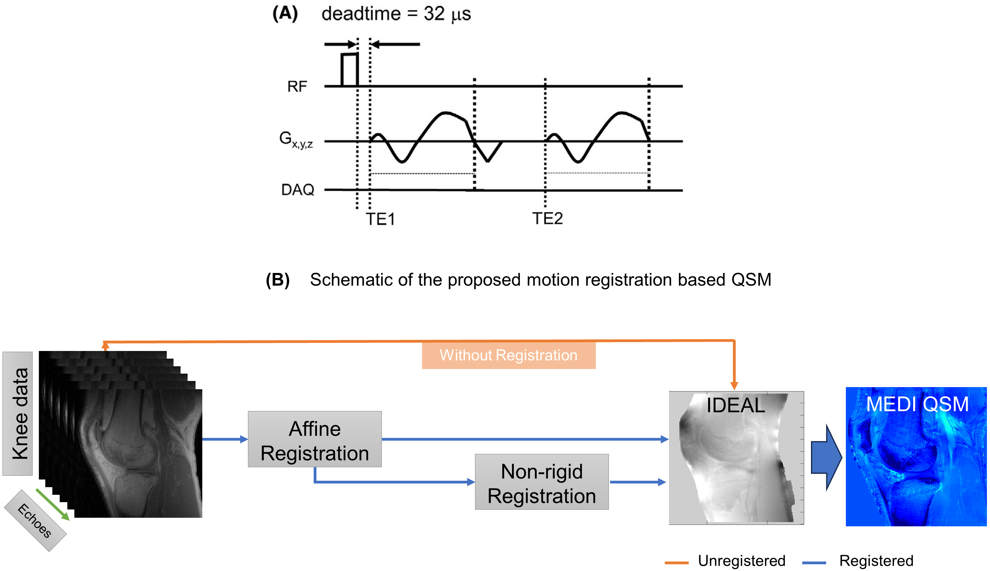
Figure 1. UTE-QSM. (A) Pulse sequence of 3D dual-echo UTE-cones used for the UTE-QSM data acquisition and (B) schematic of the motion registration-based correction for QSM input data. The dual echo imaging is repeated multiple times at different TEs (TE1 and TE2) by delaying the readout gradient. The raw data is reconstructed as complex images and registered via affine or non-rigid registration techniques before IDEAL and MEDI-QSM process.
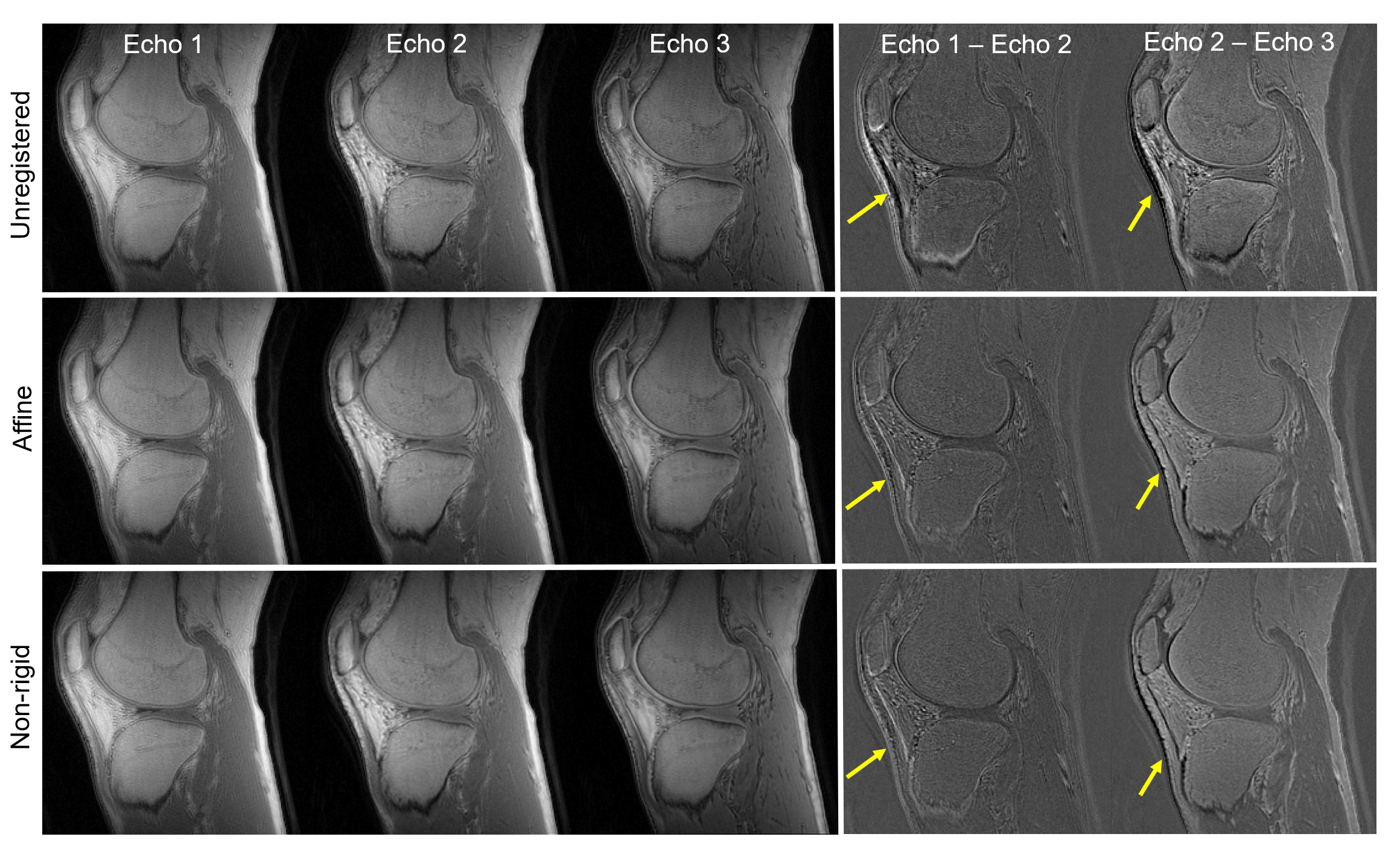
Figure 2. The efficiency of motion registration technique (a 32-year-old male volunteer). The echo-subtracted images demonstrate that the unregistered images are susceptible to strong motion, while the registration reduces the motion artifacts, therefore exhibiting less error near tissue boundaries (pointed by yellow arrows).
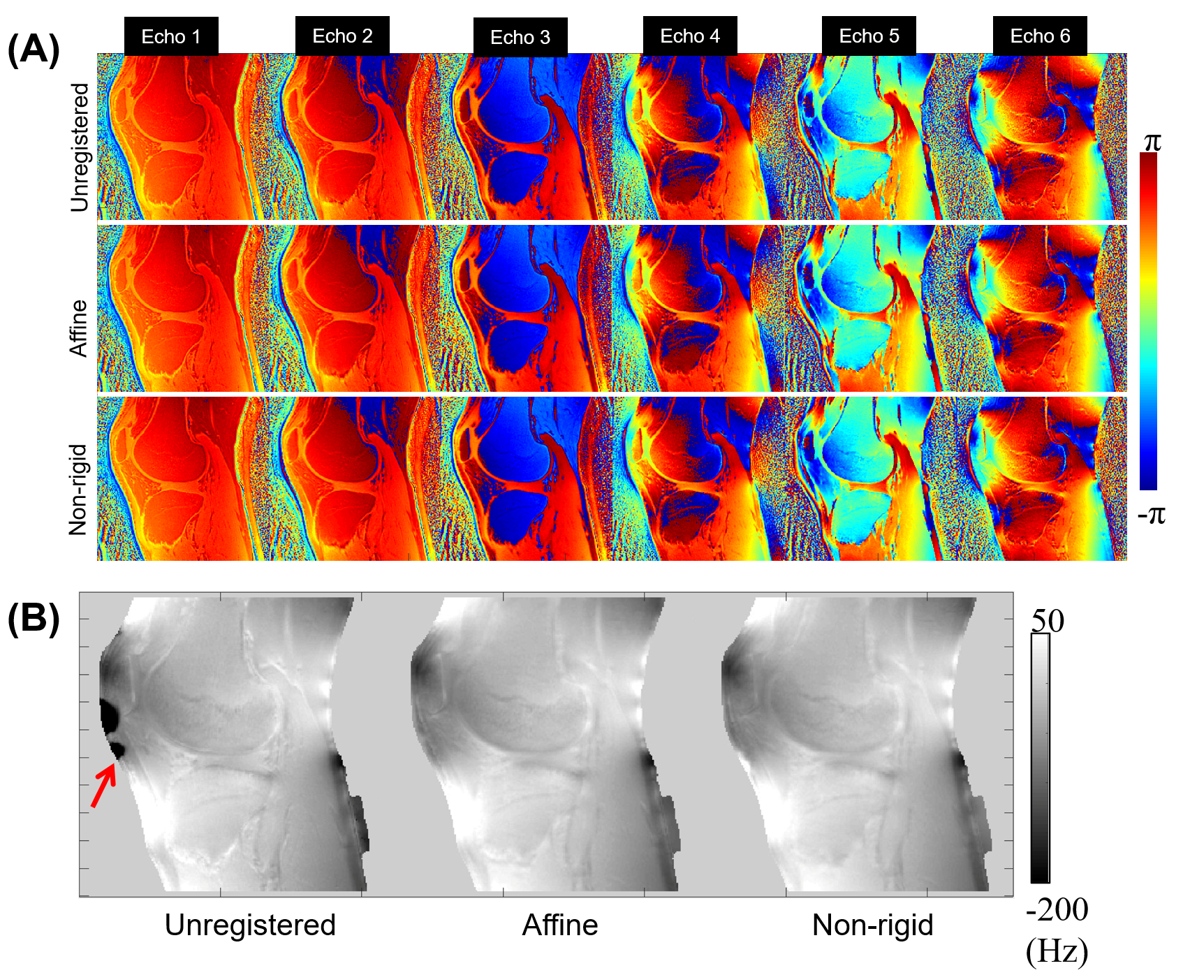
Figure 3. (A) The phase evolution over six different echoes obtained from a 32-year-old, male volunteer without or with motion registration, and (B) the resultant B0 field maps. The strong error shown in the B0 field map (a red arrow) is presumably due to the strong inter-scan motion that complicates graph cut-based field map estimation in IDEAL.
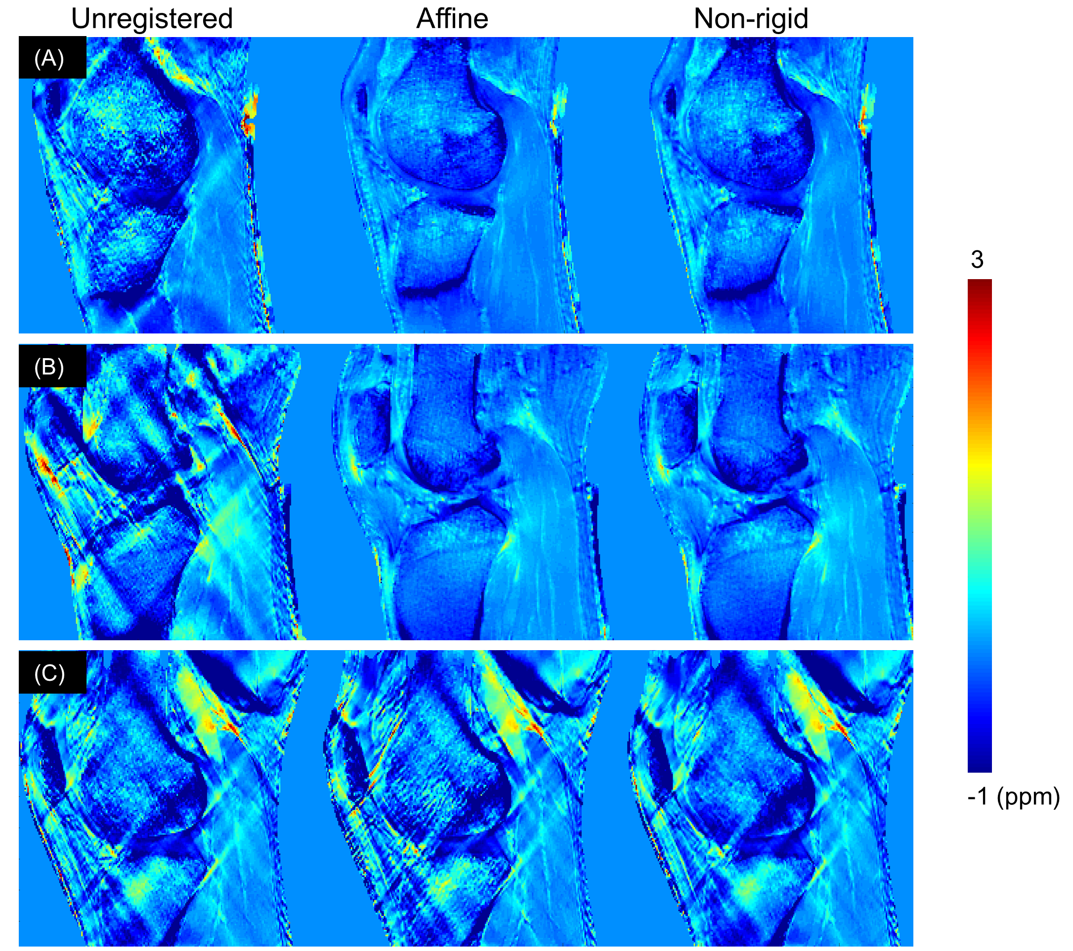
Figure 4. The feasibility of UTE-QSM for human knee joint via motion registration is presented here. (A) shows maps of 22-year-old, control with mild inter-scan motion. The susceptibility map generated from registered images shows substantial improvement with suppressed streaking artifacts. (B) presents another subject (36-year-old), showing streaking artifacts in the unregistered QSM, which has been corrected through registration. (C) demonstrates another case (23-year-old) with extreme motion creating strong artifacts in QSM, which is mitigated by registration.