4551
Improving Cardiac Phase-Resolved T2*-Mapping of the Murine Heart: Artifact Reduction and Enhanced Accuracy1Berlin Ultrahigh Field Facility (B.U.F.F.), Max Delbrueck Center for Molecular Medicine in the Helmholtz Association, Berlin, Germany, 2DZHK (German Centre for Cardiovascular Research), Berlin, Germany, 3Experimental and Clinical Research Center, Charité—Universitätsmedizin Berlin, Berlin, Germany
Synopsis
Keywords: Quantitative Imaging, Animals, Relaxometry, Gradient Moment Nulling, T2*, Retrospective gating, Mouse
Motivation: Rapid blood flow in mice heart imposes severe phase shift error in k-space data, resulting ghosting artifact in the image. These artifacts notably affect the accuracy of myocardial T2* relaxometry, as they introduce variable ghosting artifacts into echo images.
Goal(s): To investigate the effect of Flow Compensation (FC) on myocardial T2*-map acquired by retrospective-gated Multi-Gradient-Echo (MGE) sequence.
Approach: First-order GMN were added to MGE sequence. Retrospective ECG-gating was performed on murine heart.
Results: FC technique can potentially reduce T2* estimation error by mitigating the flow artifact on echo images in mice heart with very fast heart rate.
Impact: First-order Gradient Moment Nulling minimizes image artifacts and enhances the accuracy of myocardial T2* measurements.
Introduction
There is a strong demand for non-invasive characterization of myocardial remodeling, including fibrosis and microvascular diseases. T2*-mapping offers a trajectory into the non-invasive assessment of myocardial pathophysiology. Previous research demonstrated that transient T2* changes across the cardiac cycle provide valuable information about Hypertrophic Cardiomyopathy (HCM)1. However, artifacts introduced by blood flow in the heart challenge image quality and prohibit accurate estimation of myocardial T2*. This work demonstrates artifact reduction and enhanced accuracy of cardiac phase-resolved T2*-mapping in the murine heart using first-order Gradient Moment Nulling (GMN).Method
First-moment nulling gradient waveforms (1st-order GMN) were added to Bruker conventional 2D MGE sequence to mitigate the phase-shift induced by blood motion. This approach reduces ghosting artifacts introduced by blood flow. To examine the applicability of this approach in vivo, we used wild-type C57BL/6J mouse (male, 21 weeks old, in accordance with local animal welfare guidelines). Figure 1 shows the data collection and reconstruction framework. To validate our GMN implementation, a phantom study was performed with acquisition parameters provided in Table 1. For this purpose, MGE data were acquired with and without flow-compensation. Cardiac-phase resolved MGE imaging of the murine heart was performed for mid-level SAX-view with and without flow compensation. Retrospective ECG-gating was applied and the data sorted into 10 cardiac phases2. CINE T2*-maps were obtained from an exponential fit of the T2* decay.Result
Figure 2 illustrates multi-echo images obtained from conventional MGE and flow-compensated MGE (fcMGE) for the phantom study and for the in vivo study. Conventional MGE yielded flow-induced ghosting artifacts in the phase encoding direction. These artifacts were effectively mitigated with fcMGE. Phase shift correction of flowing spins in fcMGE facilitates enhanced blood myocardium contrast which supports better segmentation of the epi- and endocardial boarders. Figure 3 demonstrates improved image quality for mid-level SAX CINE T2*-maps obtained for fcMGE versus conventional MGE. Enhanced image quality is documented by the difference maps, as well as the mean ± standard deviation across the map.Discussion
GMN facilitates artifact reduction and enhanced accuracy of cardiac phase-resolved T2*-mapping of the murine heart. It is a recognized limitation of our work that first-order GMN primarily addresses the constant velocity component of blood flow and does not account for acceleration and acceleration-rate components. GMN was applied to the first echo of the MGE sequence but also exerts a corrective effect on subsequent echoes, resulting in fewer artifacts compared to conventional MGE. To further improve the accuracy of T2* mapping, we will next apply flow compensation for all echoes.Conclusion
Our framework provides a technical foundation for explorations into the (patho)physiological mechanisms underlying HCM using cardiac phase-resolved T2*-mapping of the healthy and genetically modified murine heart.Acknowledgements
No acknowledgement found.References
1. O. Laghzali, S. Lehmann, J. Periquito, A. Pohlmann, L. Carrier, T. Niendorf, S. Waiczies and M. Ku, "Full cardiac cycle coverage T2* mapping detects early myocardial changes in hypertrophic cardiomyopathy", International Society of Magnetic Resonance in Medicine (ISMRM), 2023.
2. S. Lehmann, M. Ku, A. Pohlmann, J. Periquito and T. Niendorf, "Flexible and efficient cardiac cine magnetic resonance imaging in fast beating heart," International Society of Magnetic Resonance in Medicine (ISMRM), 2020.
Figures
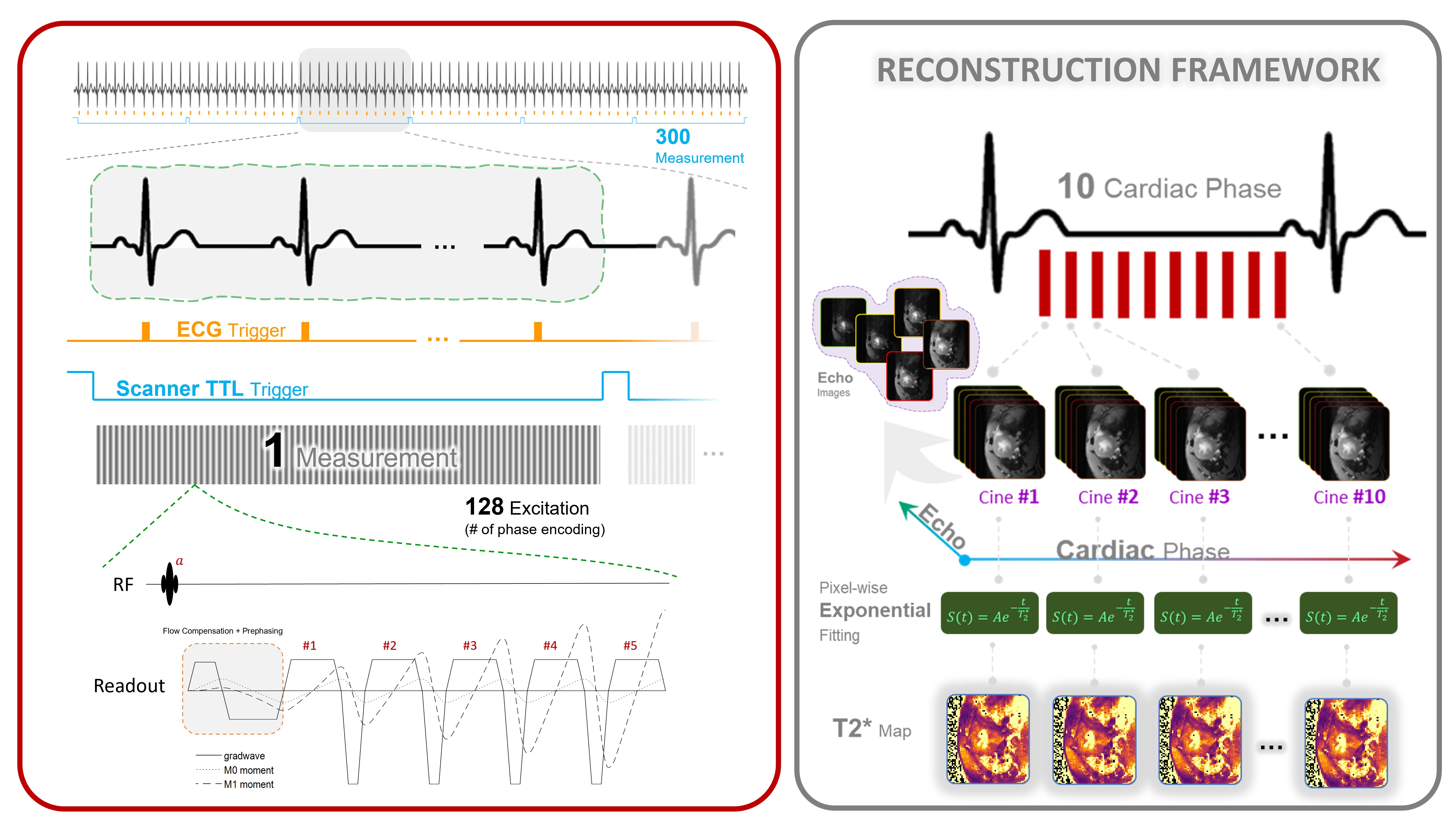
Figure 1. Data Acquisition and Reconstruction Workflow for T2*-mapping. GMN was added to MGE to suppress gradient first moment. This approach reduces blood flow artifacts and improves T2* estimation. MRI data are collected continuously along with the ECG and the scanner’s TTL trigger signal. The collected data are sorted retrospectively based on recorded signal. Each k-space line is stored in the corresponding cardiac phase and averaged to provide 10 cardiac-phases with 5 echoes each. T2* maps are calculated by pixel-wise using mono-exponential fitting of the T2* signal decay.
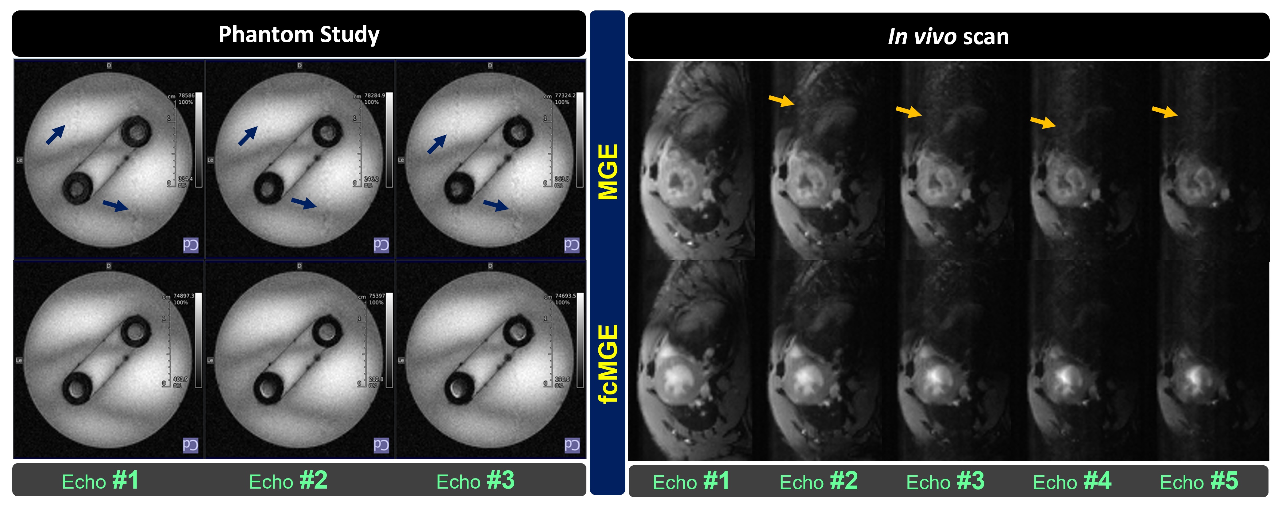
Figure 2. Effect of Flow-Compensation on echo images. Left: In conventional MGE, through-plane flow in an aquatic-solution phantom caused flow artifacts (shown by arrows) in the phase-encoding direction (the first 3 out of 6 echo images). The artifacts are removed with fcMGE. Right: Single-phase multi-echo cardiac images of the murine heart acquired with and without flow-Compensation. The flow-compensated images demonstrate artifact reduction and enhanced blood-myocardium contrast.
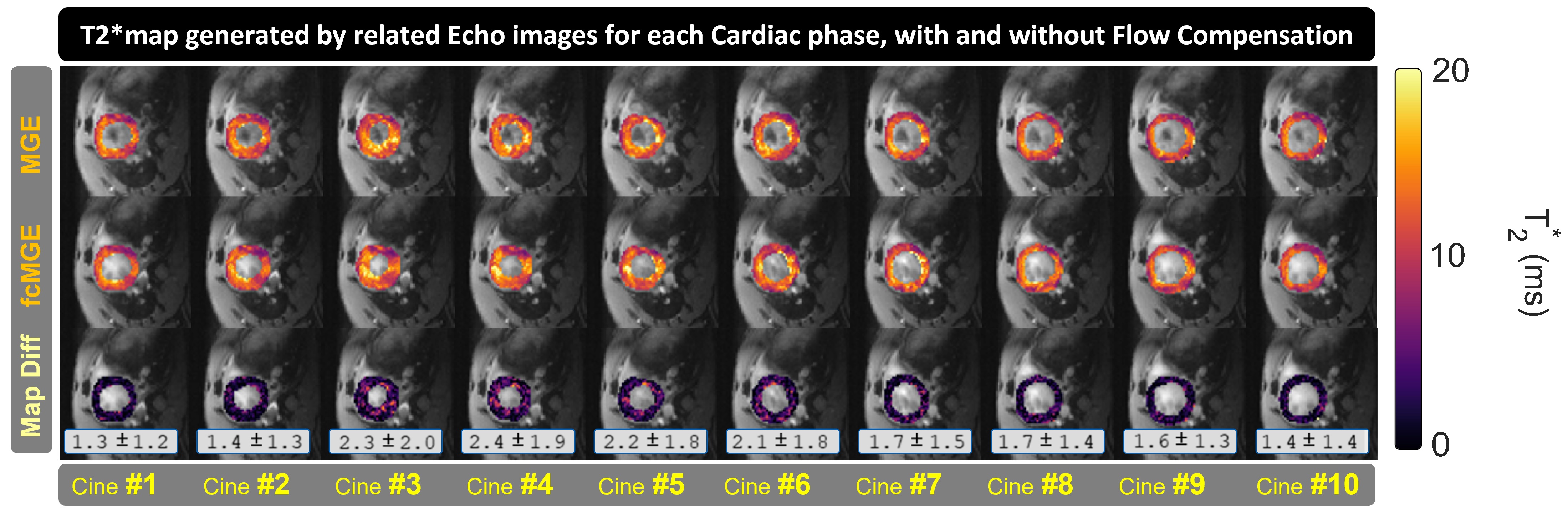
Figure 3. Cardiac motion resolved myocardial SAX-T2*-maps of the murine heart obtained w/o (top) and with (middle) flow-compensation. Color-coded T2* maps are overlaid on 2D anatomical images. The bottom row highlights the absolute T2* difference between MGE and fcMGE in terms of mean ± SD.
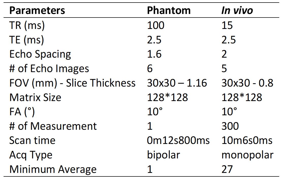
Table 1: Scan parameters used for the phantom and for the in vivo study.