4544
Single-Shot Propeller Turbo Spin Echo EPI with Model-Based Distortion Correction for Ultra Fast Abdominal T2 Mapping1Department of Radiology, Northwestern University, Chicago, IL, United States, 2Department of Radiology, Stanford University, Palo Alto, CA, United States
Synopsis
Keywords: Quantitative Imaging, Relaxometry, T2 Mapping, EPI, Distortion Correction, Liver
Motivation: T2 mapping is limited by slow acquisition speed and/or are susceptible to motion artifacts, this study aims to develop a ultra fast T2 mapping technique for abdominal imaging.
Goal(s): This study explores the possibility of using a single-shot propeller TSE EPI sequence for abdominal T2 mapping with model-based reconstruction technique for EPI distortion correction with a self-generated B0 map.
Approach: The distortion technique is tested both on the brain and abdominal contrast images. T2 mapping with distortion correction is validated in the abdomen.
Results: The proposed technique is able to generate distortion-corrected images, and provide accurate T2 maps of the liver.
Impact: This preliminary study showed the feasibility to provide distortion-corrected T2 maps from a single-shot TSE EPI sequence with a speed of <0.5 seconds/slice. The early results showed that this technique can significantly reduce distortion and provide accurate liver T2 maps.
Introduction
Spin-spin relaxation time (T2) mapping stands as a notable quantitative MRI approach that garners substantial clinical interest[1-3]. However, spin echo(SE)-based techniques face major road-block for clinical translation due to their extended acquisition time. To alleviate this issue, various strategies have been explored to expedite T2 mapping acquisition, including the adoption of a turbo spin echo (TSE) instead of the traditional SE, as well as the integration of the TSE with model-based reconstruction methods[4-5]. These techniques have made considerable reductions of the acquisition time for T2 mapping, but they still require multiple shots in order to collection sufficient data for reconstruction and are susceptible to motion artifacts.The proposed method leverages the efficient T2 relaxation curve sampling of TSE and the fast readout of EPI. With a model-based reconstruction method, this new technique can acquire T2 data in a rate of less than 0.5 seconds/slice.
Methods
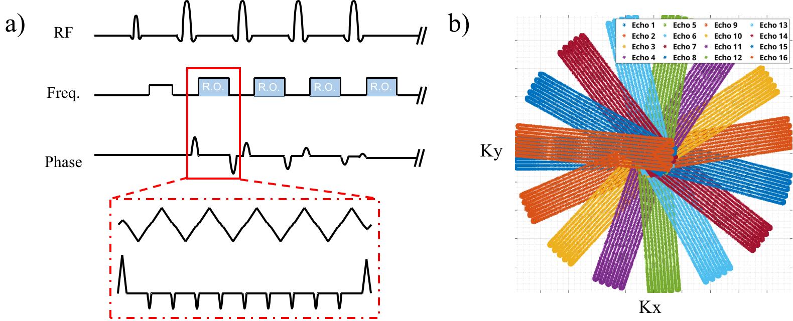
Distortion-corrected Single-Shot Propeller TSE EPI SequenceThe proposed technique uses a single-shot TSE sequence, the trajectory used is a rotated center portion of a 4-time undersampled EPI sequence. To further accelerate the acqusition, partial Fourier sampling was used in readout direction. The rotation of the EPI readout is carried out throughout the echo train length of the TSE sequence. To minimize the Eddy current artifacts on the resulting image, a tiny golden angle is used to rotate the EPI readout (Figure1). The pulse sequence was implemented in a GE 3.0T Scanner (GE Healthcare Milwaukee)
In the image reconstruction, each rotated TE image was first reconstructed with zero-filled kspace, using PICS method from BART[6], this low resolution image, labeled with it’s rotation angle information, is then passed through FSL’s topup[7] to generate a B0 map based on its degree of distortion. This B0 map is then used to correct the distortion in the original kspace data in a SENSE reconstruction with a structured low-rank regularization term.
Brain Experiment
A traditional single echo SE sequence with a rotated Cartesian trajectory was used to validate the distortion correction method. Partial Fourier in the phase-encoding direction was used. A total of 8 echoes were acquired.
Abdomen Experiment
The proposed sequence was used for abdominal T2 mapping. The parameter used are as follows: ETL=12, FOV=400mm, Slice thickness=5mm, base-resolution=330, receiver-bandwidth=976.6Hz/Pixel. A model-based reconstruction was used to correct for the distortion and T2 mapping.
Results
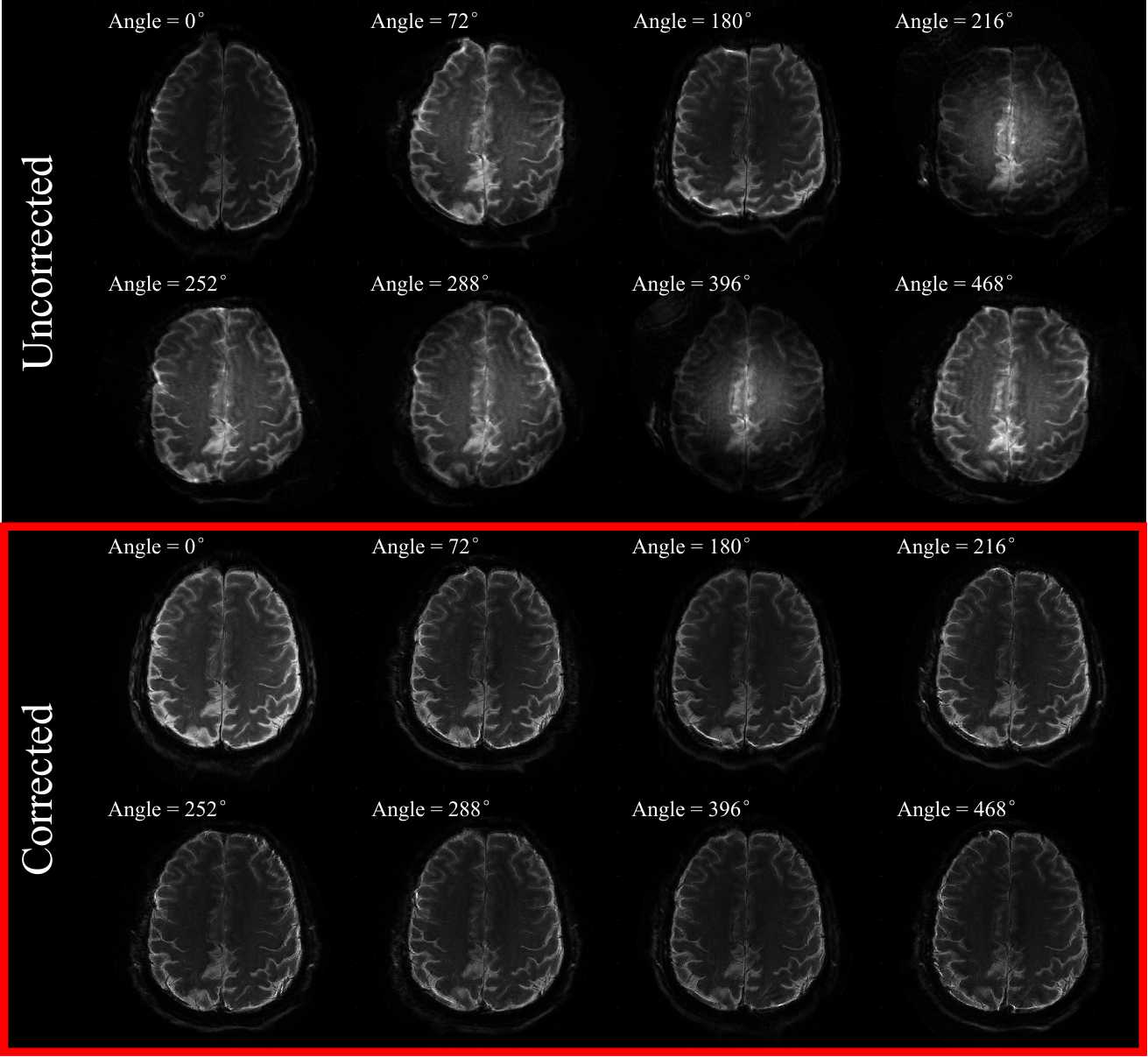
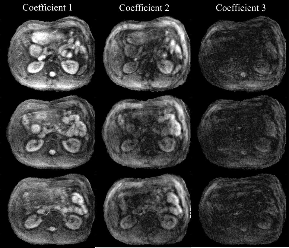
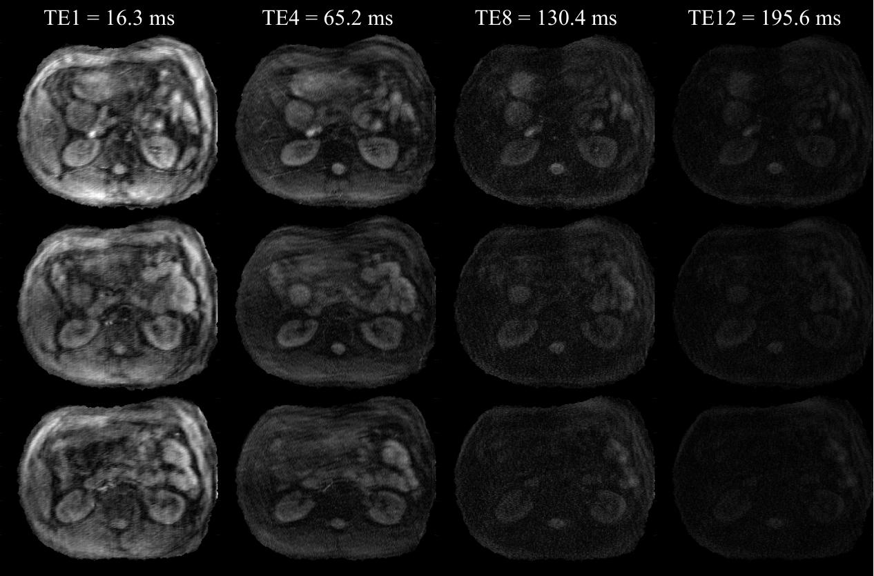
Figure2 shows the results from the brain experiment. The upper 2 rows shows a PICS reconstruction of the collected data, as can be seem the B0 inhomogeneity introduced large distortions, and this distortion changes as the trajectory rotates. The lower 2 rows shows the proposed model-based reconstruction method. When compared to the uncorrected data, the model-based reconstruction has corrected most of the distortion artifacts.For the abdomen experiment, an additional T2 model was added to the reconstruction on top of the distortion correction. Figure3 shows the reconstructed coefficient maps. We can then use the same T2 model to project the coefficient maps back to the image space, the 4 of the 12 resulting TE images are shown in Figure4.
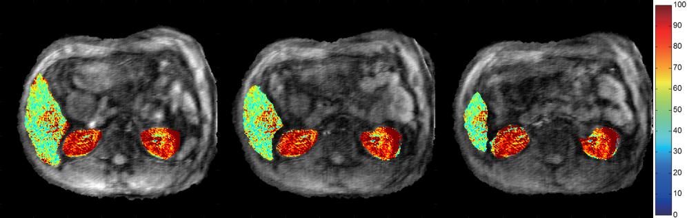
In Figure5, the T2 maps acquired from the TE image series are overlaied on top the first TE image. ROI analysis reveals the average T2 for the liver is 49.0±8.0ms, which is consistent with literature report[3-4].
Discussion
In this study we have demonstrated that the single-shot propeller TSE EPI can effectively remove EPI distortion artifacts, When coupled with a model-based reconstruction, this approach proves to be a promising tool for ultra-fast T2 mapping. Traditionally the model-based distortion correction rely on a blip-up and blip-down scans, essentially this acquires 2 phase-encoding angles[8-9]. In contrast, the propeller trajectory allows for the rapid acquisition of multiple phase encoding directions enhancing distortion correction efficiency. Additionally, the rotational nature of this trajectory results in a narrower point spread function (PSF), leading to sharper images.While our initial findings highlight some of the advantages of this novel technique, the full potential of the single-shot propeller TSE EPI sequence can be harnessed through further optimization of the sampling trajectory and refinements in the reconstruction process.
Acknowledgements
No acknowledgement found.References
[1] Deng J, Larson AC. Modified PROPELLER approach for T2-mapping of the abdomen. Magn Reson Med 2009;61:1269–1278.
[2] Kim D, Jensen JH, Wu EX, Sheth SS, Brittenham GM. Breathhold multiecho fast spin-echo pulse sequence for accurate R2 measurement in the heart and liver. Magn Reson Med 2009;62:300–306.
[3] Chung YW, Park MS, Kim MS, Kim E, Park J, Song HT, Choi JY, Kim MJ, Kim KW. Quantification of superparamagnetic iron oxide-mediated signal intensity change in patients with liver cirrhosis using T2 and T2* mapping: a preliminary report. J Magn Reson Imaging 2010; 31 (6):1379–1386.
[4] Huang C, Graff CG, Clarkson EW, Bilgin A, Altbach MI. T2 mapping from highly undersampled data by reconstruction of principal component coefficient maps using compressed sensing. Magnetic Resonance in Medicine. 2012;67(5):1355-1366.
[5] Huang C, Bilgin A, Barr T, Altbach MI. T2 relaxometry with indirect echo compensation from highly undersampled data. <i>Magnetic Resonance in Medicine</i>. 2013;70(4):1026-1037.
[6] https://mrirecon.github.io/bart/index.html
[7] Woolrich MW, Jbabdi S, Patenaude B, Chappell M, Makni S, Behrens T, Beckmann C, Jenkinson M, Smith SM. Bayesian analysis of neuroimaging data in FSL. NeuroImage, 2009;45:S173-86.
[8] Liao C, Bilgic B, Tian Q, Stockmann JP, Cao X, Fan Q, Lyer SS, Wang F, Ngamsombat C, Lo W, Manhard MK, Huang SY, Wald LL, Setsompop K, (2021). Distortion‐free, high‐isotropic‐resolution diffusion MRI with gSlider BUDA‐EPI and multicoil dynamic B0 shimming. Magnetic resonance in medicine, 86(2), 791-803.
[9] Liao C, Yarach U, Cao X, Lyer SS, Wang N, Kim TH, Tian Q, Bilgic B, Kerr AB, Setsompop K. High-fidelity mesoscale in-vivo diffusion MRI through gSlider-BUDA and circular EPI with S-LORAKS reconstruction. NeuroImage. 2023 Jul 15;275:120168.
Figures