4543
Targeting Extradomain-B Fibronectin to Monitor Immune Checkpoint Therapy with MRI in Head and Neck Squamous Cell Carcinoma1Biomedical Engineering, Case Western Reserve University, Cleveland, OH, United States, 2Biomedical Engineering, Case Western Reserver University, Cleveland, OH, United States
Synopsis
Keywords: Probes & Targets, Cancer
Motivation: Only 15-20% of patients with advanced head and neck squamous cell carcinoma (HNSCC) benefit from immunotherapy, with no reliable strategies to quickly predict or determine a patient’s response to treatment.
Goal(s): We aim to evaluate a therapeutic monitoring strategy using MRI by targeting extradomain B fibronectin (EDB-FN) in the tumor stroma.
Approach: Using mouse models bearing HNSCC allografts, we treated and monitored therapeutic efficacy of anti-PDL1 immunotherapy with MRI using MT218, our EDB-FN-targeted contrast agent.
Results: Treatment-responsive tumors exhibited unique expression patterns of EDB-FN compared to controls that were visualized with MT218 MRI, demonstrating the potential for predicting and monitoring HNSCC immunotherapy response.
Impact: We demonstrate that EDB-FN is a potential biomarker for immunotherapy response in HNSCC, and our MRI strategy targeting EDB-FN offers clinicians a potential method for predicting or monitoring immunotherapeutic outcomes to improve the clinical management of HNSCC.
Introduction
Upwards of 40% of patients with advanced head and neck squamous cell carcinoma (HNSCC) exhibit treatment failure. Immunotherapy has recently emerged as a therapeutic strategy to combat this, and immune checkpoint inhibition with PD1 and PDL1 inhibitors has achieved clinical success in HNSCC [2-5]. However, only 15-20% of patients with advanced HNSCC ultimately benefit from PD1-PDL1 blockade [6]. As such, identifying patients who have a higher likelihood of response to future treatment or are exhibiting a positive response to active treatment is of the utmost importance to improve the clinical management and treatment outcomes of HNSCC [7].We have identified an oncofetal form of fibronectin, extradomain-B fibronectin (EDB-FN), as a potential marker for monitoring the success of immune checkpoint inhibition in HNSCC. Fibronectin is a key constituent of the TGF-β pathway, making it an important regulator of TGF-β-mediated tumor growth and therapy resistance [8]. Fibronectin has also been implicated in damage-associated molecular patterns via TLR4 activation to initiate inflammatory responses, suggesting an important role in modulating the tumor response to immunotherapy [9]. We have previously developed a small peptide-targeted MRI contrast agent – ZD2-N3-Gd(HP-DO3A) [MT218] – that selectively binds to EDB-FN in the tumor extracellular matrix (ECM) and have demonstrated its ability to differentially enhance HNSCC based on EDB-FN expression levels (Figure 1) [10-13]. In this study, we demonstrate the ability of MT218 to monitor changes in EDB-FN in the tumor ECM with anti-PD-L1 immunotherapy in HNSCC.
Methods
Animal experiments were performed under an animal protocol approved by the Institutional Animal Care and Use Committee (IACUC) at Case Western Reserve University (CWRU, Cleveland, OH, USA). Mice were inoculated with MOC1 (responder) or MOC2 (non-responder) cells into the right flank. Once tumors reached 100 mm3, mice were randomly divided into control and anti-PD-L1 treatment groups (n=3 per treatment group) receiving three intraperitoneal injections per week for 4 weeks.MRI experiments were performed with a 3T MRS-3000 small animal system (MR Solutions, Surrey, UK). Mice were imaged before, during, and at the end of treatment. At each MRI time point, images were acquired before and at 10 and 20 minutes post-injection of the contrast agent MT218 (Molecular Theranostics, LLC, Cleveland, OH, USA) at a dose of 0.04 mmol/kg with a T1-weighted axial fast spin echo sequence with respiratory gating (TR = 305 ms, TE = 11 ms, FOV = 40 x 40 mm, slice thickness = 1 mm, number of slices = 15, Nav = 4, matrix = 256 x 248).
MR images were processed using FIJI software. After measuring signal values, the change in signal (δsignal), was calculated as follows for the tumors of each mouse at every time point: δsignal=Stime point - Spre-contrast.
For mouse experiments, all measurements at the different imaging and treatment time points were paired by mouse. When comparing two groups, a T-test was used. When comparing more than one variable, a Two-Way ANOVA with Fisher's Least Significant Difference test was used. All data is presented as mean ± standard error.
Results
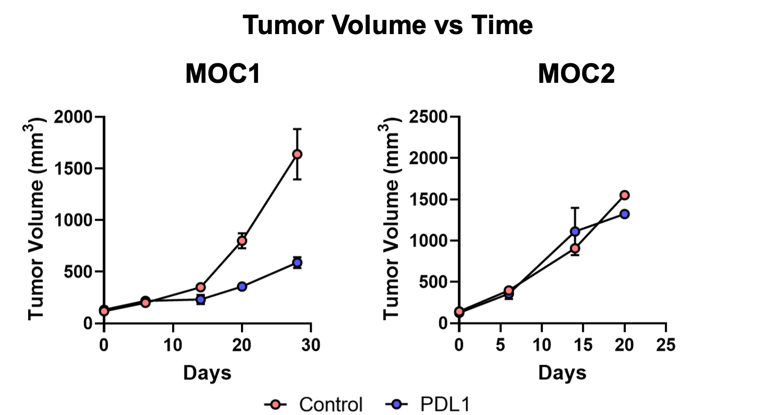
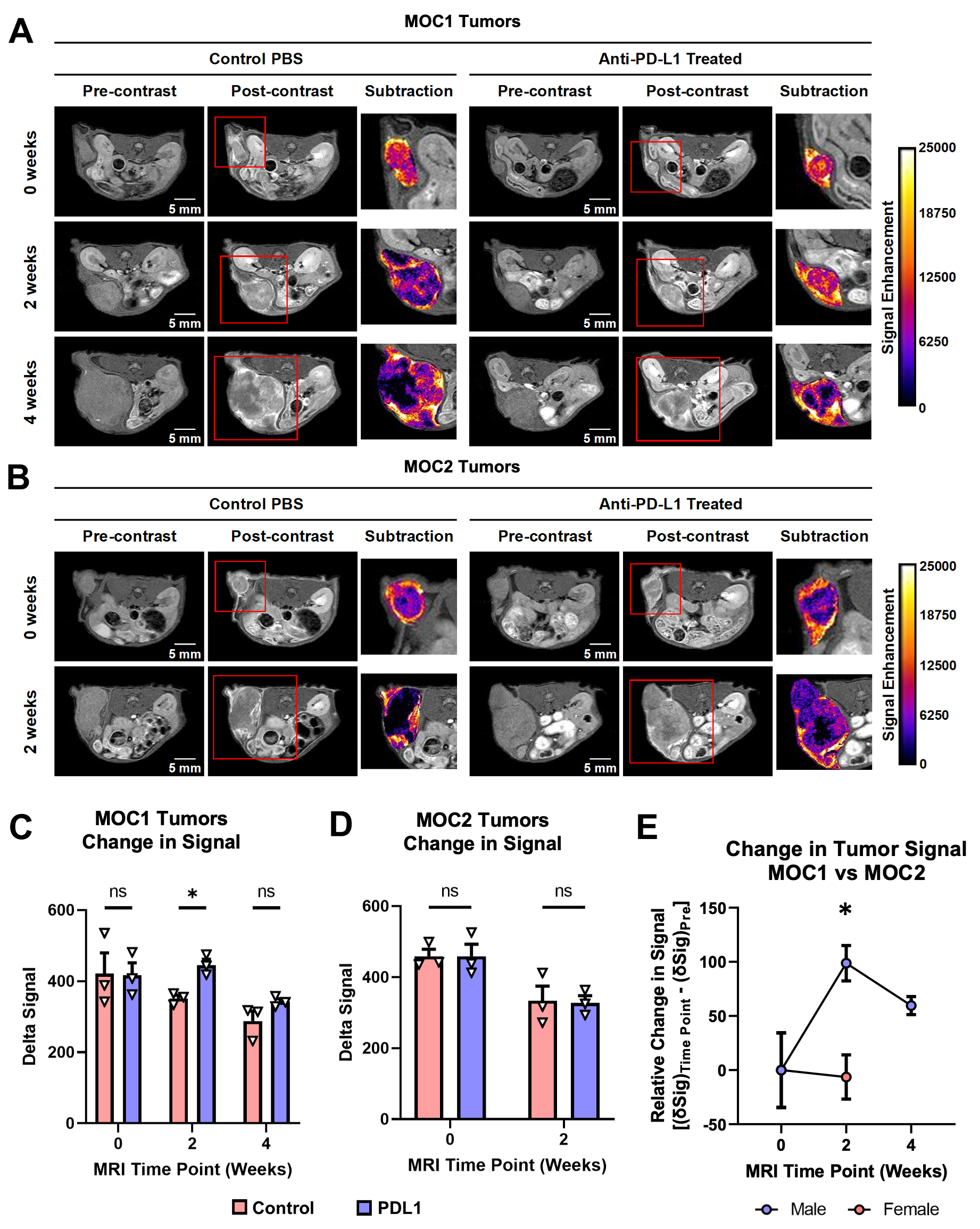
MOC1 tumor models are considered an “immune hot” tumor model, meaning MOC1 tumors respond to immune checkpoint inhibition. MOC2 tumor models, on the other hand, are considered “immune cold” tumors, meaning they do not respond to checkpoint inhibition [22, 23]. This was confirmed by tumor volume measurements, demonstrating that MOC1 tumors exhibited slower tumor growth with anti-PD-L1 treatment compared to controls, while MOC2 tumors saw no changes (Figure 2).Pre-treatment MRI of MOC1 tumors showed strong enhancement throughout the tumor. Signal remained strong throughout the tumors in the anti-PD-L1 treated mice at the 2-week time point, and was weaker in control tumors, demonstrating an observable difference in signal between the treatment groups (Figure 3A). This was confirmed by measuring the change in signal after contrast injection, with the anti-PD-L1 group showing significantly stronger enhancement at the 2-week time point than controls (Figure 3C). On the other hand, pre-treatment MRI of MOC2 tumors showed notably strong rim enhancement. Rim enhancement in the MOC2 tumors remained strong at the 2-week time point in both treatment groups, with little core enhancement (Figure 3B). There were no significant differences in signal enhancement between the treatment groups (Figure 3D). When comparing the MOC1 and MOC2 models, the change in signal at the 2-week time point is significantly higher in MOC1 tumors compared to MOC2 tumors (Figure 3E). These results suggest that targeting EDB-FN with MT218 can visualize changes in EDB-FN in the tumor during treatment, representing a potential strategy for monitoring the efficacy of immunotherapy in HNSCC.
Acknowledgements
This research was funded by NIH grants R01CA211762 and R44CA265626.References
[1] D. E. Johnson, B. Burtness, C. R. Leemans, V. W. Y. Lui, J. E. Bauman, and J. R. Grandis, "Head and neck squamous cell carcinoma," Nat Rev Dis Primers, vol. 6, no. 1, p. 92, Nov 26 2020.
[2] D. Pulte and H. Brenner, "Changes in survival in head and neck cancers in the late 20th and early 21st century: a period analysis," Oncologist, vol. 15, no. 9, pp. 994-1001, 2010.
[3] Z. Mei, J. Huang, B. Qiao, and A. K. Lam, "Immune checkpoint pathways in immunotherapy for head and neck squamous cell carcinoma," Int J Oral Sci, vol. 12, no. 1, p. 16, May 28 2020.
[4] A. D. Waldman, J. M. Fritz, and M. J. Lenardo, "A guide to cancer immunotherapy: from T cell basic science to clinical practice," Nat Rev Immunol, vol. 20, no. 11, pp. 651-668, Nov 2020.
[5] J. M. Bauml, C. Aggarwal, and R. B. Cohen, "Immunotherapy for head and neck cancer: where are we now and where are we going?," Ann Transl Med, vol. 7, no. Suppl 3, p. S75, Jul 2019.
[6] H. Shibata, S. Saito, and R. Uppaluri, "Immunotherapy for Head and Neck Cancer: A Paradigm Shift From Induction Chemotherapy to Neoadjuvant Immunotherapy," Front Oncol, vol. 11, p. 727433, 2021.
[7] S. Chen, Y. Yang, S. He, M. Lian, R. Wang, and J. Fang, "Review of biomarkers for response to immunotherapy in HNSCC microenvironment," Front Oncol, vol. 13, p. 1037884, 2023.
[8] S. Spada, A. Tocci, F. Di Modugno, and P. Nistico, "Fibronectin as a multiregulatory molecule crucial in tumor matrisome: from structural and functional features to clinical practice in oncology," J Exp Clin Cancer Res, vol. 40, no. 1, p. 102, Mar 17 2021.
[9] J. S. Roh and D. H. Sohn, "Damage-Associated Molecular Patterns in Inflammatory Diseases," Immune Netw, vol. 18, no. 4, p. e27, Aug 2018.
[10] Z. Han et al., "EDB Fibronectin Specific Peptide for Prostate Cancer Targeting," Bioconjug Chem, vol. 26, no. 5, pp. 830-8, May 20 2015.
[11] Z. Han et al., "Targeted Contrast Agent Specific to an Oncoprotein in Tumor Microenvironment with the Potential for Detection and Risk Stratification of Prostate Cancer with MRI," Bioconjug Chem, vol. 28, no. 4, pp. 1031-1040, Apr 19 2017.
[12] N. R. Ayat et al., "Optimization of ZD2 Peptide Targeted Gd(HP-DO3A) for Detection and Risk-Stratification of Prostate Cancer with MRI," ACS Med Chem Lett, vol. 9, no. 7, pp. 730-735, Jul 12 2018.
[13] R. C. Hall et al., "Preclinical Assessment of the Effectiveness of Magnetic Resonance Molecular Imaging of Extradomain-B Fibronectin for Detection and Characterization of Oral Cancer," Mol Imaging Biol, vol. 22, no. 6, pp. 1532-1542, Dec 2020.
Figures
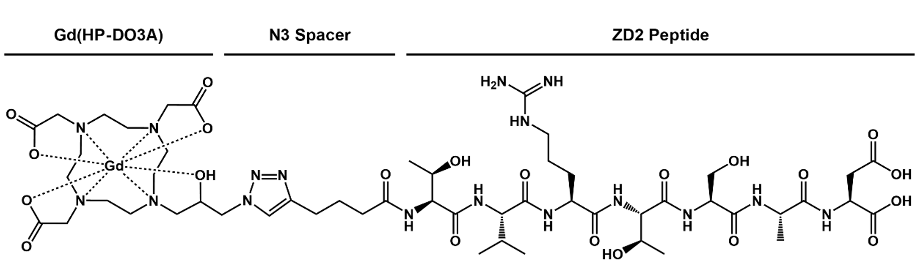
Figure 1. Chemical structure of MT218, our EDB-FN-targeted contrast agent.