4538
EPR imaging assessment of reduced tumor oxygenation induced by HIF-2a inhibitor Belzutifan in VHL-deficient renal cell carcinoma models1NCI, Bethesda, MD, United States, 2NINDS, Bethesda, MD, United States
Synopsis
Keywords: Preclinical Image Analysis, Electron Paramagnetic Resonance
Motivation: In the previous study, EPR oximetry unveiled physiological responses to a VEGF inhibitor, but the potential of EPR oximetry in upstream inhibition of the HIF pathway remained unclear.
Goal(s): To determine if Belzutifan, a HIF-2α inhibitor, affects tumor oxygenation as measured by EPR oximetry and to offer a non-invasive biomarker of its therapeutic efficacy.
Approach: Three xenograft models of clear cell carcinoma were treated with Belzutifan and changes in tumor oxygen levels were assessed using EPR oximetry.
Results: We observed significant alterations in tumor oxygenation exclusively in responding tumors, which we attributed to diminished VEGF expression.
Impact: This research underlines the significance of EPR oximetry as a non-invasive tool to detect early responses to HIF-2α inhibitors like Belzutifan, which could profoundly affect the management and therapeutic approach for cancer treatment, guiding personalized medicine.
Introduction
HIF is a transcription factor that helps regulate the body's response to hypoxia1. In von Hippel-Lindau (VHL) disease-associated renal cell carcinoma, mutations in the VHL gene lead to the stabilization and accumulation of HIF, even in normal oxygen conditions2. This abnormal HIF activity can contribute to the growth of blood vessels and promote VHL-nagative RCC tumor. Belzutifan, a novel HIF-2a inhibitor, is primarily used for the treatment of VHL-negative RCC3,4. Belzutifan specifically targets HIF-2α, one of the subunits of the HIF complex, reducing the production of proteins that drive tumor growth, nutrient uptake, and blood vessel formation, ultimately suppressing the progression of VHL-negative RCC. Belzutifan's mechanism of action may result in tumor shrinkage or the prevention of tumor growth, which can improve the clinical outcome for patients with VHL-negative RCC. In the earlier research, EPR oximetry revealed physiological reactions to a VEGF inhibitor5, but the effect of inhibiting the HIF pathway upstream was not investigated. Consequently, in this present study, our objective is to identify the early responses to Belzutifan by combining EPR oximetry with other physiological MRI imaging techniques.Method
In this study, we established xenograft models of clear cell renal cell carcinoma (RCC) in nude mice by implanting 5x10⁶ tumor cells (786-O, UOK220, and UOK121 cell lines) subcutaneously into the hindleg. The mice were divided into two groups: the Belzutifan treatment group and the control group. The treatment group received a daily dose of 3 mg/kg of Belzutifan, while the control group was administered a 1% DMSO/PBS solution over a 14-day period.To assess tissue oxygen levels (pO2), we utilized Electron Paramagnetic Resonance Imaging (EPRI). This involved administering the exogenous trityl radical probe, Ox071, via tail vein injection (1.125mmol/kg bolus). Signals were collected through free induction decay (FID) following radiofrequency excitation pulses, with data encoded in three dimensions (x, y, and z gradients) to create 3D spatial maps. T2* maps were generated by analyzing the FIDs, providing EPR line width maps, which were then used to estimate tissue pO2 on a pixel-wise basis. Imaging parameters included an 8.0 μs repetition time and 4000 averages.Dynamic Contrast-Enhanced MRI (DCE-MRI) was carried out using a 1 T scanner, capturing T1-weighted fast low-angle shot (FLASH) images. Imaging parameters consisted of TR = 117.2 milliseconds, TE = 6 milliseconds, flip angle = 30˚, 0.22 × 0.22 mm resolution, 15-second acquisition time per image, and 45 repetitions. Gadolinium-DTPA (4 mL/g of body weight, 50 mmol/L concentration) was injected via a tail vein cannula one minute into the dynamic FLASH sequence. Local Gd-DTPA concentrations were determined using T1 maps derived from six sets of Rapid Imaging with Refocused Echoes (RARE) images acquired with different TR values (320, 400, 600, 1,000, 2,000 and 3,000 ms) before the FLASH sequence.Both EPRI and DCE-MRI scans were performed at the start of the study (Day 0) and repeated on the fourth day of treatment (Day 4).Results and Discussion
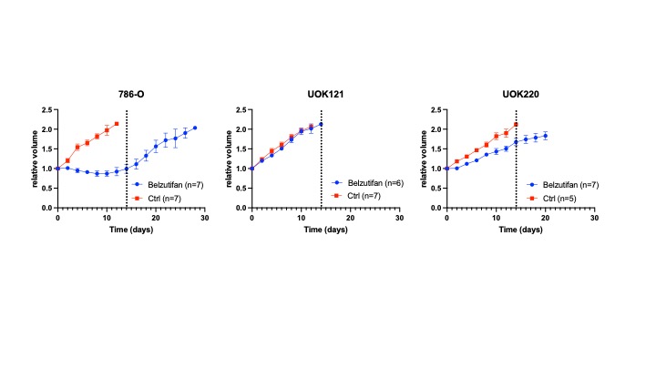
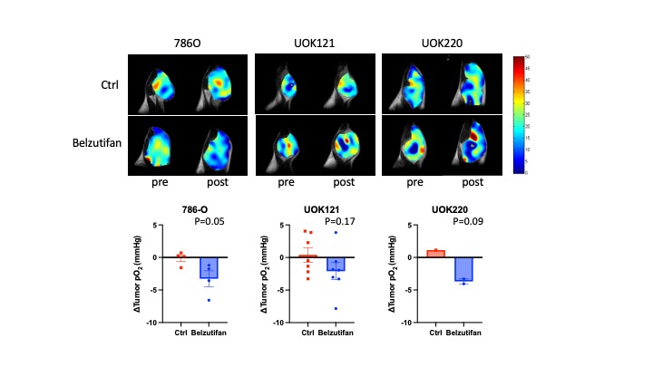
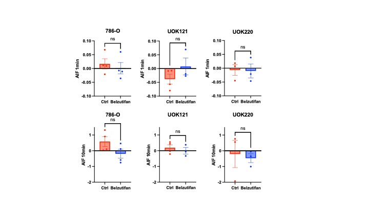
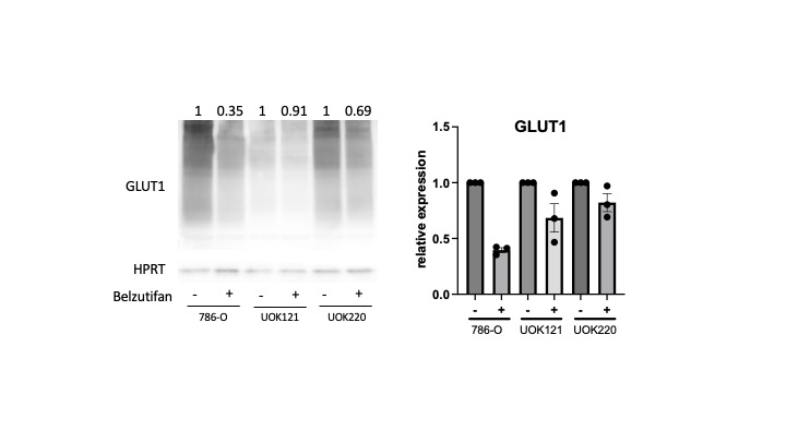
All three xenograft models exhibited varying responses to Belzutifan treatment. Among them, 786-O tumors displayed the most pronounced treatment effect, while UOK220 tumors showed a comparatively weaker response, and UOK121 tumors did not respond at all (Figure 1). Using these models, we assessed the reduction in pO2 after four days of treatment in the Belzutifan treatment group compared to the vehicle treatment group. (Figure 2) As expected, a significant difference was observed in 786-O tumors (Belzutifan group: -3.27 mmHg, vehicle group: -0.13 mmHg; p = 0.05), indicating a substantial impact. In contrast, no significant difference was detected in the UOK121 group (Belzutifan group: -2.11 mmHg, vehicle group: 0.41 mmHg; p = 0.17). However, the anti-angiogenic impact was not verified through perfusion and permeability evaluations conducted with DCE MRI. (Figure 3) The discrepancy was considered to be the result of limited sensitivity of DCE MRI in detecting the weak anti-angiogenic effect, and there may be other factors contributing to the suppression of tumor growth. We assessed the impact of Belzutifan on glucose uptake by analyzing Glut-1 expression through Western blotting. The results indicated reduced Glut-1 levels in both Belzutifan-sensitive 786-O tumors and Belzutifan-resistant UOK121 tumors. (Figure 4) This indicates that the observed tumor growth suppression effect cannot be solely attributed to reduced glucose uptake. Apart from its role in angiogenesis and nutrient uptake, the HIF-2 pathway is also associated with promoting tumor proliferation by upregulating cyclin D1 and genes related to stemness and anti-apoptotic properties. We plan to investigate the contribution of these mechanisms in our ongoing research.Acknowledgements
This research was supported by intramural funds from the Center for Cancer Research of the National Institutes of Health. The authors declare no conflicts of interest.References
- Ivanova IG, Park CV, Kenneth NS. Translating the Hypoxic Response-the Role of HIF Protein Translation in the Cellular Response to Low Oxygen. Cells. 2019 Feb 1;8(2):114.
- Hoefflin, R., Harlander, S., Schäfer, S. et al. HIF-1α and HIF-2α differently regulate tumour development and inflammation of clear cell renal cell carcinoma in mice. Nat Commun 11, 4111 (2020)
- Jonasch E, Donskov F, Iliopoulos O, Rathmell WK, Narayan VK, Maughan BL, Oudard S, Else T, Maranchie JK, Welsh SJ, Thamake S, Park EK, Perini RF, Linehan WM, Srinivasan R; MK-6482-004 Investigators. Belzutifan for Renal Cell Carcinoma in von Hippel-Lindau Disease. N Engl J Med. 2021 Nov 25;385(22):2036-2046.
- Toledo RA, Jimenez C, Armaiz-Pena G, Arenillas C, Capdevila J, Dahia PLM. Hypoxia-Inducible Factor 2 Alpha (HIF2α) Inhibitors: Targeting Genetically Driven Tumor Hypoxia. Endocr Rev. 2023 Mar 4;44(2):312-322.
- Matsumoto S, Saito K, Takakusagi Y, Matsuo M, Munasinghe JP, Morris HD, Lizak MJ, Merkle H, Yasukawa K, Devasahayam N, Suburamanian S, Mitchell JB, Krishna MC. In vivo imaging of tumor physiological, metabolic, and redox changes in response to the anti-angiogenic agent sunitinib: longitudinal assessment to identify transient vascular renormalization. Antioxid Redox Signal. 2014 Sep 10;21(8):1145-55.
Figures