4536
MRI Assessment of Sensitivity to Carbon Ion Irradiation in Rat C6 Glioma1Department of Radiology, Lanzhou University Second Hospital, Lanzhou, Gansu, China, 2Lanzhou University Second Hospital, Lanzhou, Gansu, China
Synopsis
Keywords: Tumors (Post-Treatment), Animals
Motivation: What is the specific efficacy of carbon ion radiation in the treatment of brain gliomas?
Goal(s): To assess the specific effect of carbon ion irradiation in inhibiting the growth and peritumor infiltration of gliomas.
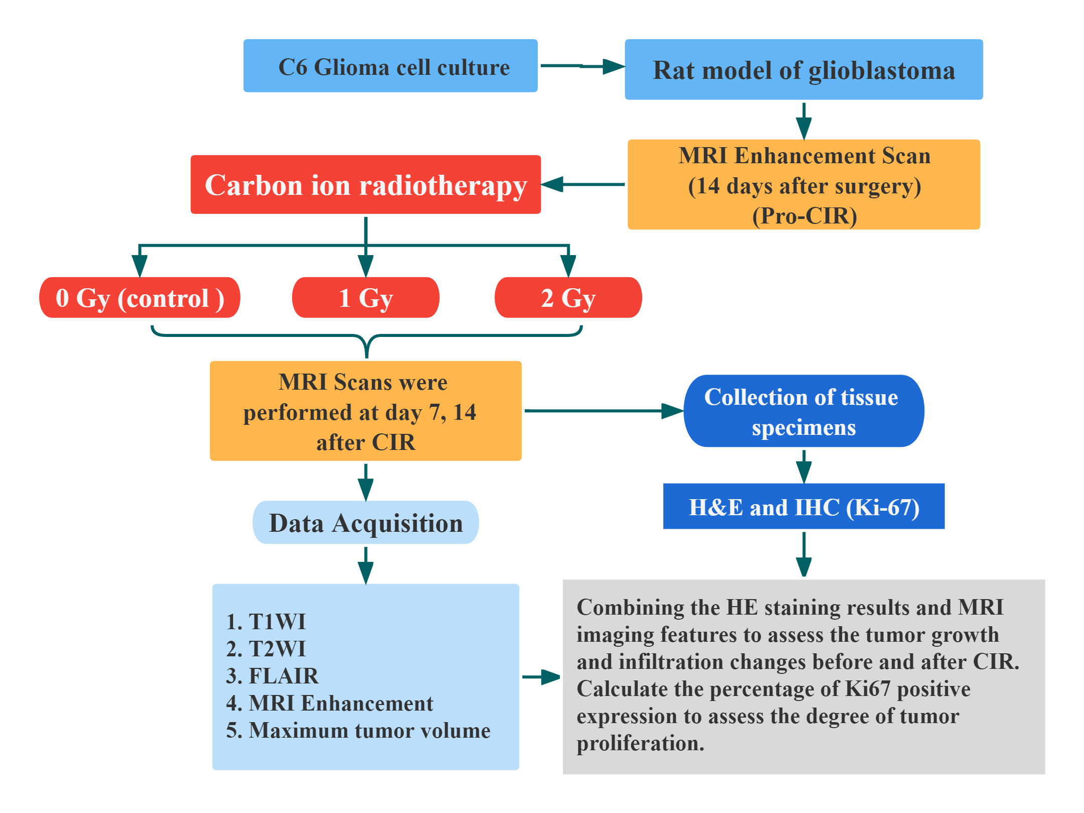
Approach: Establishment of a rat C6 brain glioma model with carbon ion irradiation and assessment of the efficacy response of brain glioma after carbon ion irradiation using MRI multiparametric imaging
Results: MRI multiparametric imaging can reflect the inhibitory effect of carbon ion irradiation on the growth and invasion of gliomas.
Impact: CIRT for glioma is still in the stage of clinical trials, the number of subjects is limited, and its specific efficacy still needs to be explored. Our study provides an experimental basis for the clinical application of CIRT for gliomas.
Purpose
Application of MRI multiparametric imaging to assess the response to carbon ion irradiation (CIR) in rat gliomas and to explore the specific efficacy of carbon ion radiotherapy (CIRT) on gliomas.Methods
42 rat C6 glioma models were established and 3 tumor-bearing rats were randomly selected as pathological control before CIR. The rest were irradiated with carbon ions (1 G y or 2 G y) and 15 rats served as controls (0 G y ). 6 were randomly selected from each group for survival analysis. MRI scans were performed before CIR and 7, 14 days after CIR. Quantitative comparisons of tumor image features, as well as tumor volume and animal survival were performed. After the terminal MRI scanning, brain tissues of tumor-bearing rats were collected and stained with HE staining and Ki-67 immunohistochemical staining.Results
Survival time of rats after CIR was significantly longer than that of the control group. The tumor volumes of the three groups still increased at 7d after CIR, but there was a statistical difference between the 0 G y and 2 G y groups. Patchy liquefied necrotic area can be seen in the center of the tumor, with T2WI high-signal edema around the peritumor, and the enhancement of the tumor is obvious on enhancement scan. 14d after CIR, the tumor volume in the 0 G y group further increased, the extent of peritumoral edema increased, the occupying effect was obvious, the tumor cells in the central necrotic area were regenerated and the enhancement scan showed a wreath shape. Tumor volume was significantly reduced in the 1 G y group and 2 G y group, and the tumor entity was reduced with cystic necrosis in the center, and the surrounding edematous area was significantly reduced and a patchy liquefied area was seen.Compared with the pre-irradiation period, the percentage of Ki-67 positive expression gradually decreased after carbon ion irradiation, indicating that carbon ion irradiation could effectively inhibit the proliferation of tumor cells.$$Conclusion
MRI multiparametric imaging could respond to the inhibitory effect of carbon ion irradiation on the growth and peritumor infiltration of rat C6 gliomas, and carbon ion irradiation significantly prolonged the survival time of tumor-bearing rats.Clinical Relevance
CIR has the advantages of inverted deep dose distribution and high relative biological effect, and can effectively treat malignant and recurrent brain tumors. CIRT for glioma is still in the stage of clinical trials, the number of subjects is limited, and its specific efficacy still needs to be explored. MRI can dynamically respond to the efficacy of the tumor after treatment. We applied MRI multiparametric imaging to evaluate the sensitivity of C6 gliomas in rats after CIR, which can provide an experimental basis for the clinical application of carbon ion radiation therapy for glioma.Acknowledgements
I am very thankful to my supervisor for his great support for my experiment and also to all the teachers and seniors in the lab for their guidance and help.References
No reference found.Figures