4530
Detection Efficiency of 18F-PSMA-1007 PET/CT and Multi-parameter MRI in Prostate Cancer and Its Correlation with Pathological Grade1Clinical medicine school of Ningxia Medical University, Yinchuan, China, 2Department of Radiology, The First Hospital Affiliated to Hainan Medical College, Haikou, China, 3Department of Radiology, General Hospital of Ningxia Medical University, Yinchuan, China, 4GE Healthcare MR Research, Beijing, China
Synopsis
Keywords: Preclinical Image Analysis, Tumor, prostate cancer
Motivation: The combination of MRI imaging and PSMA PET/CT imaging has attracted more and more attention.
Goal(s): Therefore, this study will compare the diagnostic value of 18F-PSMA-1007 PET/CT imaging and multi-parameter MRI alone and in combination with prostate cancer.
Approach: This study retrospectively collected the clinical, imaging and pathological data of 50 patients with prostate disease who underwent mp-MRI and 18F-PSMA-1007PET/CT examination in our hospital.
Results: We found that the combination of 18F-PSMA-1007 PET/CT and multi-parameter MRI can improve the diagnostic efficiency of prostate cancer. ADC value, SUVmax and SUVmax/ADC can distinguish between low-risk and medium-high-risk prostate cancer.
Impact: The diagnostic efficacy of combined multi-parameter MRI (ADC) and 18F-PSMAPET/CT (SUVmax) is better than that of ADC, SUVmax and SUVmax/ADC with higher sensitivity and specificity. The combination of these two imaging modalities can complement each other and improve the accuracy.
Introduction
Multi-parameter MRI including T2-weighted imaging (T2WI), diffusion-weighted imaging (DWI) and dynamic contrast-enhanced (DCE) MRI, has been introduced as a criterion for the diagnosis of primary prostate lesions and is helpful to guide prostate biopsies[1-3]. However, a significant number of prostate cancer patients are still missed diagnosis, and biopsies are still necessary[4]. Previous studies[5-6] have suggested that the combination of these two imaging modalities can provide complementary information and improve the overall diagnostic accuracy for prostate cancer. Therefore, we aim to explore the diagnostic value of two imaging methods for prostate cancer.Methods
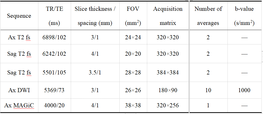
Retrospectively, we collected clinical, imaging, and pathological data from 50 patients (67.4 ± 8.0 years) with prostate diseases who underwent multi-parameter MRI and 18F-PSMA-1007 PET/CT examination in our hospital between April 2020 and September 2022. According to the pathological results, they were categorized into a prostate cancer group and a non-prostate cancer group, which included cases of benign prostatic hyperplasia and/or prostatitis.The examinations were performed on a 3.0T MR scanner (SIGNATM Architect, GE Healthcare, Milwaukee WI, USA) equipped with a 32-channel phased array surface coil and a Discovery VCT PET/CT scanner (GE Healthcare). MR scan protocols and the main scan parameters were listed in Table 1.
Two radiologists examined the pathological results, MRI images and PET images and reached agreement. On the AW4.7 workstation (GE Healthcare), the lesions were delineated layer by layer on the reconstructed ADC image, and the average ADC value was calculated. Similarly, on the workstation, a three-dimensional volume of interest (VOI) was delineated for prostate cancer, and the maximum standardized uptake value (SUVmax) within the VOI was measured. The Spearman correlation coefficient was used to assess the association between each parameter and the ISUP classification. The diagnostic performance of each parameter in detecting prostate cancer was evaluated using ROC curves, and the area under the curve (AUC) was compared among the parameters.
Results
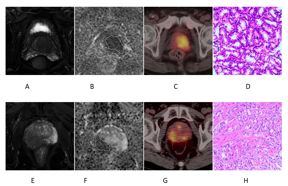
A total of 42 cases of prostate cancer were included in the study. Significant differences were observed in ADC, SUVmax, and SUVmax/ADC values between the high-grade and low-grade groups (P < 0.05). The ADC values were lower in the high-grade prostate cancer group compared to the low-grade group, while the SUVmax values were higher in the high-grade group. Additionally, the SUVmax/ADC values were higher in the high-grade group. Figure 1 illustrates the multi-parameter MRI and 18F-PSMAPET/CT images of both the high-grade and low-grade groups, along with the corresponding pathological maps.Correlation analysis revealed a negative correlation between ADC and SUVmax in the overall cohort of 50 prostate disease cases (r = 0.516, P < 0.05). Similarly, a negative correlation was observed between ADC and ISUP in the 42 diagnosed prostate cancer cases (r = 0.616, P < 0.05). On the other hand, SUVmax and SUVmax/ADC showed a positive correlation with ISUP (r = 0.549, r = 0.639, respectively; both P < 0.05).
The combined AUC of these two parameters was 0.976, indicating excellent diagnostic performance (P < 0.05). Using a critical value of 0.849, the sensitivity of diagnosing prostate cancer was 90.48%, with a specificity of 100%.
Discussion and Conclusion
The diagnostic performance of multi-parameter MRI (ADC) in detecting prostate cancer showed a sensitivity of 95.24% and a specificity of 87.50%, which aligns with the findings of Parathithasan[7]. The sensitivity and specificity of 18F-PSMA PET/CT in prostate cancer diagnosis were 83.30% and 100%, respectively. The AUC values were 0.97 for ADC and 0.94 for SUVmax. While 18F-PSMA PET/CT demonstrated higher specificity, its overall diagnostic efficiency was not superior to multi-parameter MRI, consistent with the results reported by Bodar[8]. Furthermore, the AUC, sensitivity, and specificity of SUVmax/ADC in prostate cancer diagnosis were 0.97, 90.50%, and 100%, respectively. These diagnostic measures were similar to those reported by Chinnappan[9], with AUC of 0.966, sensitivity of 87%, and specificity of 98%. However, the combined approach of multi-parameter MRI (ADC) and 18F-PSMA PET/CT (SUVmax) exhibited superior diagnostic efficacy compared to individual parameters. The combination demonstrated higher sensitivity and specificity, suggesting that the two imaging methods complement each other and enhance diagnostic accuracy.Acknowledgements
No acknowledgement found.References
[1] Stabile A, Giganti F, Rosenkrantz A B, et al. Multiparametric MRI for prostate cancer diagnosis: current status and future directions[J]. Nat Rev Urol, 2020, 17(1): 41-61.DOI: 10.1038/s41585-019-0212-4.[2] Kumar V, Bora G S, Kumar R, et al. Multiparametric (mp) MRI of prostate cancer[J]. Progress in Nuclear Magnetic Resonance Spectroscopy, 2018, 105: 23-40.DOI: 10.1016/j.pnmrs.2018.01.001.[3] Lee C H, Tan T W, Tan C H. Multiparametric MRI in Active Surveillance of Prostate Cancer: An Overview and a Practical Approach[J]. Korean Journal of Radiology, 2021, 22(7): 1087-1099.DOI: 10.3348/kjr.2020.1224.
[4] Bratan F, Niaf E, Melodelima C, et al. Influence of imaging and histological factors on prostate cancer detection and localisation on multiparametric MRI: a prospective study[J].Eur Radiol, 2013, 23(7): 2019-2029.DOI: 10.1007/s00330-013-2795-0.
[5] Sandgren K, Strandberg S N, Jonsson J H, et al. Histopathology-validated lesion detection rates of clinically significant prostate cancer with mpMRI, <SUP>68</SUP>Ga PSMA-11-PET and <SUP>11</SUP>C Acetate-PET[J]. Nuclear Medicine Communications, 2023, 44(11): 997-1004.[6] PriveRIVE B M, IsraelSRAEL B, SchilhamCHILHAM M G M, et al. Evaluating F-18-PSMA-1007-PET in primary prostate cancer and comparing it to multi-parametric MRI and histopathology[J]. Prostate Cancer and Prostatic Diseases, 2021, 24(2): 423-430.
[7] Parathithasan N, Perry E, Taubman K, et al. Combination of MRI prostate and 18F-DCFPyl PSMA PET/CT detects all clinically significant prostate cancers in treatment-naive patients: An international multicentre retrospective study[J]. J Med Imaging Radiat Oncol, 2022, 66(7): 927-935.DOI: 10.1111/1754-9485.13382.
[8] Bodar Y J L, Zwezerijnen B G J C, Van Der Voorn P J, et al. Prospective analysis of clinically significant prostate cancer detection with F-18 DCFPyL PET/MRI compared to multiparametric MRI: a comparison with the histopathology in the radical prostatectomy specimen, the ProStaPET study[J]. Eur J Nucl Med Mol Imaging, 2022, 49(5): 1731-1742.DOI: 10.1007/s00259-021-05604-9.
[9] Chinnappan S, Chandra P, Santa Kumar J, et al. SUVmax/ADC ratio as a molecular imaging biomarker for diagnosis of biopsy-naive primary prostate cancer[J].Indian J Nucl Med, 2021, 36(4): 377-384.DOI: 10.4103/ijnm.ijnm_62_21.
Figures