4529
Evaluation of Biochemically Recurrent Prostate Cancer Using the Prostate Imaging for Recurrence Reporting Systems1department of radiology, Sun Yat-Sen Memorial Hospital, Guangzhou, China, 2Sun Yat-Sen Memorial Hospital, Guangzhou, China, 3Philips Healthcare, Guangzhou, China
Synopsis
Keywords: Preclinical Image Analysis, Preclinical, Prostate Imaging for Recurrence Reporting; Prostate cancer; Recurrence; Interreader agreement.
Motivation: PI-RR assessment is not universally validated and has not yet been extensively studied for diagnostic accuracy and interreader variability.
Goal(s): To evaluate the performance of PI-RR for detecting local recurrence in PCa patients with BCR after radiation therapy (RT) or radical prostatectomy (RP).
Approach: 110 patients who underwent MRI and FDG PET/CT were included. A group of four radiologists with varying experience was recruited for recurrence evaluation.
Results: With a PI-RR of 3 as a cutoff, the AUCs ranged from 0.56 to 0.83 after RT and 0.73 to 0.90 after RP across the four readers. The ICC values were 0.54 and 0.68, respectively.
Impact: PI-RR may serve as a clinical guide to improve the management of recurrent PCa by achieving good diagnostic performance and interreader agreement.
Introduction
Multiparametric MRI (mpMRI) has been previously validated as a robust imaging modality for the detection of local recurrence in men after radiation therapy (RT) or radical prostatectomy (RP) with biochemical recurrence (BCR)[1, 2]. The Prostate Imaging for Recurrence Reporting (PI-RR) system, proposed by an international panel of experts in urology and radiology, was established for assessing local recurrence on mpMRI after whole-gland therapy. It aimed to reduce variation in MRI acquisition, interpretation and reporting to better guide therapy. In addition to improving routine clinical practice, PI-RR facilitated data collection for research and the monitoring of results[3]. However, PI-RR assessment is not universally validated and has not yet been extensively studied for diagnostic accuracy and interreader variability.Hence, the primary goal is to evaluate the diagnostic accuracy and interreader agreement of PI-RR for detecting local recurrences following RT or RP in patients with BCR among radiologists with different experience levels.Methods
A total of 110 patients with BCR after RT or RP who underwent multiparametric MRI [including multiplanar T2WI, echo-planar DWI with b values of 0, 800 or 1400 s/mm2, axial T1WI and dynamic contrast-enhanced (DCE) imaging] and FDG PET/CT between January 2017 and December 2021 were retrospectively included. BCR is defined as an undetectable PSA level following RP followed by two or more subsequent PSA levels exceeding 0.2 ng/ml. In the case of initial RT, BCR is defined as an increase of more than 2.0 ng/mL over the nadir in PSA levels following RT[4, 5].An independent group of four prostate radiologists with varying experience was recruited for the PI-RR assessment. The reference standard was defined by a panel of experts using clinical and imaging information. The study flowchart of patient enrollment and PI-RR assessment was shown in Figure 1. An evaluation of PI-RR performance for each reader was conducted by calculating sensitivity, specificity, positive predictive value (PPV), negative predictive value (NPV), and areas under the receiver operating characteristic curve (AUC). Interreader agreement was determined using the intraclass correlation coefficient (ICC).Results
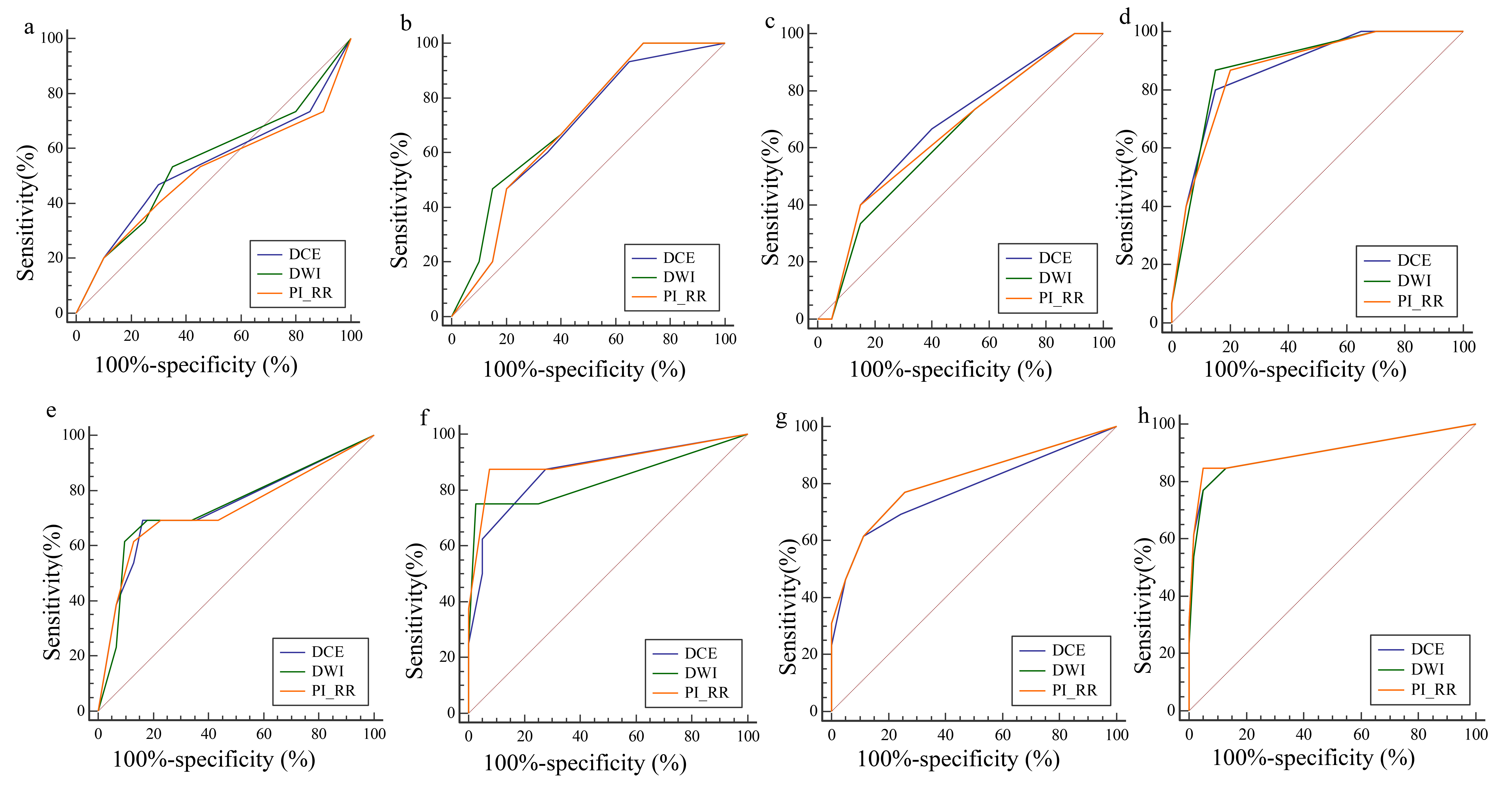
Thirty-five patients after RT and 75 patients after RP were included. With a PI-RR of 3 as a cutoff, the AUCs ranged from 0.56 to 0.83 after RT and 0.73 to 0.90 after RP across the four readers; with PI-RR 4 as the cutoff, the AUCs ranged from 0.61 to 0.84 after RT and 0.71 to 0.84 after RP. Figure 2 and Figure 3 show the ROCs of the four readers and PET/CT for the RT and RP groups, respectively.Interreader agreement using the ICC values was 0.54 (95% CI: 0.38 - 0.70) for the RT group and 0.68 (95% CI: 0.58 - 0.76) for the RP group. An example of disagreement among the four readers is shown in Figure 4. As shown in Figure 5, the per-reader AUCs for each MRI sequence and the overall PI-RRs are presented for both groups of patients.Discussion
Thirty-five patients after RT and 75 patients after RP were included. With a PI-RR of 3 as a cutoff, the AUCs ranged from 0.56 to 0.83 after RT and 0.73 to 0.90 after RP across the four readers; with PI-RR 4 as the cutoff, the AUCs ranged from 0.61 to 0.84 after RT and 0.71 to 0.84 after RP. Figure 2 and Figure 3 show the ROCs of the four readers and PET/CT for the RT and RP groups, respectively.Interreader agreement using the ICC values was 0.54 (95% CI: 0.38 - 0.70) for the RT group and 0.68 (95% CI: 0.58 - 0.76) for the RP group. An example of disagreement among the four readers is shown in Figure 4. As shown in Figure 5, the per-reader AUCs for each MRI sequence and the overall PI-RRs are presented for both groups of patients.Conclusion
PI-RR had high diagnostic accuracy and interreader agreement for recurrence assessment and may provide clinical guidelines to improve the management of men with recurrent prostate cancer by improving diagnostic performance and tailoring treatments based on the individual needs of the patient. Reader experience continues to impact the performance of prostate MRI interpretation using PI-RR.Acknowledgements
No acknowledgement found.References
1. Lindenberg L, Mena E, Turkbey B, Shih JH, Reese SE, Harmon SA, et al. Evaluating Biochemically Recurrent Prostate Cancer: Histologic Validation of (18)F-DCFPyL PET/CT with Comparison to Multiparametric MRI. Radiology. 2020,296(3):564-72.
2. Metser U, Chua S, Ho B, Punwani S, Johnston E, Pouliot F, et al. The Contribution of Multiparametric Pelvic and Whole-Body MRI to Interpretation of (18)F-Fluoromethylcholine or (68)Ga-HBED-CC PSMA-11 PET/CT in Patients with Biochemical Failure After Radical Prostatectomy. J Nucl Med. 2019,60(9):1253-8.
3. Panebianco V, Villeirs G, Weinreb JC, Turkbey BI, Margolis DJ, Richenberg J, et al. Prostate Magnetic Resonance Imaging for Local Recurrence Reporting (PI-RR): International Consensus -based Guidelines on Multiparametric Magnetic Resonance Imaging for Prostate Cancer Recurrence after Radiation Therapy and Radical Prostatectomy. Eur Urol Oncol. 2021,4(6):868-76.
4. Tanaka T, Yang M, Froemming AT, Bryce AH, Inai R, Kanazawa S, et al. Current Imaging Techniques for and Imaging Spectrum of Prostate Cancer Recurrence and Metastasis: A Pictorial Review. Radiographics. 2020,40(3):709-26.
5. Schaeffer E, Srinivas S, Antonarakis ES, Armstrong AJ, Bekelman JE, Cheng H, et al. NCCN Guidelines Insights: Prostate Cancer, Version 1.2021. J Natl Compr Canc Netw. 2021,19(2):134-43.6. Babaian RJ, Troncoso P, Bhadkamkar VA, Johnston DA. Analysis of clinicopathologic factors predicting outcome after radical prostatectomy. Cancer. 2001,91(8):1414-22.
Figures
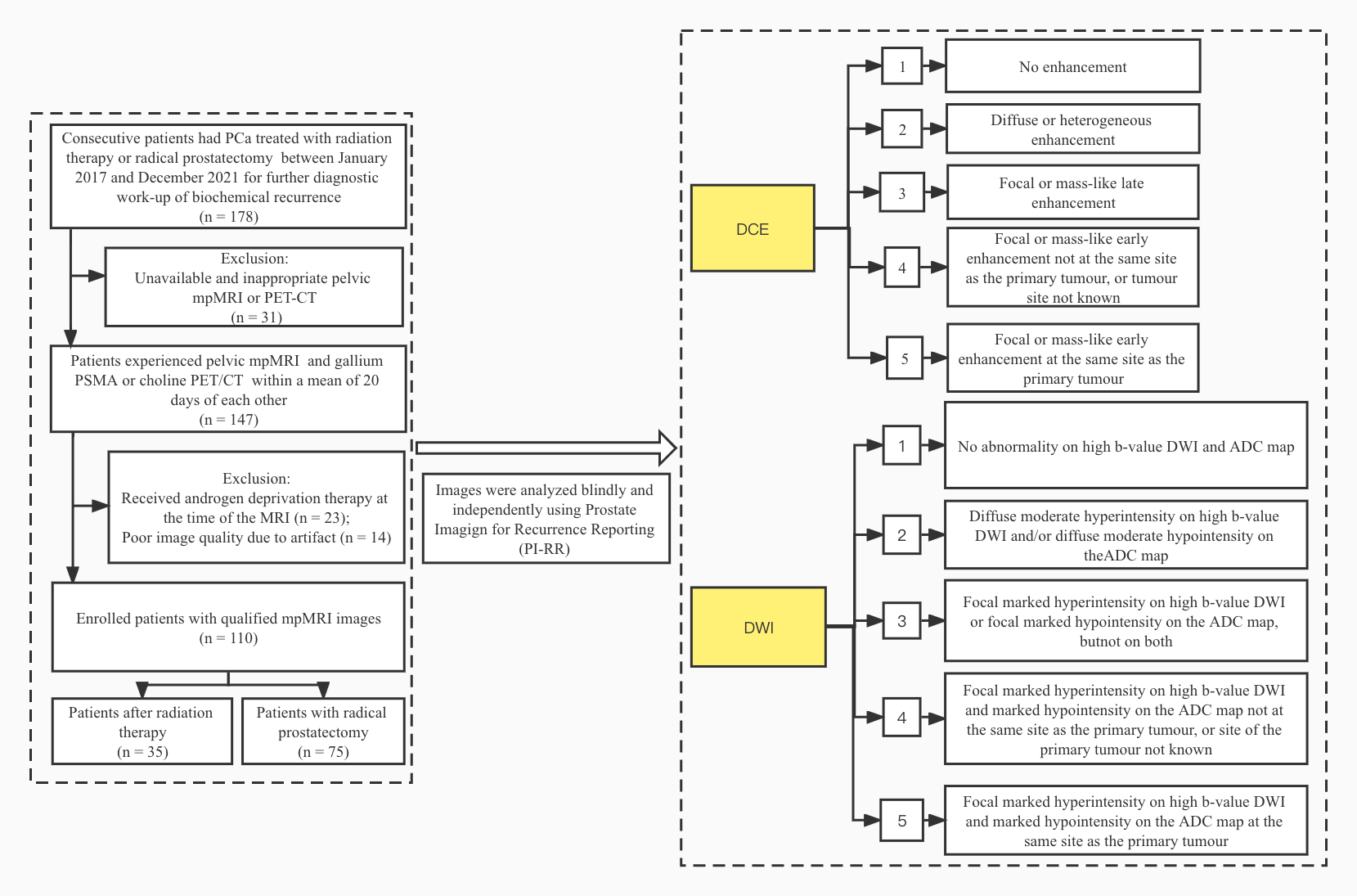
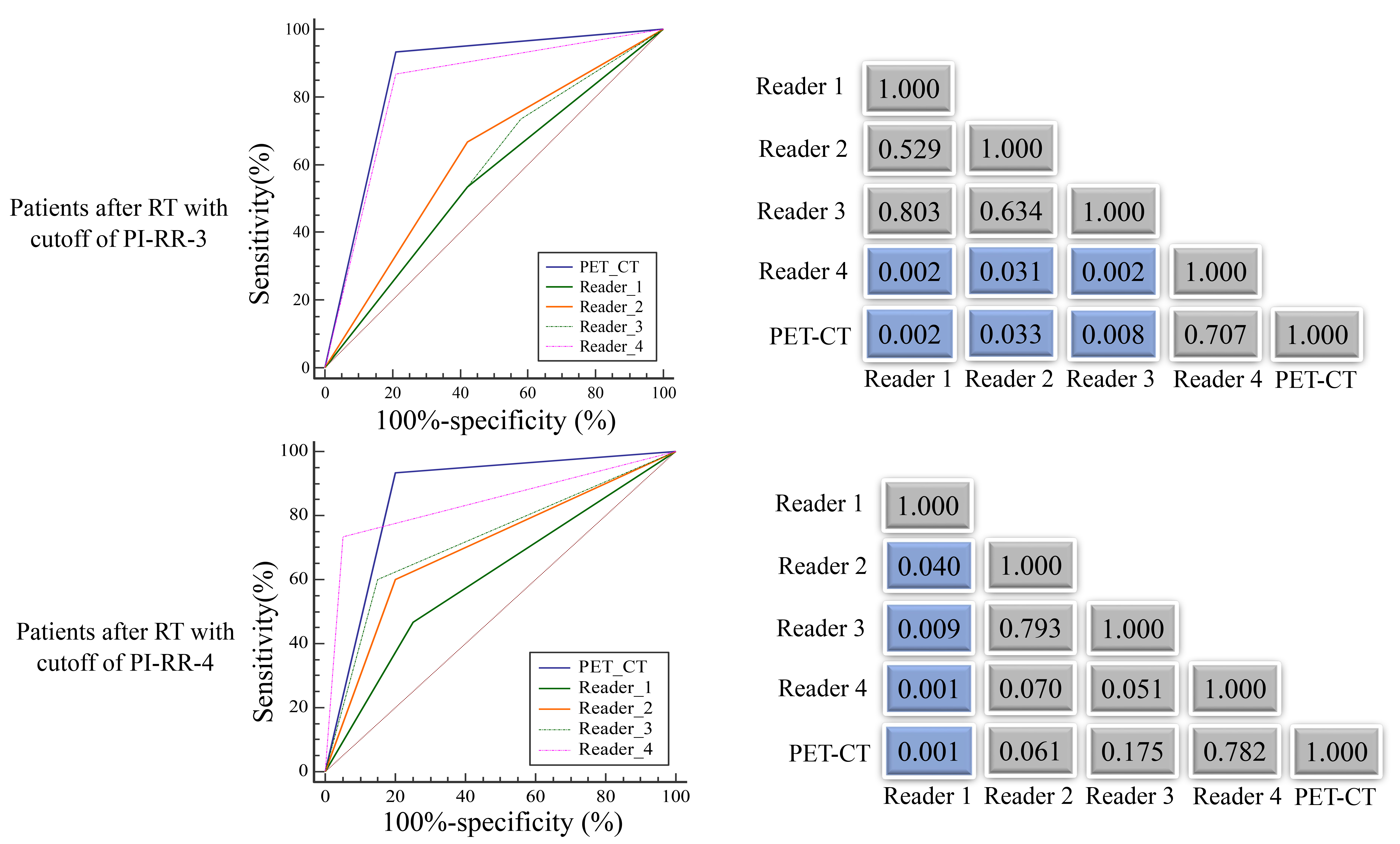
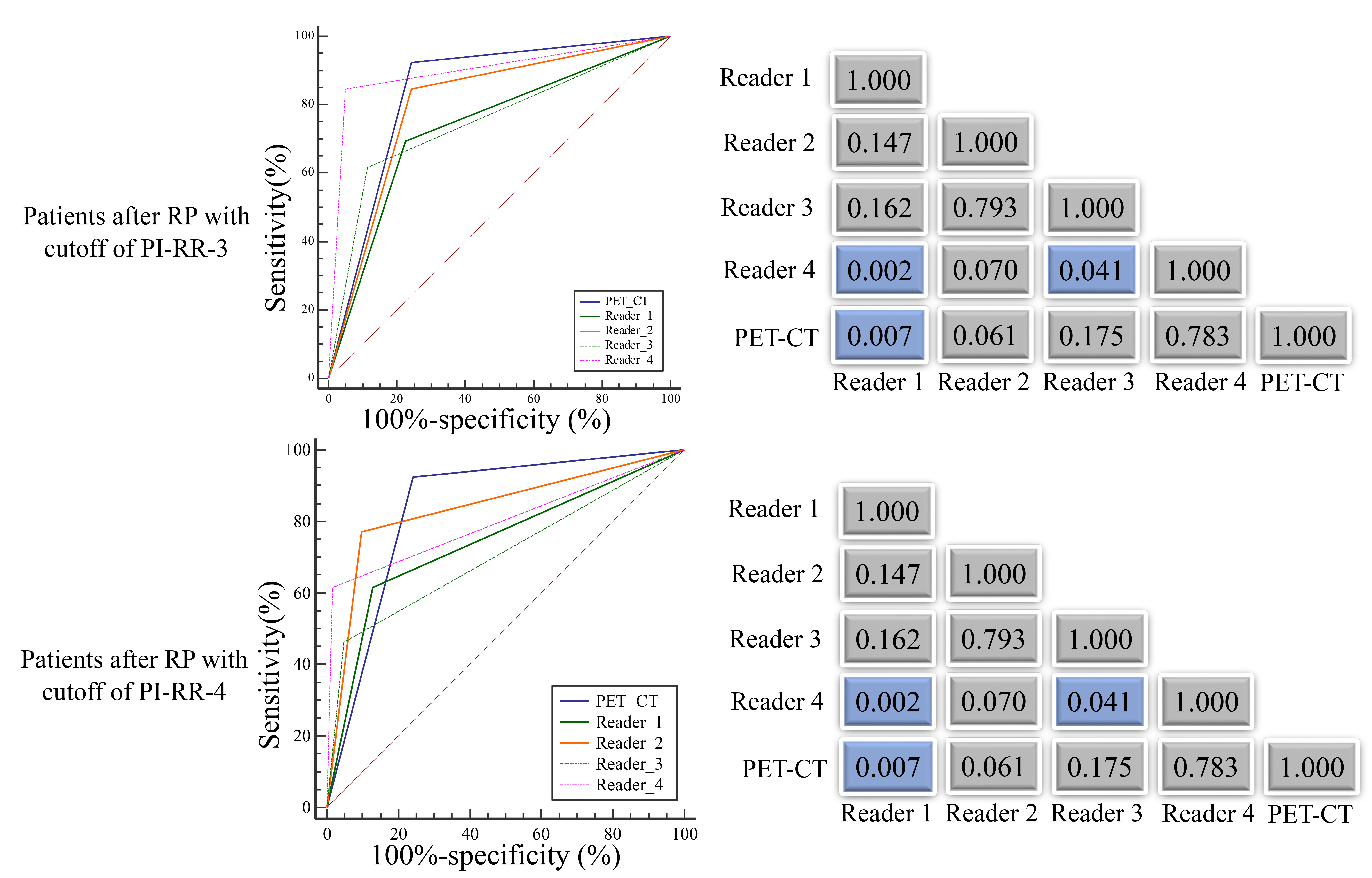
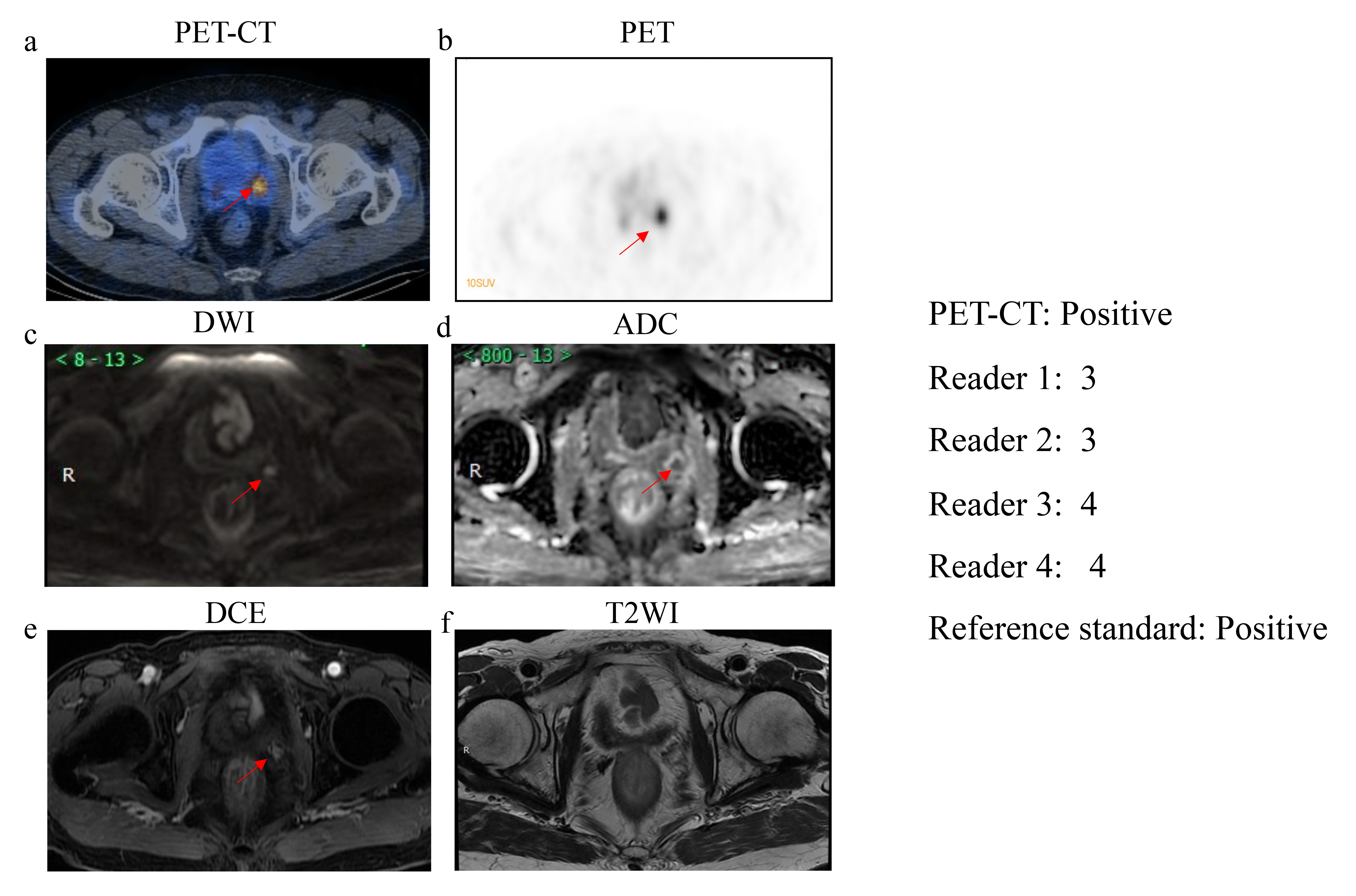
Fig 4. MRI scans in a 68-year-old man with a history of prostate cancer and prior radical prostatectomy, now with biochemical recurrence.
(a) FGD PET/CT: positive. (b) PET: positive. (c) DWI image shows hyperintensity. (d) Apparent diffusion coefficient map shows hypo-intensity. (e) Early-phase DCE image shows focal enhancement. (f) Axial T2WI image shows no abnormal signal intensity compared with the background.