4525
Brain Tumor Enhancing in Pre-contrast MRI Based on Deep Learning1Institute of Science and Technology for Brain-Inspired Intelligence, Fudan University, Shanghai, China, 2Department of Medical Imaging, Henan Provincial People's Hospital, Zhengzhou, China, 3Philips Healthcare, Shanghai, China, 4Human Phenome Institute, Fudan University, Shanghai, China
Synopsis
Keywords: Diagnosis/Prediction, Cancer
Motivation: The project was motivated by the need to reduce reliance on contrast agents in MRI for brain tumor assessment, addressing issues of cost and patient discomfort associated with their use.
Goal(s): To improve the visibility and recognition of tumors in T1-weighted MRI scans, eliminating the need for contrast agents.
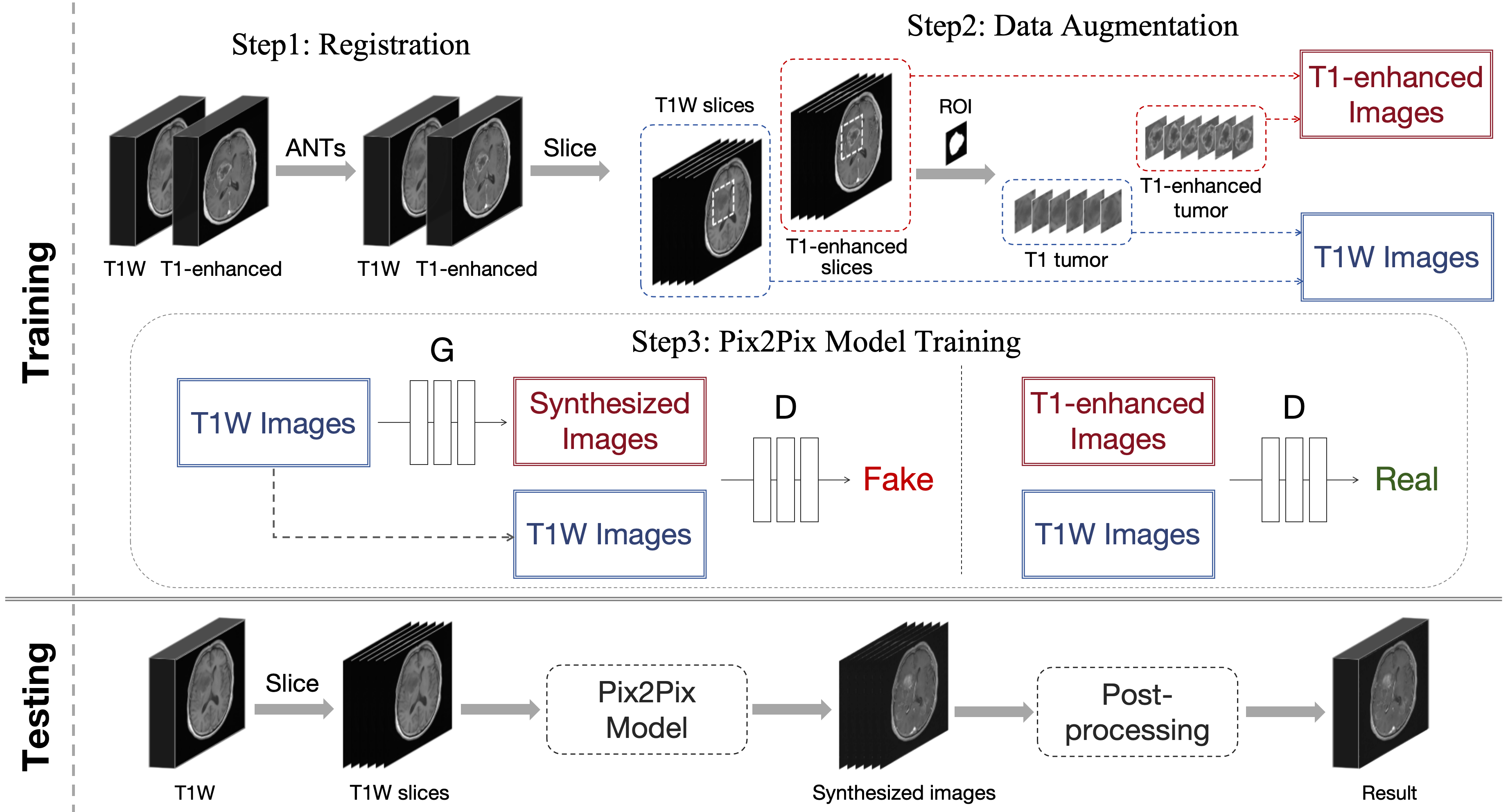
Approach: The approach involved registration, data augmentation, Pix2Pix-based image generation, and post-processing.
Results: The results demonstrated that the method significantly improved tumor discernibility in T1-weighted MRI scans, offering a valuable diagnostic tool without the need for contrast agents.
Impact: Our research provides an effective solution to enhance brain tumor visualization in MRI, directly benefiting patients and healthcare providers. By eliminating the need for contrast agents, our approach reduces costs, discomfort, and potential risks, advancing brain tumor diagnostics.
INTRODUCTION
Brain tumors are one of the deadliest cancers, threatening human health1. Clinical diagnosis of brain tumors usually relies on contrast-enhanced MRI, which can pose issues with scanning costs, patient discomfort, and other potential risks2. In response, our investigation utilizes the deep learning technique to heighten the visibility of tumors in T1-weighted MRI scans, enhancing the diagnostic value of tumors. This research endeavor introduces a cost-effective, patient-oriented, and accessible modality for tumor visualization.METHODS
Our study involved a dataset comprising paired T1-weighted and T1-enhanced from 402 patients with glioma. The dataset was randomly split into a training set (322 cases), a validation set (24 cases) and a test set (56 cases). The contours of the tumor were delineated on CEMRI by experienced radiologists.The proposed framework is illustrated in Figure 1, comprising the following salient components:
Registration: we first used the SyN registration algorithm3 to ensure correct alignment between T1-weighted and T1-enhanced MRI images, which is a symmetric image normalization method used to maximize cross-correlation within the differential isomorphism mapping space to achieve registration.
Data Augmentation: a data augmentation was executed, targeting the region of the tumor and the peritumoral area. The aim was to increase the proportion of tumor positive samples, thereby enhancing the model's focus on tumor regions.
Image synthesis: To synthesize T1-enhanced images, we utilized the Pix2Pix model4, which is a conditional GAN-based method that exhibited superior performance in multiple image-to-image translation tasks. This step involved mapping the original T1-weighted images to synthetic T1-enhanced images, emulating the appearance of real T1-enhanced images.
During the testing, T1-weighted images were fed into the trained Pix2Pix model to obtain synthetic T1-enhanced images. Afterwards, we perform post-processing to adjust the window width and window level of the synthesized image to be consistent with the T1-enhanced images in the training set. For performance evaluation, the quality of synthetic T1-enhanced images was assessed using the Peak Signal-to-Noise Ratio (PSNR) and Structural Similarity Index (SSIM).
RESULTS
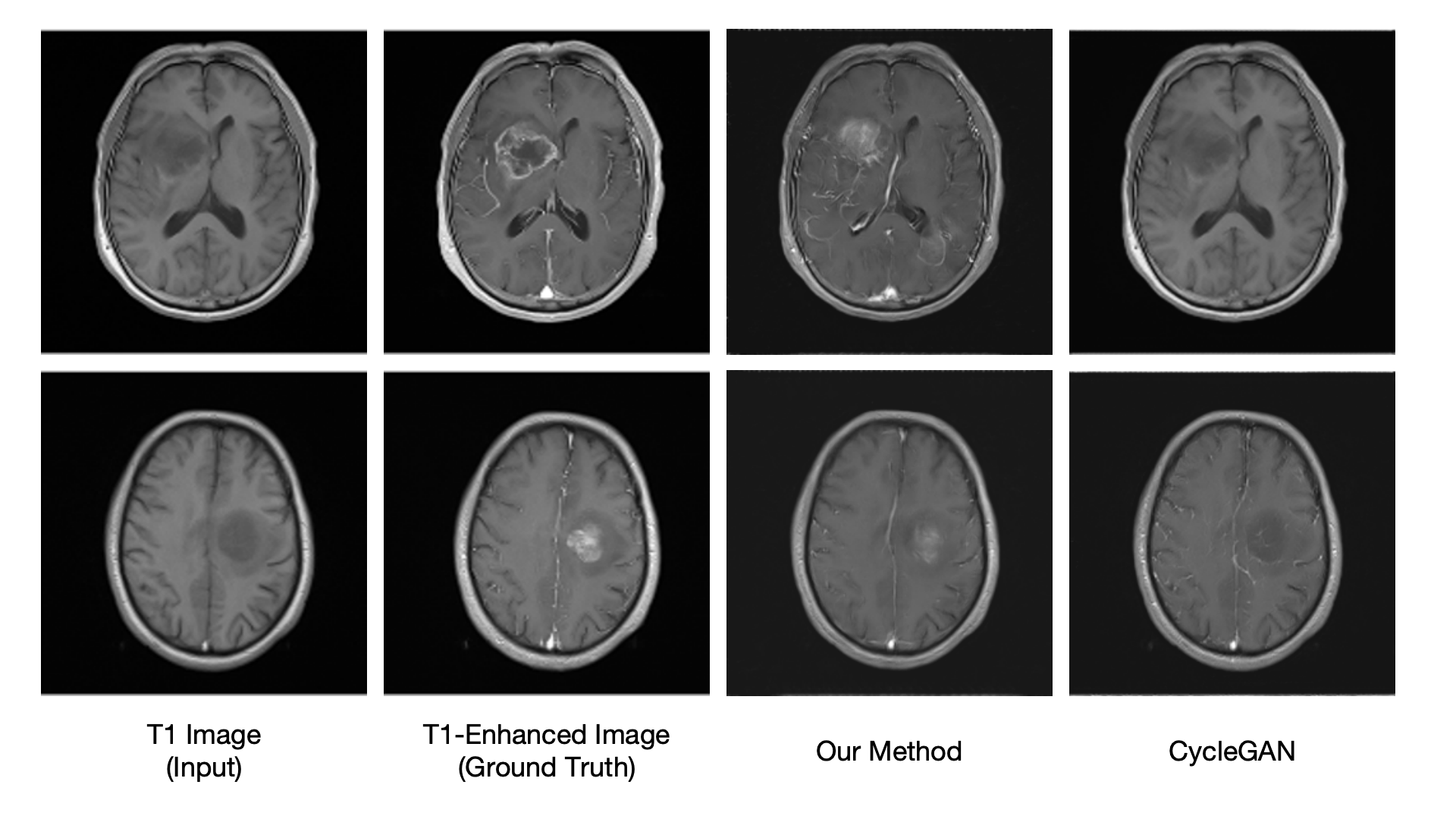
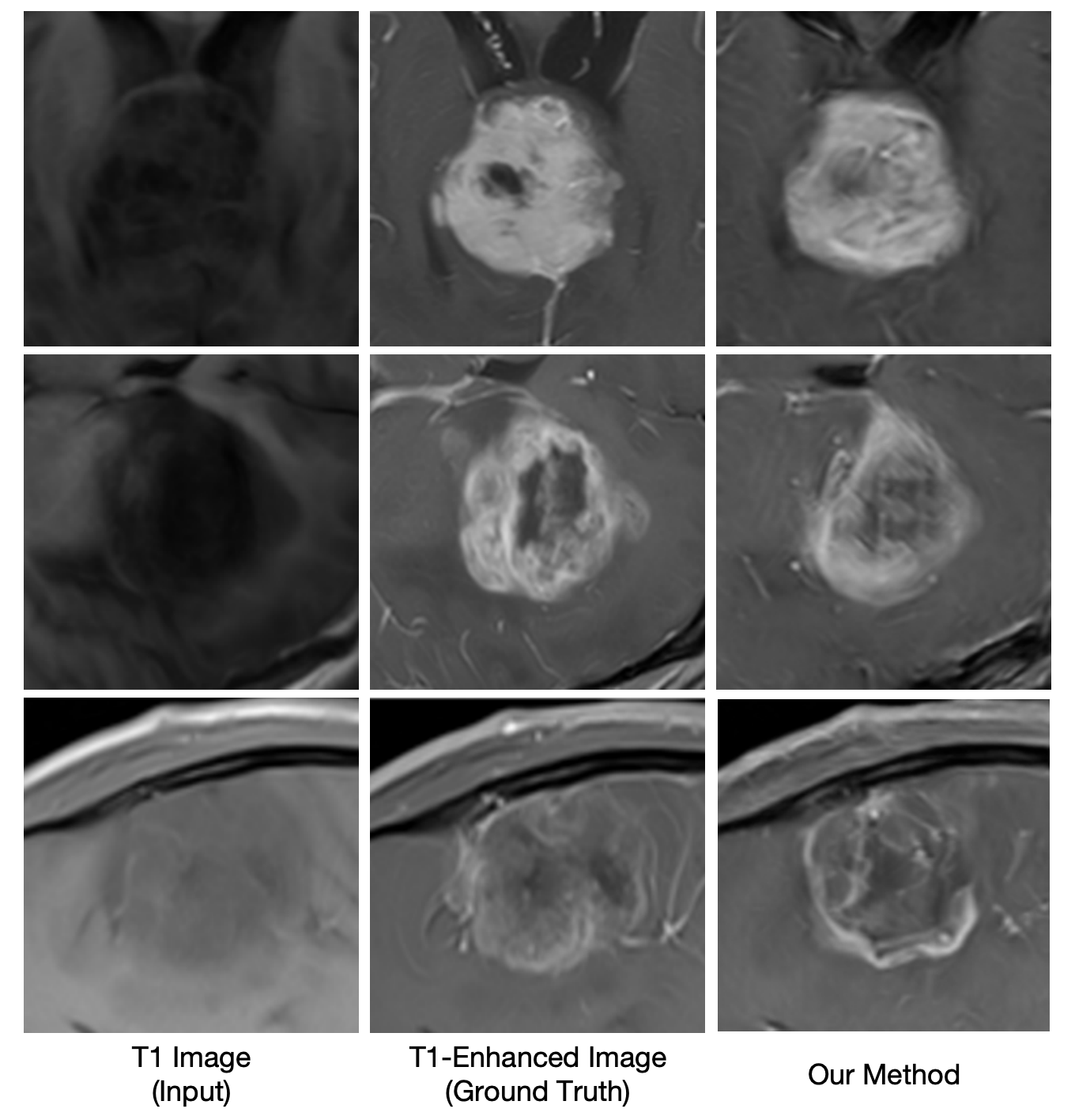
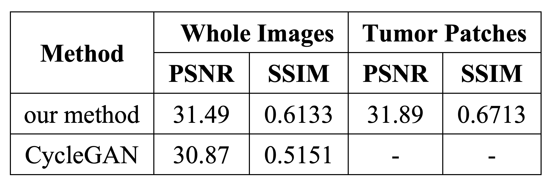
The proposed model achieved a PSNR of 31.49 and a SSIM of 0.6133 in the test set, which significantly outperformed the CycleGAN model5, indicating higher quality image generation. Specific performance metrics are listed in Table 1. In particular, we calculated the quality of the synthetic images in the tumor area, and the PSNR reached 31.89 and the SSIM reached 0.6713, which has better quality than the whole images. This shows that our model has a better synthesis effect on the tumor area and is more suitable for clinical needs.As depicted in Figure 2, the proposed model exhibits superior discernibility in tumor regions compared to T1-weighted images. This improvement in tumor visibility, without the requirement for contrast agents, offers valuable diagnostic support to clinicians. Simultaneously, in comparison with the CycleGAN model, the proposed model achieved superior recovery of tumor characteristics. The images synthesized by our method do not achieve a perfect match with real T1-enhanced images, nevertheless, it is important to emphasize that our main goal is not to simulate pseudo-T1-enhanced patterns, but to enhance the recognizability of tumor regions. Figure 3 shows the details of the tumor area in the synthesized image to better demonstrate the effect of the synthesis method. It can be observed that by providing approximate tumor region annotations, such as outlining the tumor area with a bounding box, our method achieves significant tumor region enhancement in T1-weighted images.
DISCUSSION
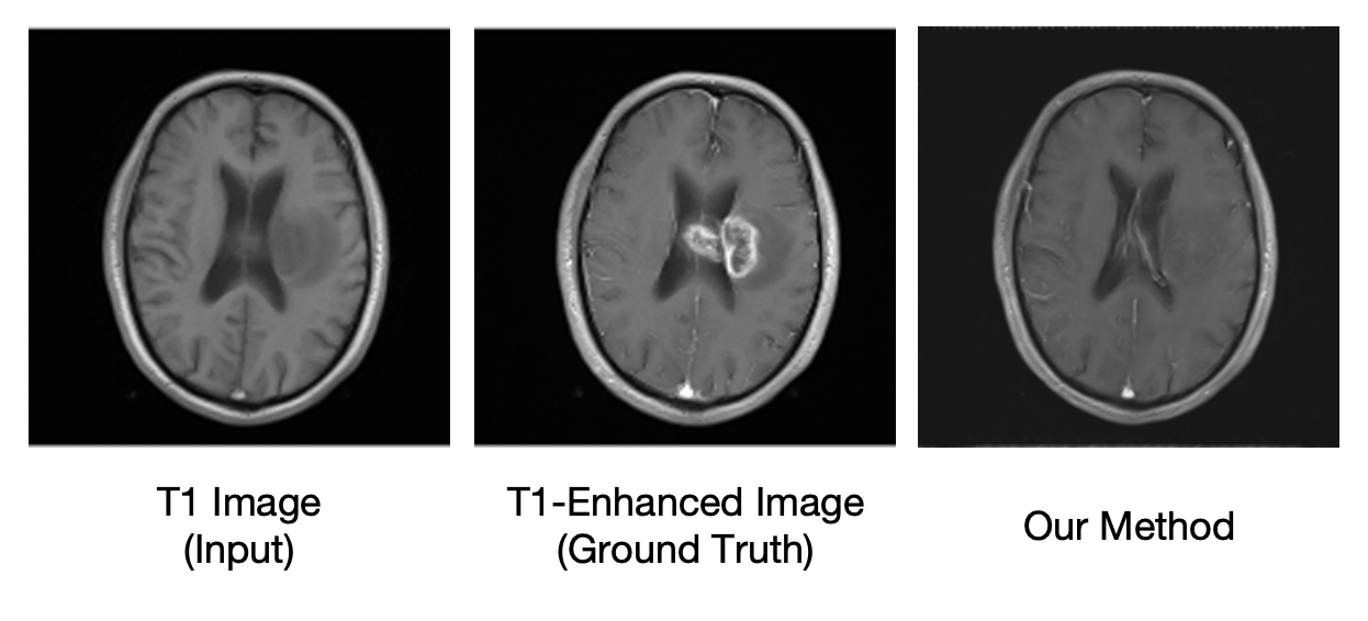
Our study presents an innovative method to enhance brain tumor visualization in T1-weighted MRI scans without contrast agents. The Pix2Pix-based approach, combined with registration and data augmentation, significantly improves tumor visibility in generated images. Our study provides a way to identify tumors under non-contrast agent conditions, reduces patient suffering and diagnostic costs, and has clinical value.While our method has shown promising results, it remains challenging to fully identify and enhance tumors in some cases (Figure 4). This limitation arises from the inherent constraints of the information provided by T1-weighted imaging alone. Future work could perform multi-modal image analysis to better assist medical professionals in identifying tumors without contrast agents.CONCLUSION
Our Pix2Pix-based approach enhances tumor visibility in T1-weighted MRI scans, offering a cost-effective and contrast agent-free modality for improved brain tumor assessment. The results demonstrate superior tumor discernibility, which provides valuable diagnostic support to medical professionals.Acknowledgements
No acknowledgement found.References
1. Miller, Kimberly D., et al. "Brain and other central nervous system tumor statistics, 2021." CA: a cancer journal for clinicians 71.5 (2021): 381-406.
2. McDonald, Robert J., et al. "Intracranial gadolinium deposition after contrast-enhanced MR imaging." Radiology 275.3 (2015): 772-782.
3. Avants, Brian B., et al. "Symmetric diffeomorphic image registration with cross-correlation: evaluating automated labeling of elderly and neurodegenerative brain." Medical image analysis 12.1 (2008): 26-41.
4. Isola, Phillip, et al. "Image-to-image translation with conditional adversarial networks." Proceedings of the IEEE conference on computer vision and pattern recognition. 2017.
5. Zhu, Jun-Yan, et al. "Unpaired image-to-image translation using cycle-consistent adversarial networks." Proceedings of the IEEE international conference on computer vision. 2017.
Figures