4523
AUTOMATED CEREBRAL CAVERNOUS MALFORMATION LESION SEGMENTATION FROM SUSCEPTIBILITY WEIGHTED MR IMAGING1Department of Radiology and Biomedical Imaging, University of California San Francisco, San Francisco, CA, United States, 2UCSF/UC Berkeley Graduate Program in Bioengineering, University of California San Francisco – University of California, Berkeley, San Francisco, CA, United States, 3Department of Radiology, University of New Mexico School of Medicine, Albuquerque, NM, United States, 4Department of Anesthesia, University of California San Francisco, San Francisco, CA, United States
Synopsis
Keywords: Analysis/Processing, Susceptibility
Motivation: Familial cerebral cavernous malformation (CCM) cases typically present with multi-focal lesions of varying size, count, and location making automatic segmentation for studying lesion burden challenging.
Goal(s): We aimed to develop a deep learning framework for automated detection and volumetric quantification for ongoing large-scale, multi-site evaluation of patients with CCM.
Approach: We implemented a framework that ensembles two-staged deep learning networks for large and small CCM lesions.
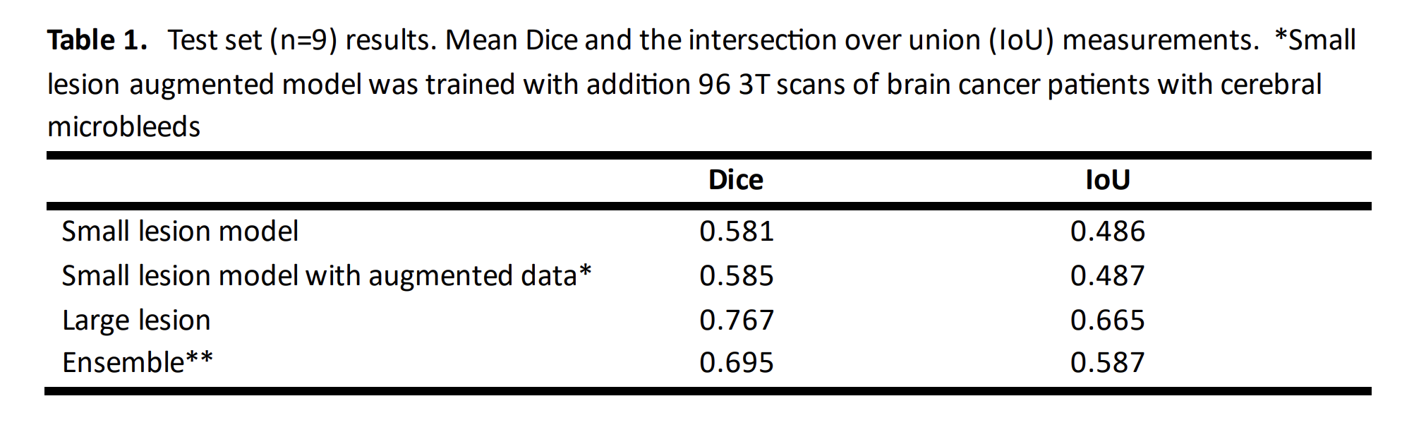
Results: Our model achieved an overall Dice-score of 70%. We demonstrated the feasibility of a deep learning approach for detecting and segmenting various-sized lesions in patients with CCM using SWI acquired from multiple sites with various parameters.
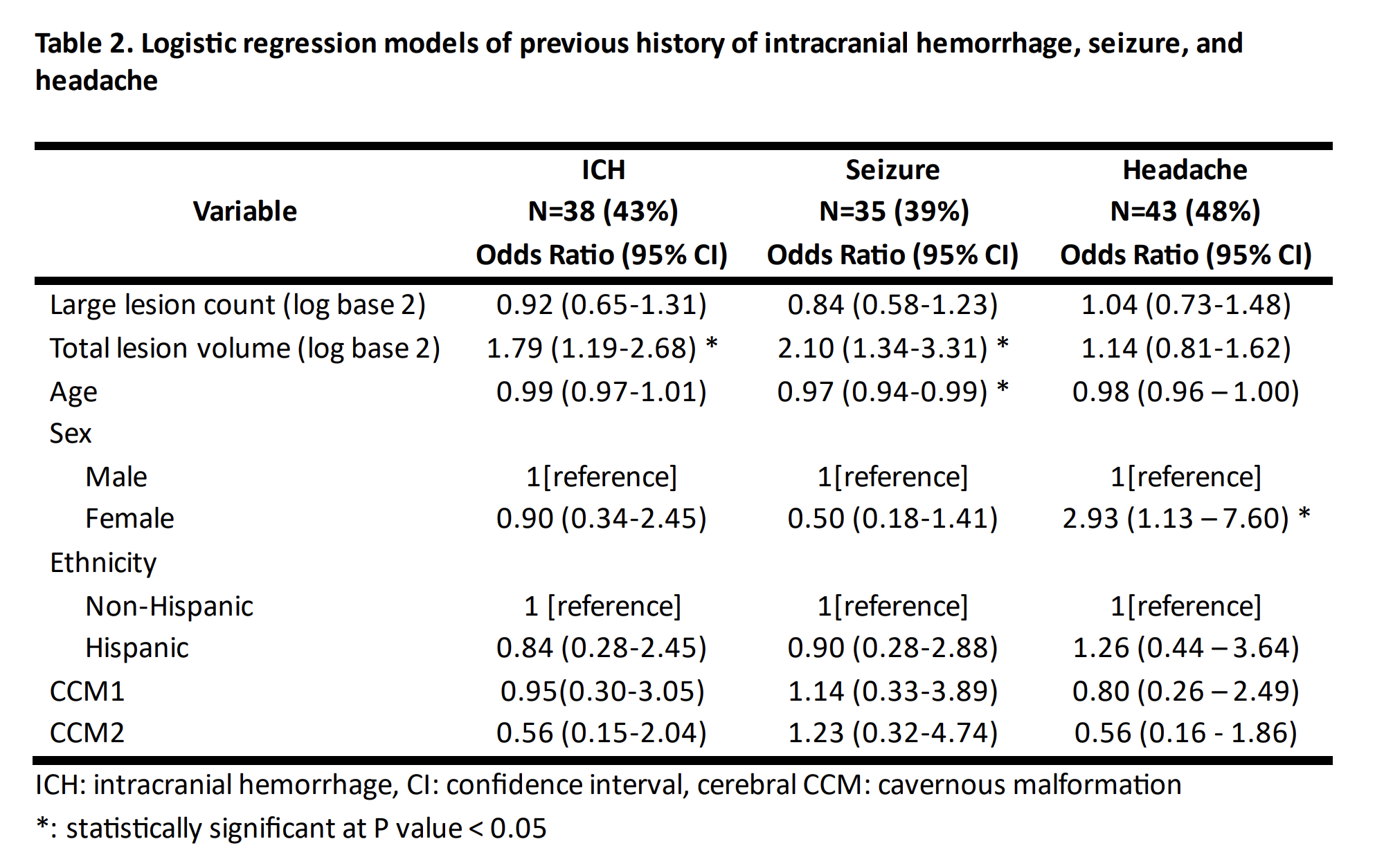
Impact: Our study found that total lesion volume quantified with our deep learning approach was statistically associated with increased odds of intracranial hemorrhage history. This demonstrates the potential benefit of our approach in providing a more accurate assessment of disease burden
INTRODUCTION
Familial cerebral cavernous malformations (CCMs) are vascular abnormalities where patients typically present with multi-focal lesions, hemorrhagic stroke, seizures, and other disabling neurological deficits1. Although CCMs are readily visible on Susceptibility-weighted MR imaging (SWI), the number and size of lesions vary widely, even among carriers of similar ages and with the same mutation, making automatic segmentation for studying lesion burden challenging. Although prior studies have attempted to automatically count lesions from a single institution2, the variability of lesion size and imaging acquisition parameters have resulted in poor performance when applied to larger multi-site datasets or focused on detection rather than lesion segmentation. The goal of this study was to develop a deep learning framework for automated lesion detection and volumetric quantification for large-scale multi-site CCM datasets and evaluate its performance on downstream tasks like association with clinical symptoms.METHODS
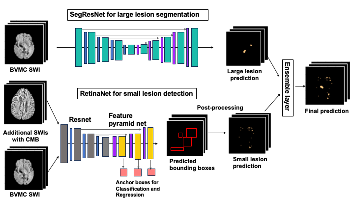
Study design We used imaging and clinical data from the Brain Vascular Malformation Consortium (BVMC) CCM project, an ongoing observational study enrolling familial CCM cases between 2010 and 2024. The current BVMC cycle (2019-2024) is enrolling familial CCM cases at seven sites across the United States using Siemens, Phillips, and GE 3T and 1.5T MRI scanners with various acquisition parameters for T2*-weighted imaging3. We selected 91 cases with SWI images readily available. Nine scans were reviewed and annotated by a neuroradiologist and used for model validation; the rest were labeled with an active learning strategy implemented in MONAI4 and randomly split into training set and validation set and used for model development.Data preprocessing and model training All SWI images were resampled to 0.70x0.70x1.25mm3, skull stripped using HD-BET software5, and standardized. Two overlapping annotated masks were empirically generated: one with lesion diameters >30mm and the other with diameters <40mm. A 3D SegResnet6 was trained for large lesion segmentation, while a RetinaNet7 for object detection was employed for small lesions, whereby the centroid of the output bounding box was then used along with region growing8 to create the final small lesion segmentation before applying a linear layer to ensemble two predictions. For small lesion segmentation, we also evaluated the effect of adding an additional 97 scans of brain tumor patients with radiation-induced cerebral microbleeds in training. Soft Dice and cross-entropy loss were used for large lesion segmentation, while binary cross-entropy and L1 loss were applied for small lesions. Both networks were independently trained with a batch size of 1, using Adam optimizer with initial learning rate of 1e-4 over 600 epochs. Our pipeline (shown in Figure 1) was implemented in PyTorch and MONAI9.
Model evaluation and statistical analysis Each 3D image with varying size and number of slices in test set was evaluated with sliding window fashion with 25 percent overlap. We report the mean values of evaluation metrics, Dice score and intersection over union (IoU), for the test set (9 scans). We investigated the association between the large and total lesion volume and clinical symptom presentations, including history of headache, ICH, and seizures, using logistic regression models adjusted for age, sex, ethnicity, and genetic mutation.
RESULTS AND DiSCUSSION
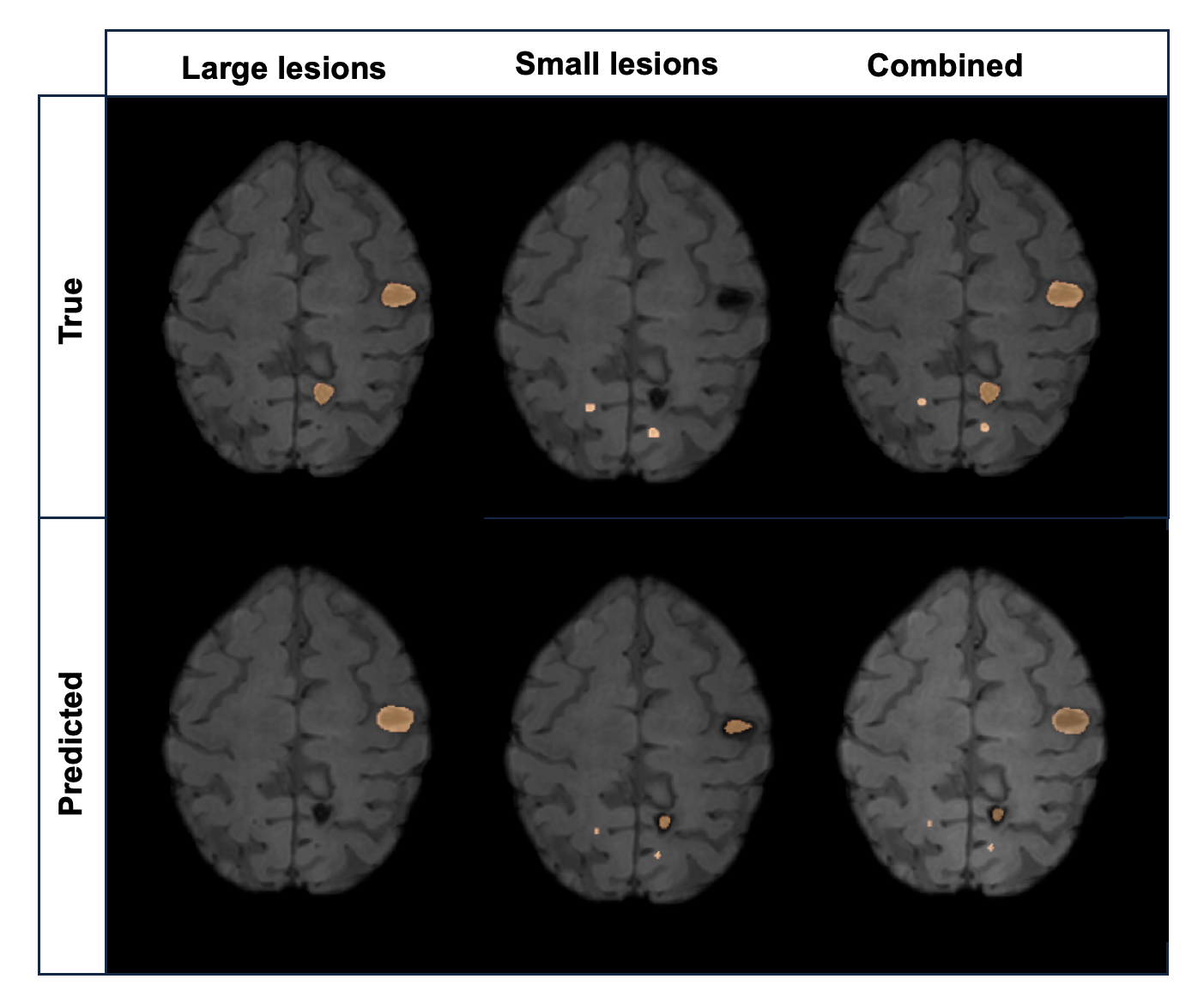
As shown in Table 1, our ensemble segmentation pipeline achieved a Dice score of 0.695, 0.585, and 0.767 in overall, small lesion, and large lesion, respectively. Figure 2 presents a typical segmentation example from a test set with true and predicted labels overlayed on SWI.The proportion of participants who had experienced ICH, seizure, and headache was 43%, 39%, and 48%, respectively. In adjusted regression analyses, the total lesion volume quantified with our deep learning approach was significantly associated with increased odds of previous ICH history (OR 1.79; 95% CI 1.19-2.68) and seizure (OR 2.10; 95% CI 1.34-3.31), respectively (Table 2). This demonstrates the potential benefit of an automated detection and segmentation approach for quantifying lesion burden with deep learning compared to lesion counts, despite prediction errors associated with the current models.
CONCLUSION
We demonstrated the feasibility of a deep learning approach for detecting and segmenting cavernous malformations in patients with familial CCM using SWI images acquired from multiple sites with various acquisition parameters. This allowed for the first time the quantification of total lesion burden based on lesion size and number, which was significantly associated with the history of prior hemorrhage and seizures.Acknowledgements
NIH-NINDS: U54-NS065705References
1 Snellings, D. A., Hong, C. C., Ren, A. A., Lopez-Ramirez, M. A., Girard, R., Srinath, A., ... & Kahn, M. L. (2021). Cerebral cavernous malformation: from mechanism to therapy. Circulation research, 129(1), 195-215.
2 Zou, X., Hart, B. L., Mabray, M., Bartlett, M. R., Bian, W., Nelson, J., ... & Kim, H. (2017). Automated algorithm for counting microbleeds in patients with familial cerebral cavernous malformations. Neuroradiology, 59, 685-690.
3 Weinsheimer, S., Nelson, J., Abla, A. A., Ko, N. U., Tsang, C., Okoye, O., ... & Kim, H. (2023). Intracranial Hemorrhage Rate and Lesion Burden in Patients With Familial Cerebral Cavernous Malformation. Journal of the American Heart Association, 12(3), e027572.
4 Monai Consortium. (2020). MONAI: Medical open network for AI. Online at https://doi. org/10.5281/zenodo, 5525502.Chicago
5 Isensee, F., Schell, M., Pflueger, I., Brugnara, G., Bonekamp, D., Neuberger, U., ... & Kickingereder, P. (2019). Automated brain extraction of multisequence MRI using artificial neural networks. Human brain mapping, 40(17), 4952-4964.
6 Adams, R., & Bischof, L. (1994). Seeded region growing. IEEE Transactions on pattern analysis and machine intelligence, 16(6), 641-647.
7 Lin, T. Y., Goyal, P., Girshick, R., He, K., & Dollár, P. (2017). Focal loss for dense object detection. In Proceedings of the IEEE international conference on computer vision (pp. 2980-2988).
8 Paszke, A., Gross, S., Massa, F., Lerer, A., Bradbury, J., Chanan, G., ... & Chintala, S. (2019). Pytorch: An imperative style, high-performance deep learning library. Advances in neural information processing systems, 32.
Figures