4522
Deep learning to predict DTI tractography labels from diagnostic CT scans in acute ICH1Division of Informatics, Imaging and Data Science, University of Manchester, Manchester, United Kingdom, 2University of Manchester, Manchester, United Kingdom, 3Brainomix, Oxford, United Kingdom, 4King's College London, London, United Kingdom, 5University Medical Center Groningen, Groningen, Netherlands, 6Isala Klinieken, Zwolle, Netherlands, 7Johns Hopkins, Baltimore, MD, United States, 8Radboud University Medical Center, Nijmegen, Netherlands
Synopsis
Keywords: Diagnosis/Prediction, Diffusion Tensor Imaging
Motivation: Assessing white matter integrity may enhance intracerebral haemorrhage (ICH) surgical trial selection. However, patients often only receive CT imaging, on which delineating white matter is challenging.
Goal(s): This study aimed to train a model to delineate white matter in CT scans, using paired DTI tractography maps, and test if the model predicted outcome in an external dataset.
Approach: Tractography maps were generated from the DTI images. A nnU-Net model was trained on paired CT and registered tractography data, and then run on an external dataset of ICH diagnostic CT scans.
Results: The model performed at 58% Dice, and significantly predicted outcome after ICH.
Impact: Our model can predict tractography labels of the corticospinal tract on diagnostic CT scans without the need for DTI, allowing an enhanced prediction of outcome after intracerebral haemorrhage, and potentially leading to more informed selection of candidates for surgical trials.
Background
Haematoma evacuation for intracerebral haemorrhage (ICH) reduces mortality, but clinical trials have yet to show improved recovery 1,2. Selection of patients with a preserved corticospinal tract (CST) may reveal a subset where haematoma evacuation may be life-saving and lead to a good functional outcome 3.Non-contrast CT scans are routinely available for all ICH patients but delineating white matter on a CT is challenging. Our aim was to use DTI based tractography maps of the CST to train a convolutional neural network to delineate the CST on diagnostic CT images. Then, combined with a model trained to segment the haematoma on the CT scan, a measure of tract integrity could be calculated.
Methods
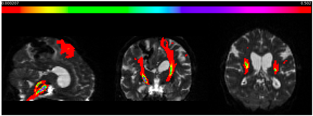
Data from the FETCH study was used, including 138 ICH patients who had DTI and CT imaging 4. At the time of this study, DTI, CT and T1 images from 57 patients were available. These were from centres in Nijmegen (n = 19, DTI acquired at 3T in 64 directions), Utrecht (n = 20, DTI acquired at 3T in 45 directions) and Leiden (n = 18, DTI acquired at 3T in 45 directions).The DTI data were acquired in one phase encoding direction, so PreQual 5 was used to apply FSL’s Topup and Eddy correction tools using a synthesised b0 image based on the T1 scan.Bayesian Estimation of Diffusion Parameters Obtained using Sampling Techniques with Crossing Fibres (BEDPOSTX) was used to determine the number of crossing fibres per voxel 6,7. FSL’s XTRACT performed probabilistic tractography on the crossing fibres data to produce a dissected CST 8-10. These CST tractography maps were then registered to CT space for each patient, and thresholded at 0.01 to produce binary labels. 42 patients with good quality tractography were selected for training (36 training, 6 testing).
The model used was nnU-Net; a self-configuring 3D U-Net variation 11. nnU-Net selects hyperparameters and network architecture according to the size, resolution and modality of the training dataset. Such a carefully designed U-Net can achieve state-of-the-art medical image segmentation 11. The model was trained for 1000 epochs using a combination Dice binary cross entropy loss, on an NVIDIA a100 GPU. The trained model was tested on the unseen testing dataset.
To assess whether our CT CST segmentations are predictive of clinical outcomes, the trained model was run over a dataset of 487 ICH diagnostic CT scans from the MISITE III clinical trial 1. Segmentations of the haematoma volume were also generated for each diagnostic CT in this dataset using an in-house nnU-Net based model. Two metrics of tract integrity were calculated; haematoma overlap, and tract dissection. Haematoma overlap was defined to be true if any of the CST segmentation overlapped with the haematoma segmentation. Tract dissection was defined to be true if either side of the CST wasn’t detected in any axial slice, implying an interruption to the tract.
Multiple linear regressions were conducted to assess the impact of CST integrity on the National Institute of Health Stroke Scale (NIHSS) in the motor domain (a sum of scores for questions 4, 5 and 6) at day 1 and day 180, and modified Rankin Scale (mRS) score at day 365. Age, natural logarithm of the ICH volume, and medical or surgical treatment group were adjusted for.
Results
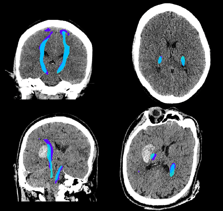
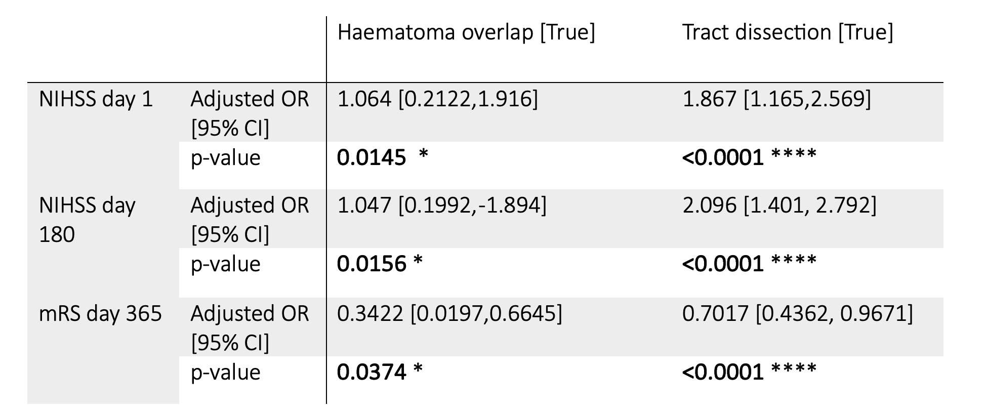
When tested, the average Dice similarity coefficient, which measures the spatial overlap between a model's predictions and the ground truth, was 58%. Figure 2 shows predictions for 2 out of 487 analysed MISTIE patients (average age: 61, average mRS at 365 days: 4). 377 patients had no haematoma overlap, 251 patients had no tract dissection. Haematoma overlap and tract dissection significantly predict an increase in NIHSS at day 1 and 180, and mRS at day 365, indicating an association with worse motor outcome after stroke. The results of the linear regression analysis are shown in Table 1.Discussion and Conclusions
The model can predict, from non-contrast CT scans, segmentations of the CST that are 58% similar to high angular resolution DTI tractography. The potential of this prediction is shown in the results of the linear regressions; whilst the DTI tractography may not be perfectly replicated, the CT generated CST prediction is significantly associated with outcome, and predicts functional recovery after stroke in both the acute and chronic timeframes.This could lead to an automated tool to predict prognosis, or to select a population of ICH patients for a surgical trial that may benefit from haematoma evacuation. Future work involves processing recently available data from the FETCH study to expand the training dataset.
Acknowledgements
ONM is supported by a Medical Research Council studentship.References
[1] Hanley D, Thompson R, Rosenblum M, et al. Efficacy and safety of minimally invasive surgery with thrombolysis in intracerebral haemorrhage evacuation (MISTIE III): a randomised, controlled, open-label, blinded endpoint phase 3 trial. Lancet. 2019; 393(10175):1021–1032.
[2] Mendelow A, Gregson B, Rowan E, et al. Early surgery versus initial conservativetreatment in patients with spontaneous supratentorial lobar intracerebral haematomas (STICH II): a randomised trial. Lancet. 2013; 382(9890):397–408.
[3] Stinear C, Barber P, Smale P, et al. Functional potential in chronic stroke patients depends on corticospinal tract integrity. Brain: A Journal of Neurology. 2007;130(Pt 1):170–180.
[4] Wiegertjes K, Voigt S, Jolink WMT, et al. Diffusion-Weighted Lesions After Intracerebral Hemorrhage: Associated MRI Findings. Front Neurol. 2022;13:882070.
[5] Cai L, Yang Q, Hansen C, et al. PreQual: An automated pipeline for integrated preprocessing and quality assurance of diffusion weighted MRI images. Magn Reson Med, 2021;86(1):456-470.
[6] Behrens T, Woolrich M, Jenkinson M, et al. Characterization and propagation of uncertainty in diffusion-weighted MR imaging. Magn Reson Med. 2003; 50(5):1077-1088.
[7] Behrens T, Johansen-Berg H, Jbabdi S,et al. Probabilistic diffusion tractography with multiple fibre orientations. What can we gain? NeuroImage. 2007;23:144-155.
[8] Warrington S, Thompson E, Bastiani M, et al. Concurrent mapping of brain ontogeny and phylogeny within a common space: Standardized tractography and applications. Science Advances. 2022; 8(42).
[9] Warrington S, Bryant K, Khrapitchev A, et al. XTRACT - Standardised protocols for automated tractography and connectivity blueprints in the human and macaque brain. NeuroImage. 2020;217(116923).
[10] de Groot M; Vernooij MW. Klein S, et al. Improving alignment in Tract-based spatial statistics: Evaluation and optimization of image registration. NeuroImage. 2013;76(1), 400-411.
[11] Isensee F, Jaeger PF, Kohl SA, et al. nnU-Net: a self-configuringmethod for deep learning-based biomedical image segmentation. Nature methods, 2021;18(2), 203-211.
Figures