4521
Meta-matching to translate phenotypic predictive models from big to small data on structural MRI1Centre for Sleep & Cognition & Centre for Translational Magnetic Resonance Research, Yong Loo Lin School of Medicine, National University of Singapore, Singapore, Singapore, 2Department of Electrical and Computer Engineering, National University of Singapore, Singapore, Singapore, 3N.1 Institute for Health & Institute for Digital Medicine, National University of Singapore, Singapore, Singapore, 4Department of Biomedical Engineering,McConnell Brain Imaging Centre (BIC), Montreal Neurological Institute (MNI), Faculty of Medicine, School of Computer Science, McGill University, Montreal, QC, Canada, 5Mila – Quebec Artificial Intelligence Institute, Montreal, QC, Canada, 6Institute for Systems Neuroscience, Medical Faculty, Heinrich-Heine University Düsseldorf, Düsseldorf, Germany, 7Institute of Neuroscience and Medicine, Brain & Behavior (INM-7), Research Center Jülich, Jülich, Germany, 8Department of Psychiatry, Brain Health Institute, Rutgers University, Piscataway, NJ, United States, 9Integrative Sciences and Engineering Programme (ISEP), National University of Singapore, Singapore, Singapore, 10Martinos Center for Biomedical Imaging, Massachusetts General Hospital, Charlestown, MA, United States
Synopsis
Keywords: Diagnosis/Prediction, Brain, Phenotypic prediction, structural MRI, transfer learning
Motivation: Small sample size on structural MRI is evitable in reality and significantly limits phenotypic prediction performance.
Goal(s): Our goal was to improve prediction performance on small datasets for structural MRI brain imaging.
Approach: We adapted the meta-matching framework from functional to structural MRI, and compared it with baseline methods (Elastic net and direct transfer learning).
Results: Our meta-matching-based approaches can greatly boost behavioral prediction performance for different small-scale structural MRI datasets.
Impact: Our meta-matching-based methods should be able to make good predictions for a variety of neurological and psychiatric disorders even if the availability of structural MRI brain imaging is quite small.
Introduction
A central goal in neuroscience is understanding how brain imaging is associated with behavior. Structural magnetic resonance imaging (MRI) is a non-invasive technique for examining the anatomy and pathology of the human brain. It produces images with high contrast between gray and white matter, providing excellent anatomical detail1. Due to its unique properties, structural MRI has been used to make individualized predictions in a variety of neurological and psychiatric disorders2-4. However, the prediction performance is strongly limited by a small sample size for many current MRI studies5-7. By transferring knowledge from large-scale source datasets (e.g. UK Biobank) to small target datasets and exploiting the underlying correlation structure between the source and target phenotype, meta-matching has greatly improved prediction performance in functional MRI8. Here, we tailored meta-matching approaches to predict new phenotypes in small boutique datasets with structural MRI.Methods
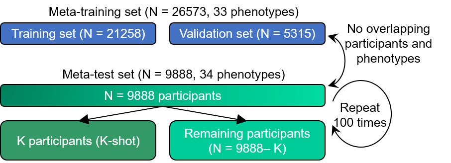
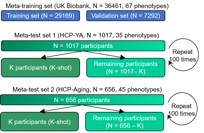
Our study departed from the UK Biobank9 (N=36,461, 67 phenotypes) HCP-YA10 (N=1017, 35 phenotypes), and HCP-Aging11(N=656, 45 phenotypes). FreeSurfer recon-all was used to derive thickness and volume measures with the DKT40 cortical atlas12 and ASEG subcortical segmentation13. Furthermore, we used FMRIB's Linear Image Registration Tool (FLIRT) to transform T1 to MNI152 standard-space T1 template with 1 mm resolution14,15.We transferred models pretrained from meta-training set to meta-test sets. Figure 1 shows the data split framework within UK Biobank dataset. For within UK Biobank analysis, we randomly split UK Biobank dataset into meta-training set (N=26573, 33 phenotypes) and meta-test set (N=9888, 34 phenotypes). There is no overlap between participants or phenotypes across meta-training and meta-test sets. On meta-test set, K participants (K-shot, where K had a value of 10, 20, 50, 100, and 200) were randomly selected to mimic traditional small sample size studies, while the remaining participants in the meta-test set served for evaluation. Each random K-shot split was repeated 100 times to ensure stability. Figure shows the data split framework for cross-dataset analysis. For cross-dataset analysis, the UK Biobank served as a meta-training set, while HCP-YA and HCP-Aging served as meta-test sets separately.
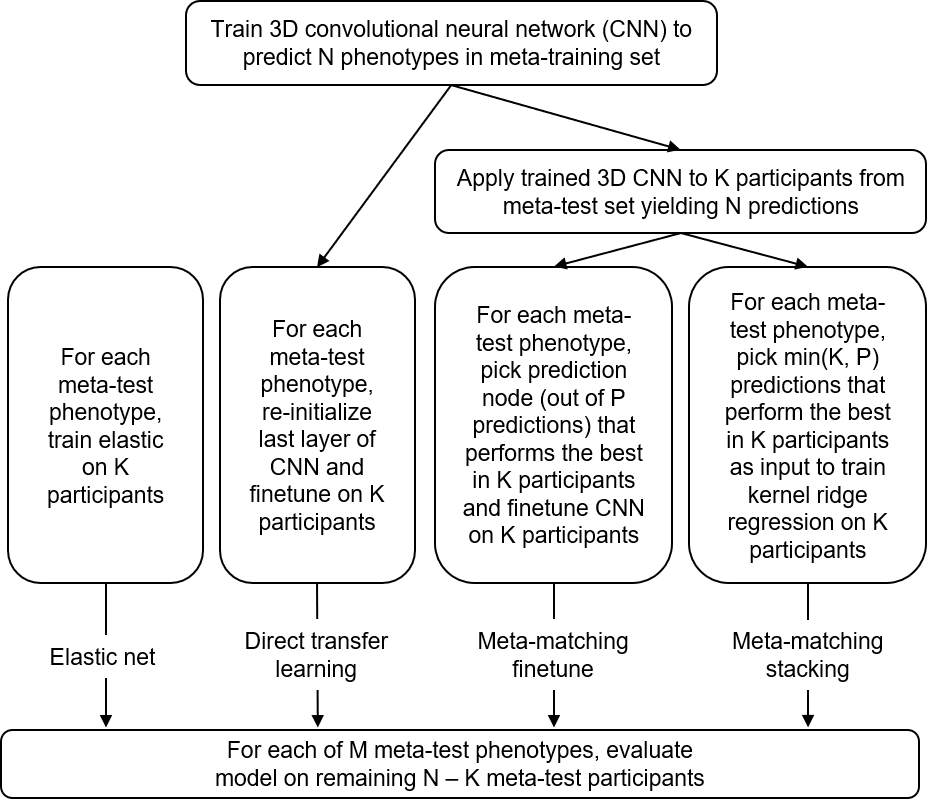
We adopted meta-matching by pretraining a 3D CNN model16 on the meta-training set structural brain imaging to improve phenotypic prediction performance on meta-test sets (Figure 3). For baseline approaches, we considered the elastic net and direct transfer learning algorithm (Figure 1). The input of elastic net was morphometric measures (volumes and thickness from cortical and/or subcortical ROIs from FreeSurfer); the input of deep learning approaches is T1 images affine transformed to MNI152 standard space.
Results
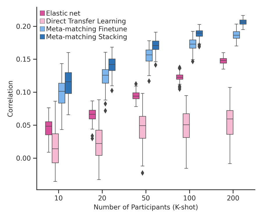
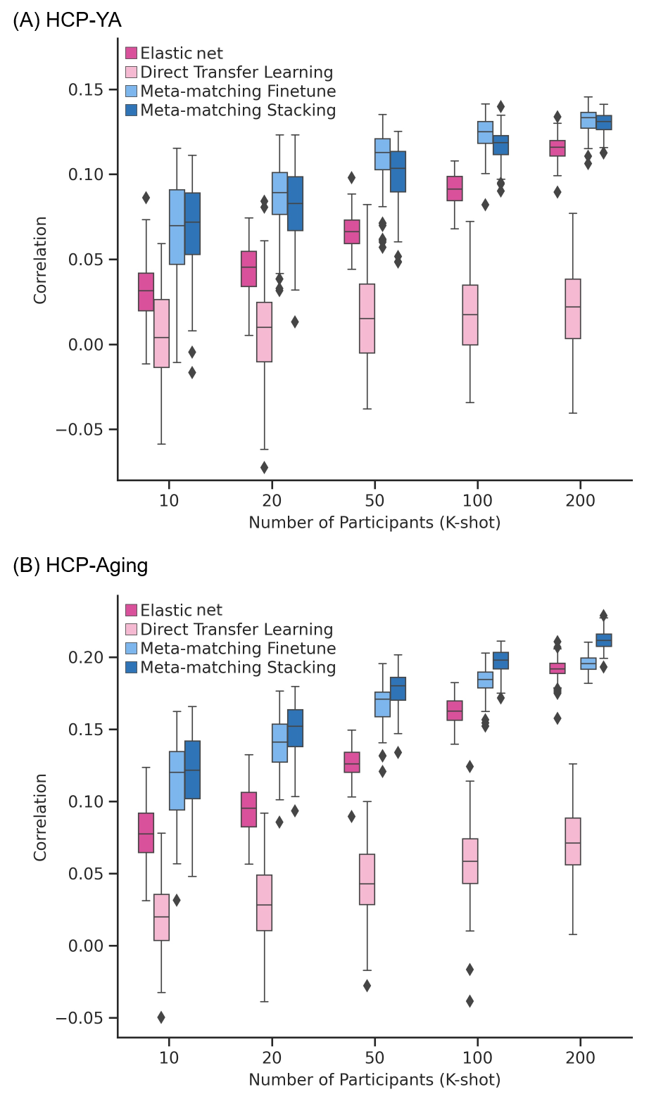
Figure 4 shows the prediction accuracy (Pearson’s correlation) across all test phenotypes on the UK Biobank meta-test set. The boxplots represent 100 repetitions for K-shot. We can observe that meta-matching-based approaches (meta-matching finetune and meta-matching stacking) can significantly outperform the Elastic net baseline and direct transfer learning methods (for every K number).The previous experiment results (Figure 4) suggested that meta-matching-based methods can perform well when transferring within the same dataset (e.g. UK Biobank). To demonstrate the generalization ability of meta-matching-based methods. approaches are also applied to the meta-test set in the HCP-YA dataset Figure 5 (A), and the HCP-Aging dataset Figure 5 (B) respectively. Figure 5 (A) and Figure 5 (B) show that the meta-matching-based methods can significantly outperform baseline methods in most cases when transferring from the meta-training dataset (UK Biobank) to the meta-test dataset (HCP-YA or HCP-Aging).
Discussion
For within UK Biobank analysis in Figure 4, we noticed that the huge improvement from meta-matching-based methods may be because the meta-training set and meta-test set are both from the same UK Biobank dataset. The same distribution of demographics and pre-processing pipeline make meta-matching-based methods effectively transfer knowledge from the meta-training set to the meta-test set.In Figure 5, when generalized to new target datasets, we noticed that although our meta-matching-based methods cannot achieve huge improvement like within the UK Biobank experiment, they can still significantly improve the phenotypic prediction. This indicates that meta-matching-based methods to some extent can handle datasets with different distributions of demographics and pre-processing pipelines during knowledge transfer to achieve better prediction performance.
Conclusions
We adopted meta-matching from functional to structural imaging and achieved superior performance over elastic net and direct transfer learning on small datasets including HCP-YA and HCP-Aging. Our results showed the great potential of meta-matching framework in structural MRI-based behavior predictions.Acknowledgements
No acknowledgement found.References
1. Gifford, G., McCutcheon, R., & McGuire, P. (2020). Neuroimaging studies in people at clinical high risk for psychosis. In Risk Factors for Psychosis (pp. 167-182): Elsevier.
2. Arbabshirani, M. R., Plis, S., Sui, J., & Calhoun, V. D. (2017). Single participant prediction of brain disorders in neuroimaging: Promises and pitfalls. NeuroImage, 145, 137-165.
3. Bhagwat, N., Pipitone, J., Voineskos, A. N., Chakravarty, M. M., & Initiative, A. s. D. N. (2019). An artificial neural network model for clinical score prediction in Alzheimer disease using structural neuroimaging measures. Journal of Psychiatry and Neuroscience, 44(4), 246-260.
4. Cohen, S. E., Zantvoord, J. B., Wezenberg, B. N., Bockting, C. L., & van Wingen, G. A. (2021). Magnetic resonance imaging for individual prediction of treatment response in major depressive disorder: a systematic review and meta-analysis. Translational psychiatry, 11(1), 168.
5. Kharabian Masouleh, S., Eickhoff, S. B., Hoffstaedter, F., Genon, S., & Initiative, A. s. D. N. (2019). Empirical examination of the replicability of associations between brain structure and psychological variables. elife, 8, e43464.
6. Poldrack, R. A., Huckins, G., & Varoquaux, G. (2020). Establishment of best practices for evidence for prediction: a review. JAMA psychiatry, 77(5), 534-540.
7. He, T., Kong, R., Holmes, A. J., Nguyen, M., Sabuncu, M. R., Eickhoff, S. B., . . . Yeo, B. T. (2020). Deep neural networks and kernel regression achieve comparable accuracies for functional connectivity prediction of behavior and demographics. NeuroImage, 206, 116276.
8. He, T., An, L., Chen, P., Chen, J., Feng, J., Bzdok, D., . . . Yeo, B. T. (2022). Meta-matching as a simple framework to translate phenotypic predictive models from big to small data. Nature neuroscience, 25(6), 795-804.
9. Alfaro-Almagro, F., Jenkinson, M., Bangerter, N. K., Andersson, J. L., Griffanti, L., Douaud, G., . . . Vallee, E. (2018). Image processing and Quality Control for the first 10,000 brain imaging datasets from UK Biobank. NeuroImage, 166, 400-424.
10. Van Essen, D. C., Smith, S. M., Barch, D. M., Behrens, T. E., Yacoub, E., Ugurbil, K., & Consortium, W.-M. H. (2013). The WU-Minn human connectome project: an overview. NeuroImage, 80, 62-79.
11. Bookheimer, S. Y., Salat, D. H., Terpstra, M., Ances, B. M., Barch, D. M., Buckner, R. L., . . . Elam, J. S. (2019). The lifespan human connectome project in aging: an overview. NeuroImage, 185, 335-348.
12. Klein, A., & Tourville, J. (2012). 101 labeled brain images and a consistent human cortical labeling protocol. Frontiers in neuroscience, 6, 171.
13. Fischl, B., Salat, D. H., Busa, E., Albert, M., Dieterich, M., Haselgrove, C., . . . Klaveness, S. (2002). Whole brain segmentation: automated labeling of neuroanatomical structures in the human brain. Neuron, 33(3), 341-355.
14. Jenkinson, M., Bannister, P., Brady, M., & Smith, S. (2002). Improved optimization for the robust and accurate linear registration and motion correction of brain images. NeuroImage, 17(2), 825-841.
15. Jenkinson, M., & Smith, S. (2001). A global optimisation method for robust affine registration of brain images. Medical Image Analysis, 5(2), 143-156.
16. Peng, H., Gong, W., Beckmann, C. F., Vedaldi, A., & Smith, S. M. (2021). Accurate brain age prediction with lightweight deep neural networks. Medical Image Analysis, 68, 101871.
Figures