4515
Semi-Nonlinear Self-Adaptation Normalization Generator (SNSAN) to Improve Generalization of White Matter Hyperintensities Segmentation1The Institute of Science and Technology for Brain-inspired Intelligence (ISTBI), Fudan University, Shanghai, China, 2Human Phenome Institute, Fudan University, Shanghai, China, 3Zhongshan Hospital Fudan University, Shanghai, China, 4Department of Biomedical Engineering, Chung Yuan Christian University, Taoyuan, Taiwan
Synopsis
Keywords: AI Diffusion Models, White Matter, WMH segmentation, Domain Generalization, Normalization
Motivation: Self-adaptation normalization (SAN) method generalizes lesion segmentation to new sites by using a linear generator to convert inputs to a site-independent style. But it fails when the domain difference is mostly nonlinear.
Goal(s): Develop a semi-nonlinear self-adaptation network(SNSAN) to generalize the White Matter Hyperintensity(WMH) segmentation model to an external site.
Approach: To replace the linear generator, the method blends two SAN results with a pseudo correlation map, later use the gradient reversal method to guide the result to a site-unrelated style.
Results: SNSAN normalizes the input data close to a Gaussian distribution and improves the generalization performance on the data from external site.
Impact: We provide a simple and efficient semi-nonlinear normalization method to enhance the domain generalization, and its performance is better than SAN when the domain gap is affected by more nonlinear factors.
INTRODUCTION
White matter hyperintensities (WMH) are common brain lesions in healthy elderly individuals. Li et al. proposed a deep fully convolutional network and ensemble models to automatically segment the WMH.1 However, in practical applications, the segmentation model often fails on the data from unseen sites. Yu et al. developed a self-adaptive normalization network (SAN) to generalize automatic lesion segmentation to unseen sites by linearly transforming the input to a site-independent style.2 However, when the difference between sites is not a simple linear transformation, SAN does not have enough generalization ability. We propose a semi-nonlinear normalization method to enhance SAN ability.METHODS
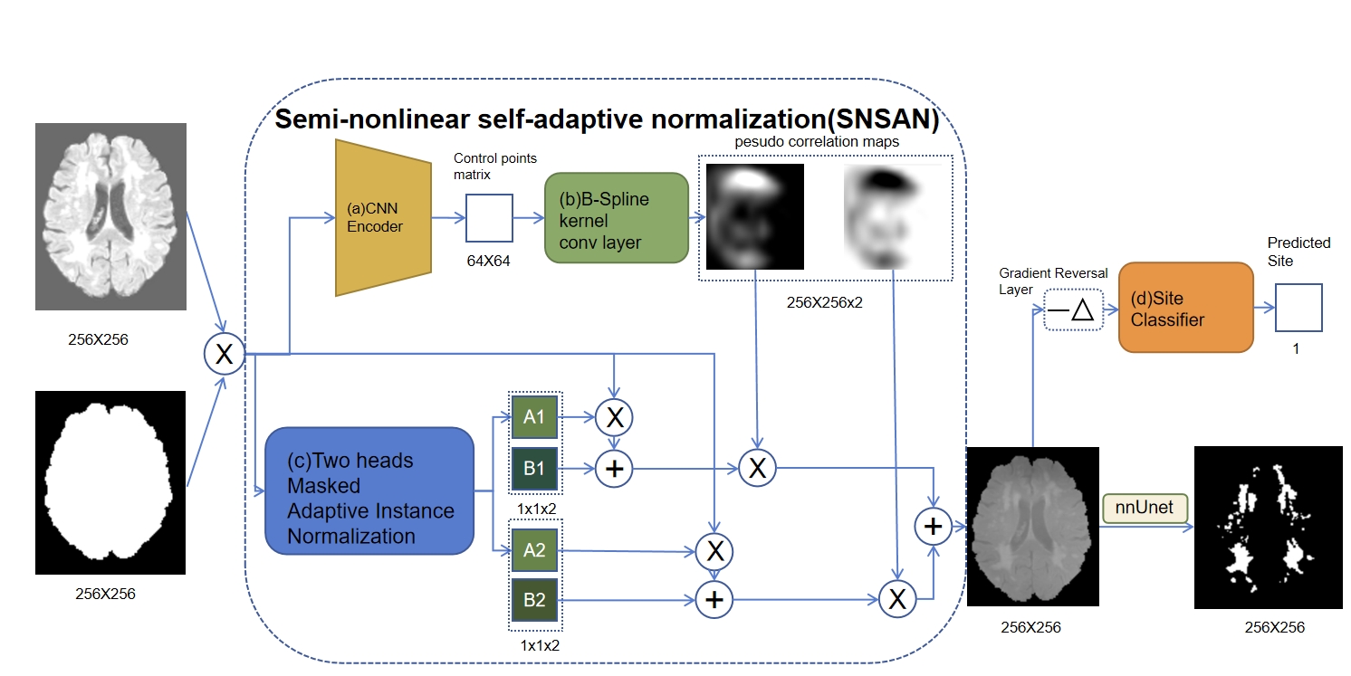
This study included 200 cases from two sources: 150 cases from MICCAI WMH challenge (MWC), and 50 cases from an external hospital3. MWC included 3 sites: 50 cases from UMC Utrecht (3T Philips Achieva), 50 cases from NUHS Singapore (3T Siemens TrioTim), and 50 cases from VU Amsterdam (3T GE Signa HDxt). The external dataset consisted of data from different unknown devices and parameters. We only used T2-weighted and fluid-attenuated inversion recovery (FLAIR) modality for WMH segmentation, since the external cases only had FLAIR modality. We assumed that we had WMH segmentation labels from the Utrecht site. Amsterdam and Singapore data contained no label but were visible in the training phase, and external data was completely invisible in the training phase.The proposed model consisted of 2 steps (Figure 1): SNSAN normalization part, and WMH segmentation part. In the SNSAN normalization part, we replaced the linear generator with a semi-nonlinear version. On the one hand, a 4-layer CNN encoder extracted a 64 by 64 control points matrix from the input image. Through a B-spline kernel deconvolution layer, the matrix became a field of continuous-valued scalars with low spatial frequency. Cheng named it as the pseudo correlation map (PCM).4 On the other hand, two contrast results of the input were generated by linear adaptive normalization with a similar approach of SAN. The two contrast images were multiplied separately with PCM and one minus PCM, and then added together to obtain the semi-nonlinear result. Finally, the site classifier with gradient reversal layer guided the blended result to a site-invariant style. In the WMH segmentation part, we adopted nnUNet to segment WMH5. Before normalization, all data was registered to standard (MNI -152) space. We used the training cases from the three sites of MWC to train the SNSAN model, and converted all the cases into a site-independent style with the trained SNSAN. Later we used nnUNet to train the WMH segmentation model with normalized training data from the Utrecht site. In terms of performance evaluation, the segmentation performance was evaluated using the Dice coefficient.
RESULTS
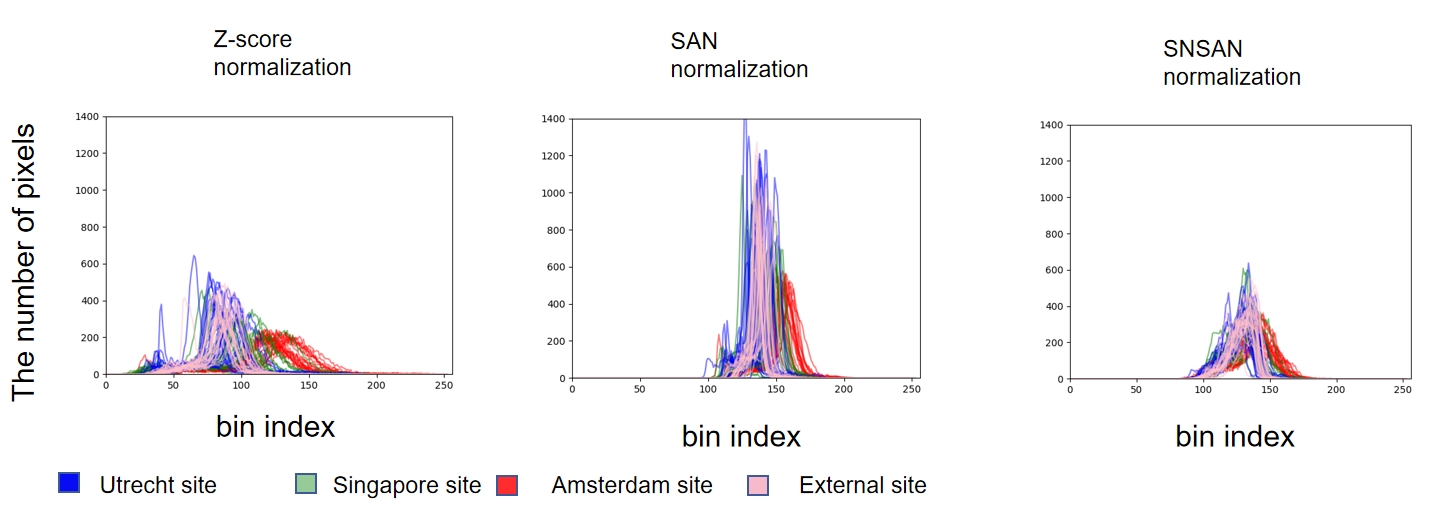
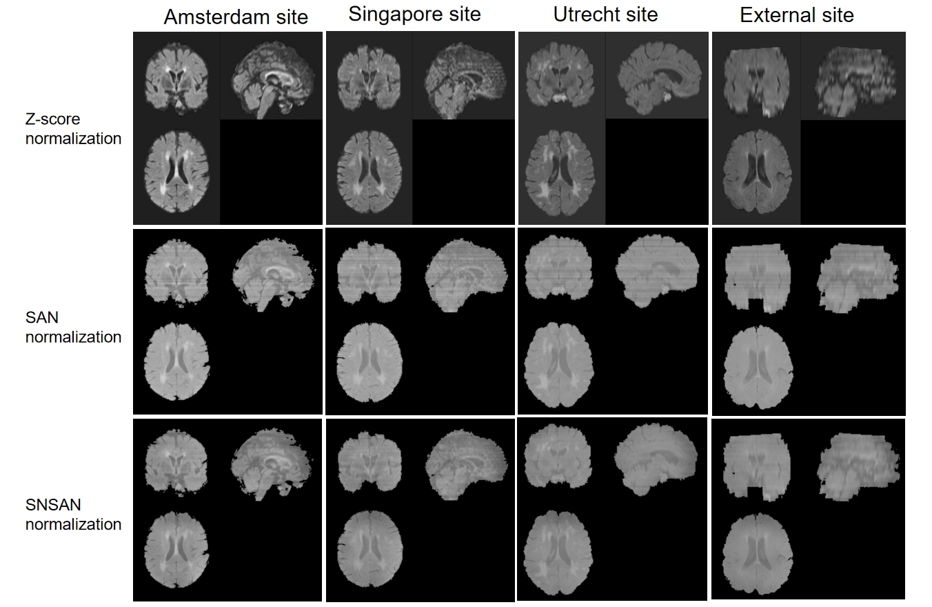
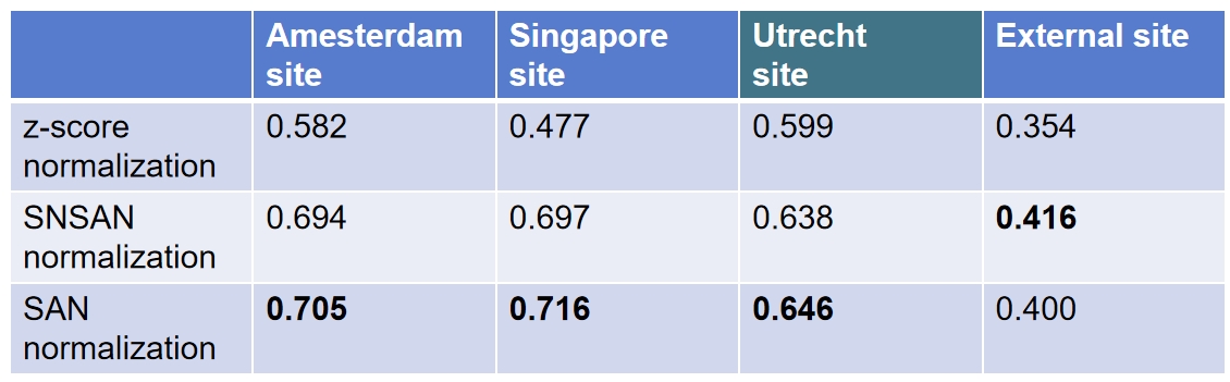
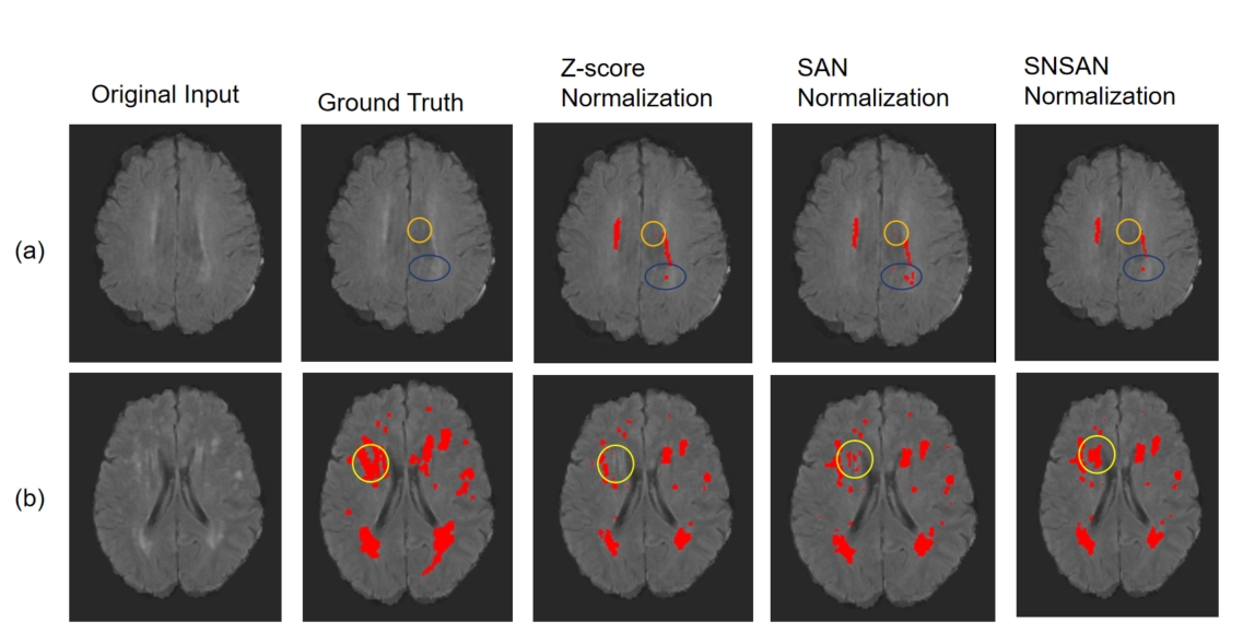
Figure 2 shows that SAN and SNSAN make the intensity distribution more consistent, and SNSAN's result is closer to a Gaussian distribution. Figure 3 shows that SNSAN reflects some nonlinear mapping, and the resulting images are smoother in the sagittal and coronal directions. We used 15 training data and 5 validation data from the Utrecht site to train the segmentation model, and validated the improvement of different normalization methods on the model's generalization ability. The Dice coefficient is not high because of the few training data, but it does not affect our validation. Figure 4 shows that SAN and SNSAN methods outperform the Z-score method. They reduce the domain gap on the three MWC datasets. SNSAN performs 1.6% higher than SAN. This may be due to the larger nonlinear difference between the external site and the Utrecht site. Figure 5 shows that SNSAN reduces the false positive area in the artifacts area and gains the true positive in the high incidence area.DISCUSSION
SNSAN method shows some potential, but the improvement over SAN is very limited. There may be several reasons: 1) The degree of nonlinear transformation is not enough. Combining deep learning and prototype learning methods to transform the models from different centers into the data distribution with segmentation labels may improve the performance. 2) A large part of the segmentation errors are caused by the artifacts unique to the external site, which cannot be solved by normalizing the contrast. It may be necessary to introduce other prior knowledge. Another problem is that it did not compare with the feature-consistent methods. In addition, cross-validation is necessary to obtain more reliable conclusions.CONCLUSION
We developed SNSAN to improve the performance of SAN, and demonstrate that nonlinear normalization methods can further improve the model's generalization ability when the external validation set has a significant nonlinear gap.Acknowledgements
No acknowledgement found.References
1. Li H, Jiang G, Zhang J, et al. Fully convolutional network ensembles for white matter hyperintensities segmentation in MR images[J]. NeuroImage, 2018, 183: 650-665.
2.Yu W, Huang Z, Zhang J, et al. SAN-Net: Learning generalization to unseen sites for stroke lesion segmentation with self-adaptive normalization[J]. Computers in Biology and Medicine, 2023, 156: 106717.
3. Kuijf H J, Biesbroek J M, De Bresser J, et al. Standardized assessment of automatic segmentation of white matter hyperintensities and results of the WMH segmentation challenge[J]. IEEE transactions on medical imaging, 2019, 38(11): 2556-2568.
4. Ouyang C, Chen C, Li S, et al. Causality-inspired single-source domain generalization for medical image segmentation[J]. IEEE Transactions on Medical Imaging, 2022, 42(4): 1095-1106.
5. Isensee F, Jaeger P F, Kohl S A A, et al. nnU-Net: a self-configuring method for deep learning-based biomedical image segmentation[J]. Nature methods, 2021, 18(2): 203-211.
Figures