4514
Deep Learning Based Key Point Detection Algorithm to Assist Accurate Vascular Centerline Extraction and Automatic Quantitative Plaque Analysis1Paul C. Lauterbur Research Center for Biomedical Imaging, Shenzhen Institute of Advanced Technology, Chinese Academy of Sciences, Shenzhen, China, 2University of Chinese Academy of Sciences, Beijing, China, 3Department of Image Advanced Analysis of HSW BU, Shanghai United Imaging Healthcare Co., Ltd., Shanghai, China, 4Key Laboratory of Biomedical Imaging Science and System, Chinese Academy of Sciences, Shenzhen, China, 5United Imaging Research Institute of Innovative Medical Equipment, Shenzhen, China
Synopsis
Keywords: Diagnosis/Prediction, Machine Learning/Artificial Intelligence, Plaque analysis;Vascular centerline extraction
Motivation: Magnetic Resonance Vessel Wall Imaging is a crucial method for assessing arterial plaques.
Goal(s): Achieving a rapid and accurate algorithm for vascular centerline extraction and automatic detection of the pituitary stalk, thereby enabling fast, automatic, and quantitative analysis of plaques.
Approach: Proposed a point detection algorithm based on V-NET, designed to achieve rapid and accurate extraction of vascular centerlines and automatic localization of the pituitary stalk.
Results: Accurate key point detection had been achieved, enhancing the precision of vascular centerline extraction and enabling fast and accurate automatic localization of the pituitary stalk.
Impact: The accuracy of vascular centerline detection has been improved, and rapid and automatic quantitative analysis of plaque enhancement has been realized. This can assist in enhancing diagnostic efficiency in clinical settings.
introduction
Atherosclerotic plaque is a major cause of ischemic strokes. Quantitative assessment of morphological characteristics and enhancement of plaques based on magnetic resonance vessel wall imaging is of great significance for the clinical accurate diagnosis and treatment of ischemic stroke [1-4]. Rapid and accurate extraction of vascular centerlines is an important part in achieving quantitative analysis of plaques [5, 6]. However, current algorithms have significant errors in tortuous vessels, leading to inaccurate centerline extraction. In this work, a point detection algorithm based on V-NET is proposed and evaluated to assist in precise vascular centerline extraction. Simultaneously, it enables fast and accurate localization of the pituitary stalk, thereby achieving precise and quantitative assessment of plaque enhancement.methods
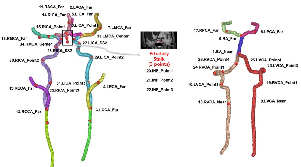
Data sources: The study was approved by the local institutional review board. Written informed consent was obtained from all patients. A total of 539 patients with cerebrovascular disease were recruited from a multiple center study. MR vessel wall images were obtained using a head and neck joint scan. Of these, 305 patients had occluded vessels.Data preprocessing: As shown in Figure 1, 35 points from common carotid artery to intracranial arterial terminus were annotated by four experienced radiologists, especially vessel bifurcation, carotid siphon, and pituitary stalk. Cropping and random mirroring were employed, expanding the total training set to 1296 cases. Zero-mean normalization and truncation were applied to eliminate the influence of outliers on input data. Centered random sampling across the entire image is used to control memory usage and ensure input data diversity.
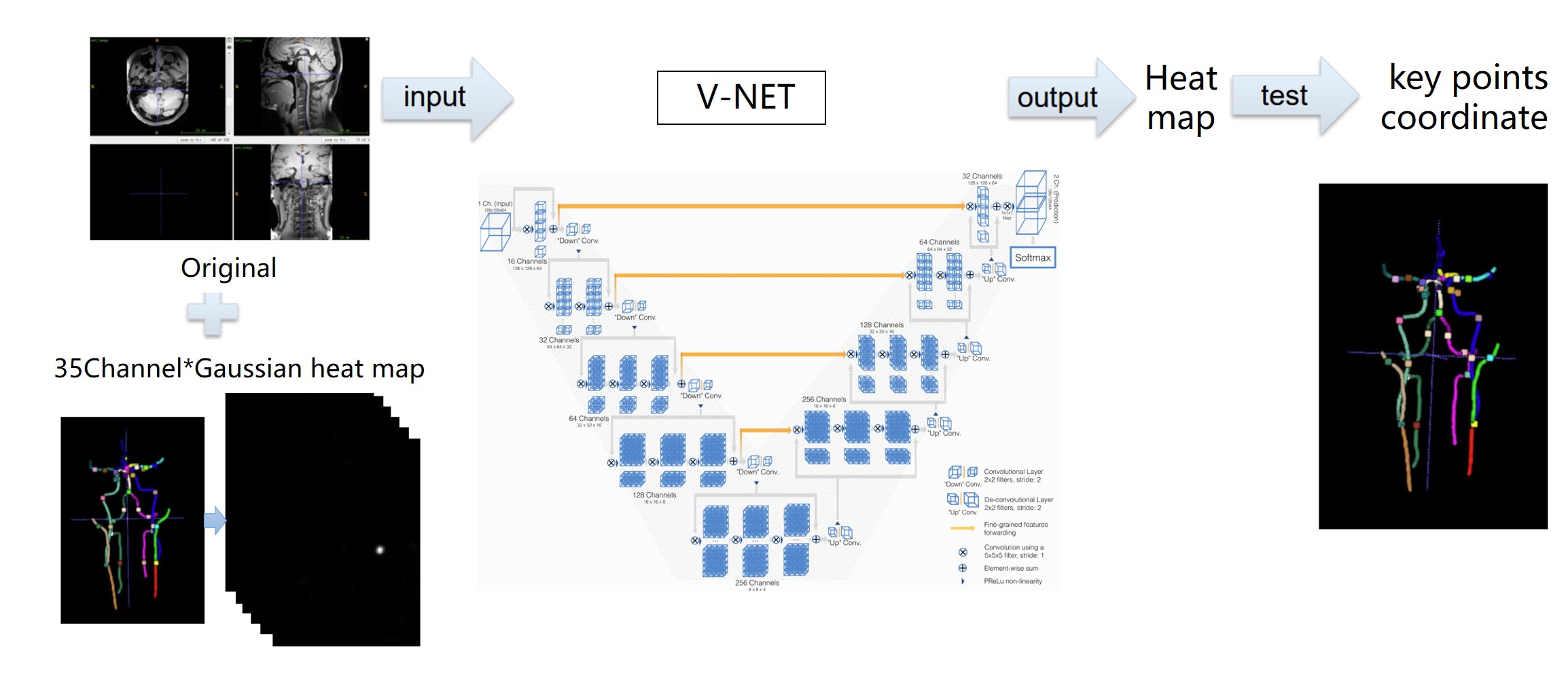
Detection framework: The main workflow of the network is illustrated in Figure 2. The input consists of the original images and a combination of Gaussian maps for 35 channels. The output were also Gaussian maps. The physical coordinates of key points are obtained through a module. The Gaussian maps are generated by placing a Gaussian kernel at the center of the ground-truth key points. The 3D V-NET network is used to effectively preserve information within the volume [7]. The network consists of a compression path and a decompression path, addressing the gradient vanishing issue through skip connections and incorporating residual units to minimize information loss. The images of 432 and 107 patients were used for training and testing the network, respectively.
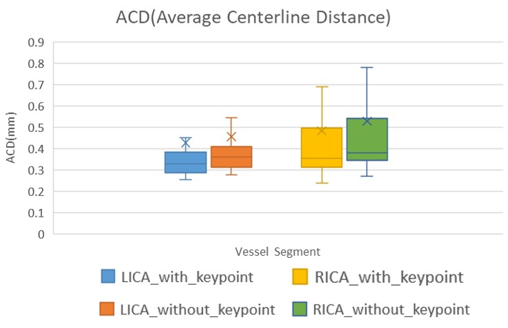
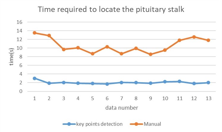
Evaluation indexes: A grayscale threshold was set on the outputs to determine whether it is detected. The Euclidean distance between the physical coordinates of key points and the ground-truth was calculated, with an appropriate threshold set to determine whether it’s error detected. The Average Centerline Distance (ACD) is employed to assess the difference between the predicted centerline and the ground-truth. Additionally, the time needed for detecting the pituitary stalk with and without this algorithm was compared.
result
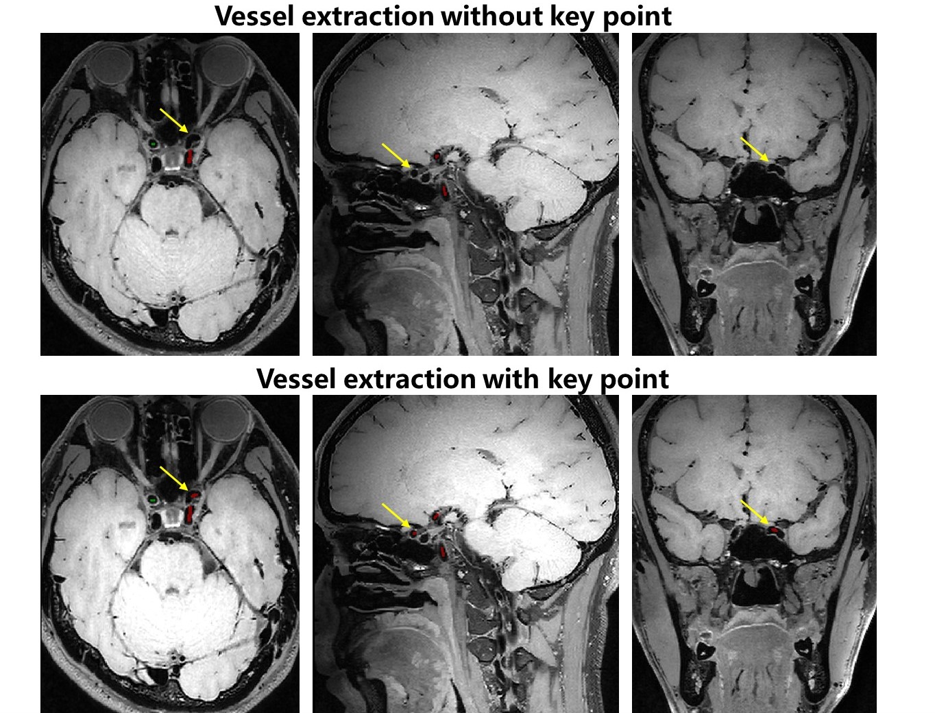
The average detection accuracy for the 35 target points was 88.99%, with 3.644 missed detections. Specifically, the detection accuracy for the upper, middle, and lower endpoints of the pituitary stalk was 99%, and the accuracy at the sharp corner of the siphon section reached 97%. Notably, 21 points had an accuracy exceeding 90%. As shown in Figure 3, the anterior curvature of the carotid siphon is effectively traced. The ACD for centerline extraction is significantly reduced, as illustrated in Figure 4. Simultaneously, the time needed for pituitary stalk localization is notably shortened, as depicted in Figure 5.discussion
The carotid siphon poses a significant challenge in vascular centerline extraction, and this algorithm has achieved a 0.97 accuracy in addressing this difficulty by accurate key points detection. The algorithm successfully annotated key points in both normal and occluded scenarios, optimizing the model's sampling methods and loss functions. It dynamically adjusted evaluation criteria while ensuring both algorithm accuracy and computational speed. The results indicated that the algorithm can assist in achieving faster and more accurate vascular centerline extraction. In addition, then aided in segmenting the lumen, analyzing the vessel wall, and quantifying plaque geometry. Additionally, through the rapid and automatic localization of the pituitary stalk, the algorithm achieved automated and quantitative analysis of plaque enhancement. This work hold significant clinical implications for the prevention and treatment of cerebrovascular diseases. The algorithm allows for flexible adaptation to clinical needs, and a wide range of potential applications exist.conclusion
The proposed algorithm has achieved fully automatic and accurate detection of 35 key points, including the head and neck vessels and pituitary stalk, which can improve the accuracy of vascular centerline extraction and realize rapid and automatic quantitative analysis of plaque enhancement. This improvement will enhance the clinical efficiency in diagnosing arterial plaques.Acknowledgements
The study was partially support by Natural Science Foundation of Guangdong Province-Outstanding Youth Project (2023B1515020002), National Key Technology Research and Development Program of China (2021YFF0501502), Key Laboratory for Magnetic Resonance and Multimodality Imaging of Guangdong Province (2023B1212060052), and Central guidance for local science and technology development project (ZYYD2023D02).References
[1] Saba, L., et al. "Carotid artery wall imaging: perspective and guidelines from the ASNR vessel wall imaging study group and expert consensus recommendations of the American Society of Neuroradiology." American Journal of Neuroradiology 39.2 (2018): E9-E31.
[2] Millon, Antoine, et al. "Clinical and histological significance of gadolinium enhancement in carotid atherosclerotic plaque." Stroke 43.11 (2012): 3023-3028.
[3] Kawahara, Ichiro, et al. "High-resolution magnetic resonance imaging using gadolinium-based contrast agent for atherosclerotic carotid plaque." Surgical neurology 68.1 (2007): 60-65.
[4] Fakih, Rami, et al. "Detection and quantification of symptomatic atherosclerotic plaques with high-resolution imaging in cryptogenic stroke." Stroke 51.12 (2020): 3623-3631.
[5] Zhou, Hanyue, et al. "VWI‐APP: Vessel wall imaging–dedicated automated processing pipeline for intracranial atherosclerotic plaque quantification." Medical Physics 50.3 (2023): 1496-1506.
[6] Diedrich, Karl T., et al. "Comparing performance of centerline algorithms for quantitative assessment of brain vascular anatomy." The Anatomical Record: Advances in Integrative Anatomy and Evolutionary Biology 295.12 (2012): 2179-2190.
[7] Milletari, Fausto, Nassir Navab, and Seyed-Ahmad Ahmadi. "V-net: Fully convolutional neural networks for volumetric medical image segmentation." 2016 fourth international conference on 3D vision (3DV). Ieee, 2016.
Figures