4512
Denoising Arterial Spin Labeling Perfusion MRI using Deep Learning Methods1Center for Biomedical Imaging Research, Tsinghua University, Beijing, China
Synopsis
Keywords: Other AI/ML, Machine Learning/Artificial Intelligence, Arterial Spin Labeling
Motivation: The low SNR of arterial spin labeling (ASL) perfusion weighted images (PWI) directly affects the accuracy of cerebral blood flow (CBF) quantification.
Goal(s): Our goal was to improve SNR of ASL PWI and access accurate CBF quantification.
Approach: We developed XtwASL, a deep learning model for denoising ASL PWI. Evaluation included SNR, peak signal-to-noise ratio (PSNR) and structural similarity index (SSIM) for PWI quality as well as ICC and Bland-Altman analysis for CBF assessment.
Results: Compared with other methods, the proposed XtwASL significantly improved SNR of ASL in the case of short acquisition time and the CBF after XtwASL denoising was more accurate.
Impact: The ASL denoising method XtwASL not only improves SNR but also has good clinical usability in CBF quantification, and this may reduce patient discomfort and artifacts caused by long scan time especially in multi-PLD ASL and high-resolution ASL field.
Introduction
Cerebral ischemia is a common pathophysiological process of many brain diseases, including stroke and neurodegenerative diseases[1]. The severity of cerebral ischemia can be quantitatively expressed by cerebral blood flow (CBF)[2]. Arterial spin labeling (ASL) is a non-invasive, non-radiation and contrast agent-free magnetic resonance imaging technique that enables quantitative measurement of CBF[3]. However, limited by various factors such as blood labeling efficiency, blood longitudinal relaxation rate (T1), and post-labeling delay time, the perfusion signal of ASL images is low, which leads to poor signal-to-noise ratio (SNR). To address this, ASL typically requires repeated scans for averaging, which unfortunately prolongs the imaging time of ASL. The longer scan time may introduce more artifacts due to head movements, which may further affect the accuracy of CBF quantification. In this study, we proposed XtwASL, a deep learning model for ASL Perfusion Weighted Image (PWI) denoising, with the goal of shortening the imaging time while ensuring high SNR and accurate CBF quantification.Methods
1.Study design and data collectionThis study utilized cerebral ASL imaging data from 52 asymptomatic subjects, consisting of pCASL source images (label & control) and corresponding M0 images. The experiment was performed on a 3T MR scanner (Philips Achieva, Philips Healthcare, Netherlands) equipped with a 32-channel head coil (TR/TE=4378.4/12.0ms; PLD=1800ms; labeling duration=1800ms; voxel size=3×3×6mm3; FOV=240×240mm2; 22 slices).
2.Denoising task and network architecture
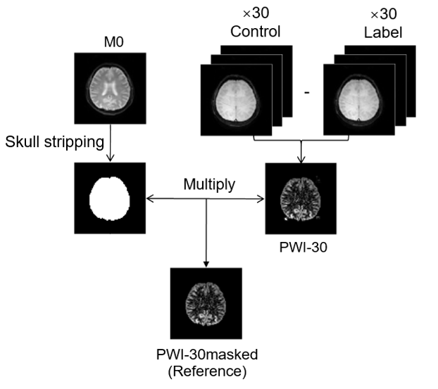
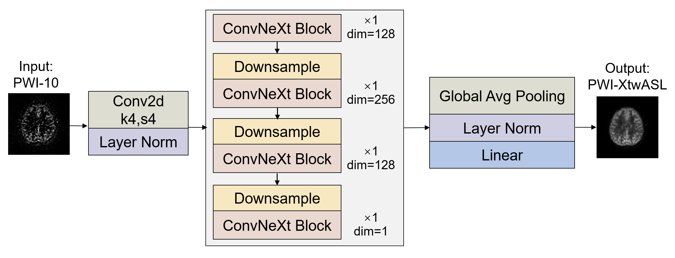
The dataset was divided into training and testing sets in a 10:3 ratio. The gold standard (PWI-30masked) was obtained by averaging 30 pairs of control-label images after skull stripping (Fig 1). For the deep learning network, 10 pairs of control-label averaged images (PWI-10) were used as input. The XtwASL network architecture, based on ConvNext[4], had a shallow depth design of [1,1,1,1] and utilized reduced channels: [128,256,128,1] (Fig 2).
3.Training details
The mean square error (MSE) served as the loss function, and the AdamW optimizer was employed for training with a learning rate of 0.004, batch size of 16, and weight decay of 0.05.
4.Denoising result evaluation
To comprehensively evaluate the denoising outcomes, XtwASL was compared with other models proposed in prior studies, namely ASLDLD[5], DeepASL[6], DAE[7], and DLASL[8]. The denoising performance of these algorithms was assessed using metrics such as SNR, peak signal-to-noise ratio (PSNR), and structural similarity index (SSIM).
5.CBF calculation and statistical analysis
CBF values were calculated for the original image (CBF-10), the XtwASL denoised image (CBF-XtwASL), and the gold standard (CBF-30)[9]. Statistical analysis was conducted using SPSS (version 16.0, SPSS Inc., Chicago, IL) and GraphPad Prism 9 (GraphPad Software Inc., San Diego, CA). Intraclass correlation coefficient (ICC) was used to measure the agreement and ICC > 0.75 was considered very high consistency. Bland-Altman analysis was employed to measure bias and estimate confidence intervals.
Results
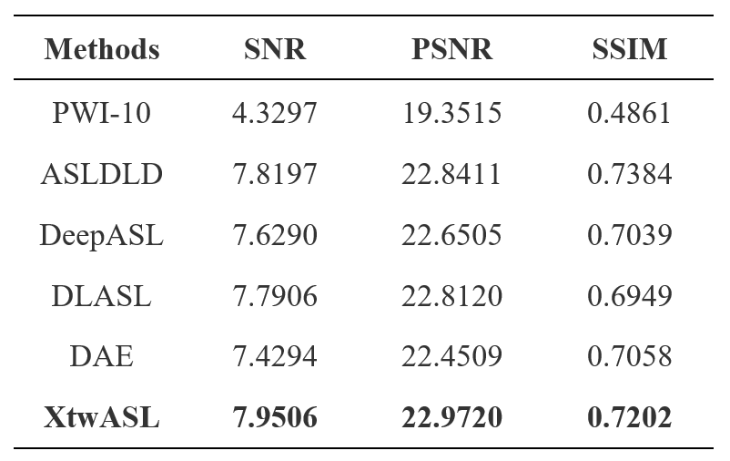
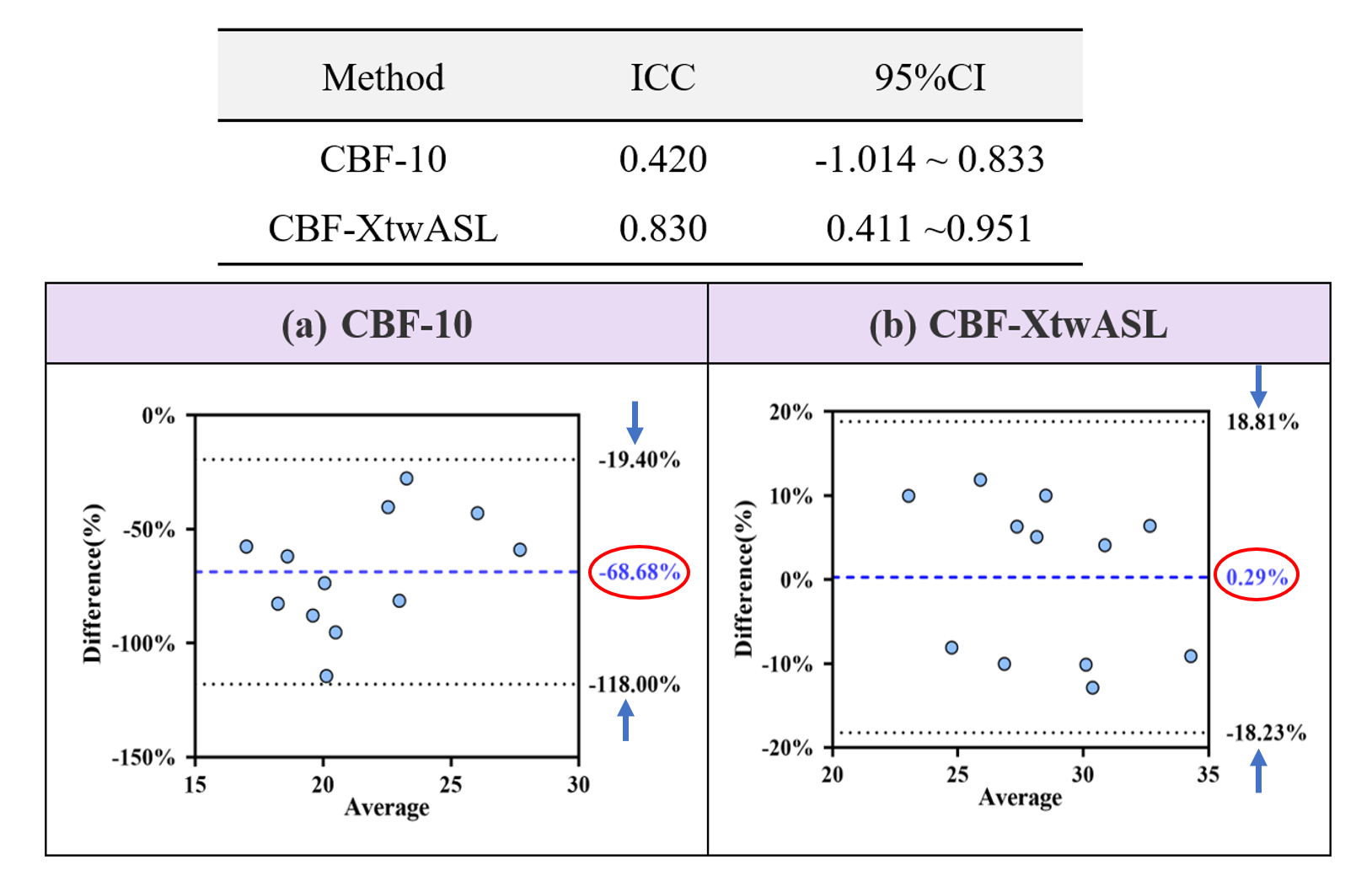
The proposed XtwASL method exhibited significant improvements in ASL PWI quality compared to ASLDLD[5], DeepASL[6], DAE[7], and DLASL[8], as demonstrated by higher SNR (7.9506), PSNR (22.9720), and SSIM (0.7402) values (Fig 3, Table 1). The PWI-XtwASL showed comparable image quality to PWI-30 but with only one-third of the acquisition time. Furthermore, the CBF map denoised by XtwASL demonstrated a higher structural similarity to the gold standard. Quantitative analysis revealed a higher agreement between CBF-XtwASL and CBF-30 (ICC: 0.830; 95% CI: 0.411-0.951) than between CBF-10 and CBF-30 (ICC: 0.420; 95% CI: -1.014~0.833), along with a smaller bias (0.29%, 95% CI: -18.23% to 18.81%) compared to CBF-10 (-68.68%, 95%CI: -118.00%~-19.40%) (Fig 4).Discussion
This was a comparative study of using deep learning method to denoise ASL PWI. The results showed that compared with previous studies, our model, XtwASL, demonstrated a superior performance and accurate CBF quantification, which is beneficial to shorten the acquisition time as well as improve the image quality. This technology may also have important potential in actual clinical applications. High-resolution ASL imaging with a resolution of 1mm or less is a time-consuming process and often yields poor image quality. XtwASL presents a promising solution by enabling high-resolution ASL imaging within a shorter acquisition time, which has significant implications for the diagnosis of small lesions and advancements in neuroscience research. Furthermore, patients requiring long PLD ASL imaging face challenges due to relaxation-induced signal attenuation, resulting in decreased signal amplitude and reduced image SNR. XtwASL demonstrates great potential in substantially improving SNR and facilitating accurate CBF measurements, thereby providing more precise data support for patient diagnosis and treatment.Conclusion
The proposed XtwASL model exhibits superior performance in ASL denoising compared with four models proposed in previous studies, providing images with enhanced SNR characteristics and maintaining high agreement with the gold standard CBF estimation.Acknowledgements
No acknowledgement found.References
[1] Durukan A, et al. Pharmacology Biochemistry and Behavior 2007; 179-197.[2] Botteri M, et al. Crit Care Med 2008 ;36(11): 3089-92.
[3] Jezzard P, et al. 2018 ; 38(4):603-626.
[4] Liu Z, et al. IEEE 2022; 11976-11986.
[5] Xie D, et al. arXiv 2018; arXiv: 1801.09672.
[6] Xie D, et al. Magn Reson Imaging 2020; 68:95-105.
[7] Hales P W, et al. Journal of Magnetic Resonance Imaging, 2020, 52(5): 1413-1426.
[8] Ulas C, et al. MICCAI 2018; 30-38.
[9] Alsop D C et al. Magn Reson Imaging 2015; 73(1): 102-116.
Figures