4502
Aliasing Artefact Suppression in Machine Learning MRI Reconstruction for Random Phase-Encode Undersampling1College of Electronics and Information, Qingdao University, Qingdao, China, 2College of Mechanical and Electrical Engineering, Qingdao University, Qingdao, China
Synopsis
Keywords: AI/ML Image Reconstruction, Image Reconstruction
Motivation: Random phase-encode undersampling of Cartesian k-space trajectories is widely implemented in magnetic resonance imaging. However, its one-dimensional randomness inherently introduces large coherent aliasing artefacts along the undersampled direction in the reconstruction, which need to be suppressed.
Goal(s): Our goal is to introduce a novel reconstruction scheme to reduce the one-dimensional undersampling-induced aliasing artefacts.
Approach: We propose an intermediate-domain network tailored for operation in image-Fourier space, which utilizes the superior non-coherent properties of decoupled one-dimensional signals to reduce aliasing artifacts.
Results: Experiments illustrate that the proposed method is particularly well-suited for regular Cartesian undersampling scenarios.
Impact: The intermediate-domain network tailored to operate in the Image-Fourier space, can efficiently reduce aliasing artefacts by utilizing the superior incoherence property of the decoupled one-dimensional signals. This could further inspire the development of MRI reconstruction technology based on machine learning.
Introduction
To accelerate MRI scans, a common method involves under-sampling the k-space data. Since the pioneering work of Wang et al. 1, the application of machine learning (ML) algorithms in fast MRI reconstruction has attracted widespread attention. In MRI scans where conventional phase-only random under-sampling patterns are applied to Cartesian k-space, images reconstructed directly through inverse Fourier transformation will exhibit global aliasing artifacts throughout the entire spatial space. In theory, utilizing global information of the MR images can reduce these aliasing artifacts2. Due to the narrow receptive field of the convolutional kernel in a neural network makes convolutional neural networks sub-optimal in capturing global information of an image3.In this study, we present ID-Net, an intermediate domain-oriented network that utilizes one-dimensional (1-D) global convolution to estimate incomplete intermediate domain signals represented via 1-D inverse Fourier transform (IFT). The estimated intermediate domain signals are transformed to the image domain via another 1D IFT. ID-Net improves the quality of reconstructed images and suppresses aliasing artifacts, achieved with lower computational complexity and fewer parameters. Experiments illustrate that the proposed method is particularly well-suited for regular Cartesian undersampling scenarios.
Methods
Let $$$ x=C^{H\times W}$$$ be a fully sampled k-space data. 2-D IFT is given by:$$$ y(h,w)=\sum_{v=0}^{W-1}\sum_{u=0}^{H-1}x(u,v)e^{j2\pi\left(\frac{hu}{H}+\frac{wv}{W}\right)} $$$ (1)
2-D IFT can be easily split into two 1-D IFTs in sequence:
$$$ y=IFT_{W}(IFT_{H}(x)) $$$ (2)
where, $$$ IFT_{H}(x)=\sum_{u=0}^{H-1}x(u,v)e^{j2\pi\left(\frac{hu}{H}\right)} $$$ or $$$IFT_{W}(x)=\sum_{v=0}^{W-1}x(u,v)e^{j2\pi\left(\frac{wv}{W}\right)}$$$ represent 1-D IFT along the H or W direction, respectively.
In the phase-encoded undersampling pattern, the intermediate domain data obtained from 1-D IFT along the H direction has the superior incoherent property, which allows the rows (W direction) of data to be processed independently during the recovery of the intermediate domain data. 1-D data operation reduces the requirement for the convolutional sensing field from 2-D to 1-D. For this reason, we propose ID-Net, shown in Fig.1. ID-Net consists of five cascading blocks, each of which uses 1-D global convolution to recover data in the intermediate domain, a feature enhancement module composed of complex convolutions with different expansion rates to enhance features in the image domain, and a data consistency unit (DC)4 to impose constraints from the raw MR measurement and ensure data fidelity. We used CReLU5 as the choice of non-linearity.
Experiments
The proposed ID-Net is compared with four state-of-the-art MRI reconstruction approaches on the fastMRI single-coil knee dataset and Calgary-Campinas brain dataset: dAUTOMAP6, DOTA7, Recon-former8 and DIMENSION9. The undersampling pattern is 1-D Cartesian random sampling along the phase-encoding direction with four-fold acceleration.All networks were trained with 50 epochs using the Adam optimizer (lr=10-3). The reconstructions were evaluated by peak signal-to-noise ratio (PSNR), structural similarity (SSIM) and normalized mean square error (NMSE). Additionally, the number of parameters (#Params) and floating-point operations per second (FLOPs) are used as additional evaluation metrics.
As shown in Table 1, ID-Net achieves optimal performance while maintaining a relatively small amount of computation and parameters. Fig.2 shows knee slices reconstructed from the fastMRI test dataset. We notice that: 1) In the first and second rows, except for the proposed ID-Net, other methods cannot effectively suppress artifacts. 2) The area in the red box (third row) indicates only the proposed model provides the most distinct and improved visualization of image quality. Fig.3 shows brain slices reconstructed from the Calgary-Campinas test dataset. As observed from the first and second rows, the reconstructed image from dAUTOMAP appears smooth and lacks contrast and fine texture details. While DOTA, Recon-former and DIMENSION contribute to an overall improvement in image quality, detailed information remains missing. Only the proposed model can reconstruct the image with high contrast and rich texture details.
Discussion
Overall, the intermediate domain data obtained from 1-D IFT has the superior incoherent property. ID-Net recovers the intermediate domain data using 1-D global convolution. This technique addresses the challenge posed by the current receptive field of the convolutional neural network, allowing for the capturing of global information effectively. The experimental results indicate that ID-Net offers the best performance Since ID-Net focuses on direct data recovery in the intermediate domain, the resulting reconstructed images may still have room for improvement.Conclusion
This study investigates the ID-Net estimates the intermediate-domain signals decoupled by a 1-D IFT using a low-dimensional global convolution. This approach improves the quality of the reconstructed images and suppresses aliasing artifacts with lower computational complexity and fewer parameters. This study focuses on single-coil datasets, and in the future, we plan to extend our approach to parallel MRI with multi-coil datasets, further improving the reconstruction quality of the proposed neural network.Acknowledgements
The authors are grateful for the financial support from Shandong Provincial Natural Science Foundation, China, Grant/Award Number: ZR2021MF025.
References
1. Wang S, Su Z, Ying L, et al. Accelerating magnetic resonance imaging via deep learning. In Proceedings of the 13th IEEE International Symposium on Biomedical Imaging, Prague, Czech Republic, 2016:514-517.
2. Li Y, Yang J, Yu T, Chi J, Liu F. Global attention‐enabled texture enhancement network for MR image reconstruction. Magn Reson Med. 2023;90(5):1919-1931.
3. Cole E, Cheng J, Pauly J, Vasanawala S. Analysis of deep complex‐valued convolutional neural networks for MRI reconstruction and phase‐focused applications. Magn Reson Med. 2021;86(2):1093-1109.
4. Schlemper J, Caballero J, Hajnal JV, Price AN, Rueckert D. A deep cascade of convolutional neural networks for dynamic MR image reconstruction. IEEE Trans Med Imaging. 2017;37(2):491-503.
5. Shang W, Sohn K, Almeida D, Lee H. Understanding and Improving Convolutional Neural Networks via Concatenated Rectified Linear Units. In Proceedings of the 33rd International Conference on Machine Learning. 2016:2217-2225.
6. Schlemper J, Oksuz I, Clough JR, Duan J, King AP, Schnabel JA, Hajnal JV, Rueckert D. dAUTOMAP: Decomposing AUTOMAP to achieve scalability and enhance performance. 2019. arXiv preprint arXiv:1909.10995.
7. Eo T, Shin H, Jun Y, Kim T, Hwang D. Accelerating Cartesian MRI by domain-transform manifold learning in phase-encoding direction. Med Image Anal. 2020;63:101689.
8. Guo P, Mei Y, Zhou J, Jiang S, Patel VM. ReconFormer: Accelerated MRI reconstruction using recurrent transformer. IEEE Trans Med Imaging. 2023.
9. Wang S, Ke Z, Cheng H, Jia S, Ying L, Zheng H, Liang D. DIMENSION: dynamic MR imaging with both k‐space and spatial prior knowledge obtained via multi‐supervised network training. NMR Biomed. 2022;35(4): e4131.
Figures
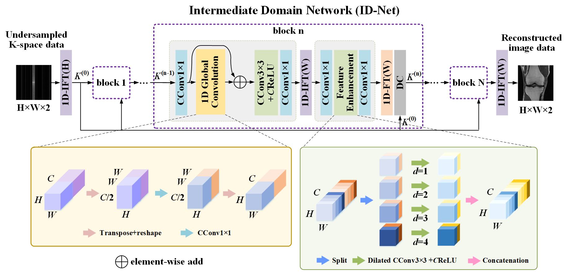
Fig. 1. Overview proposed model ID-Net.
Abbreviations: IFT, Inverse Fourier Transform; DC, Data consistency; CConv1×1, complex convolution layer with kernel size 1; CReLU, Concatenated ReLU.
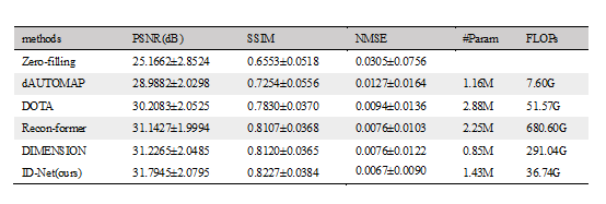
Table 1. Evaluation results from different methods on fourfold fastMRI test dataset, where the data represents the mean ± SD.
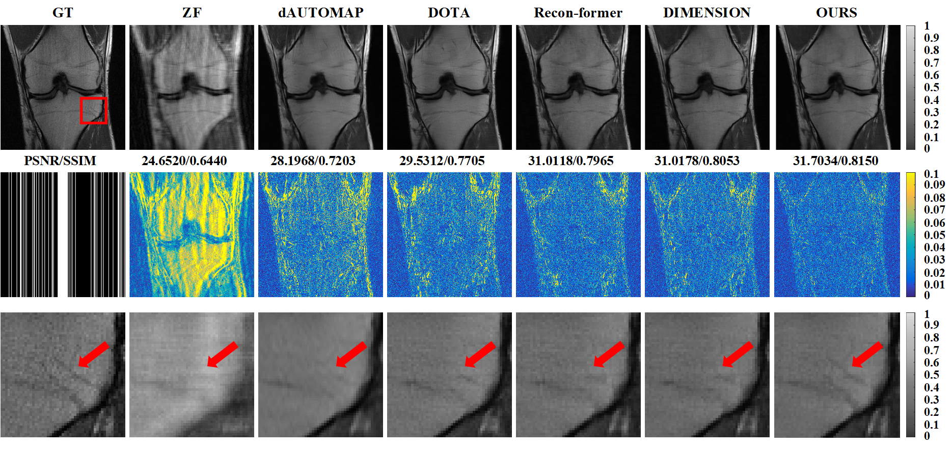
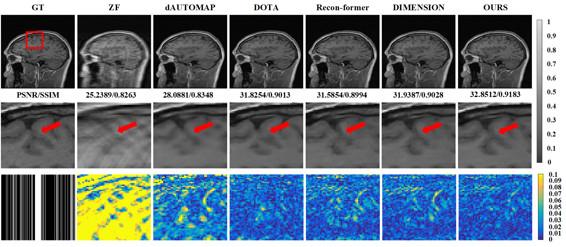
Fig. 3. Brain slices reconstructed from the Calgary-Campinas test dataset under 4-fold acceleration rate (row 1) . The regions of interest (red patches) and their corresponding error images are zoomed in side-by-side in rows 2-3. At the beginning of the third row, the under-sampled mask is given. We direct the reader’s attention to the red arrows shown in the reconstructed images, which indicate fine anatomical features.