4500
DL based reconstruction method for an undersampled PROPELLER MRI data1GE HealthCare, Bangalore, India
Synopsis
Keywords: AI/ML Image Reconstruction, Image Reconstruction, PROPELLER
Motivation: Periodically Rotated Overlapping Parallel Lines with Enhanced Reconstruction (PROPELLER) is a popular MRI acquisition scheme used for clinical and research MRI data acquisition due to its robustness to motion. However, it is known to have long scan times.
Goal(s): Reduce scan time for PROPELLER scans to make it feasible for usage in regular clinical settings.
Approach: An unrolled algorithm based deep learning reconstruction method for PROPELLER scans has been proposed, which performs reconstruction at the blade level.
Results: Proposed method has been demonstrated to perform good reconstruction on single coil data for multiple anatomies and contrasts.
Impact: This method has the potential to reduce PROPELLER scan times and make it a popular choice for acquisition in clinical settings.
Introduction
PROPELLER1 is a popular MRI acquisition scheme used for clinical and research MRI data acquisition and is known for its robustness to motion. By virtue of the acquisition scheme PROPELLER scans have higher scan times as compared to similar Cartesian MRI acquisition scheme. In this work, we propose a reconstruction method to reduce scan time for PROPELLER acquisitions. There are several approaches to reduce scan time for PROPELLER acquisition2. The scan time reduction approach which we use for this work is to undersample each PROPELLER blade. The problem of reconstruction in such cases can be solved completely in the non-cartesian space3,4. However, the PROPELLER acquisition can be considered to be a set of blades acquired in a Cartesian manner which are then arranged at certain angles in the non-cartesian grid. In this work, we utilize the Cartesian representation of the individual blades in a PROPELLER acquisition to reconstruct undersampled PROPELLER MRI data.Method
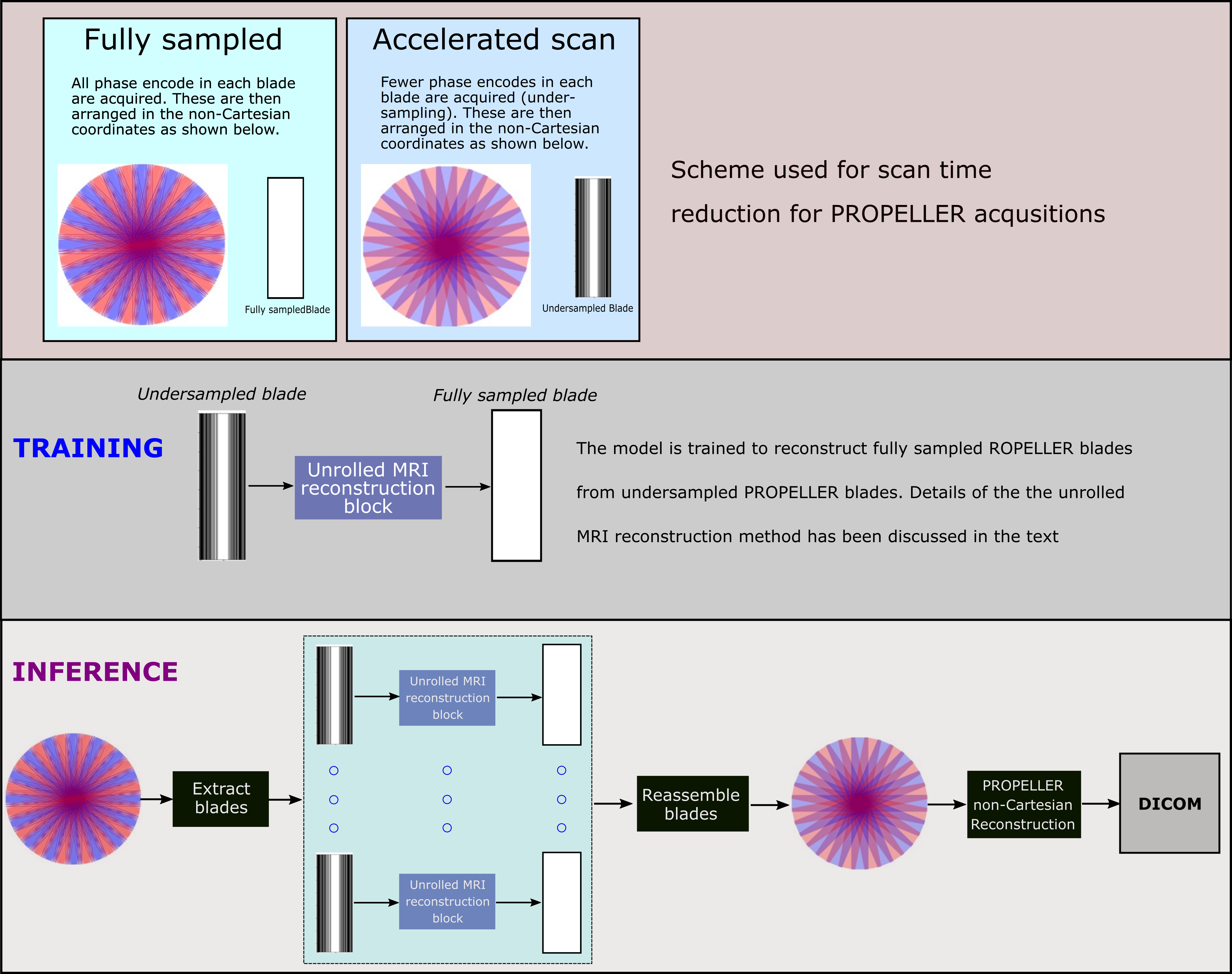
Reconstruction ApproachScan time reduction approach for PROPELLER MRI data acquisition has been illustrated in Figure-1 (top row). Since each blade is a Cartesian representation, an unrolled algorithm-based DL network is trained to reconstruct fully sampled PROPELLER blades from undersampled PROPELLER blades. For this work, we limit our training and results to single channel data reconstruction. Hence, the MR image formation can be explained as $$$y=MFx$$$, where $$$y$$$ is the acquired undersampled data and $$$x$$$ is the fully sampled image to be estimated, $$$M$$$ is the binary acquisition mask, $$$F$$$ is the Fourier transform operation. The problem for optimization is setup as one where we use proximal mapping data consistency5,6. Hence, the optimization problem to be solved is $$$\min_x \left \| y-Ax \right \|_2^2 + \lambda \left \| x-f_\theta\left(x\right) \right \|_2^2$$$, where, $$$A=MF$$$, $$$f_\theta(\cdot)$$$ is the DL network with $$$\theta$$$ trainable parameters, and $$$\lambda$$$ is the regularization weight. As shown in Ravishankar et al.5, this has a closed form update step in k-space which has been used for our method. The training and inference approaches have been shown in Figure-1. As shown in the figure, post blades are reconstructed, final DICOM data is obtained by passing the assembled reconstructed blades through regular PROPELLER reconstruction chain.
Model training and data details
A residual channel attention network (RCAN)8 was used for realizing the data fidelity term. A choice of hyper-parameters as 5 blocks, 5 groups, and CNNs with kernel size of 3 were used for the RCAN network. A total of 10 unrolls were performed for reconstruction. Across unrolls, weights are shared for the DL network (as suggested in Aggarwal et al.7). Here, $$$\lambda$$$ was provided an initial value of 0.01 and set as a trainable parameter. Loss function: SSIM and MAE were used with weights of 1.0 and 0.5 respectively. The reconstruction network was trained using in-house dataset. A total of 14542 blades were extracted from the PROPELLER datasets which were used for training.
All data was acquired on a 1.5T commercial GE HealthCare MRI scanner using the body array coil. Informed consent was obtained from the volunteers in the IRB approved study. For testing the proposed method, the following data was acquired using single array coil (body coil): sagittal pelvis T2-w, coronal knee T1-w, axial abdomen T2-w, sagittal C-spine T2-w, sagittal knee T2-w PROPELLER MRI.
Results
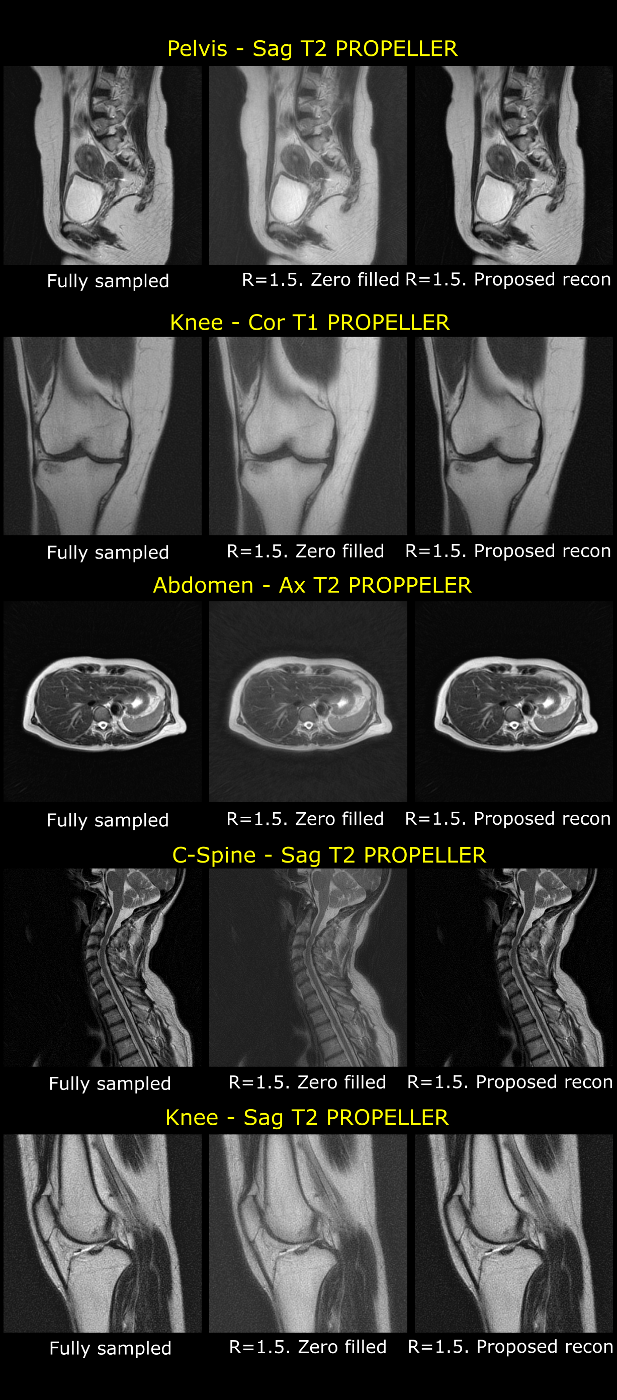
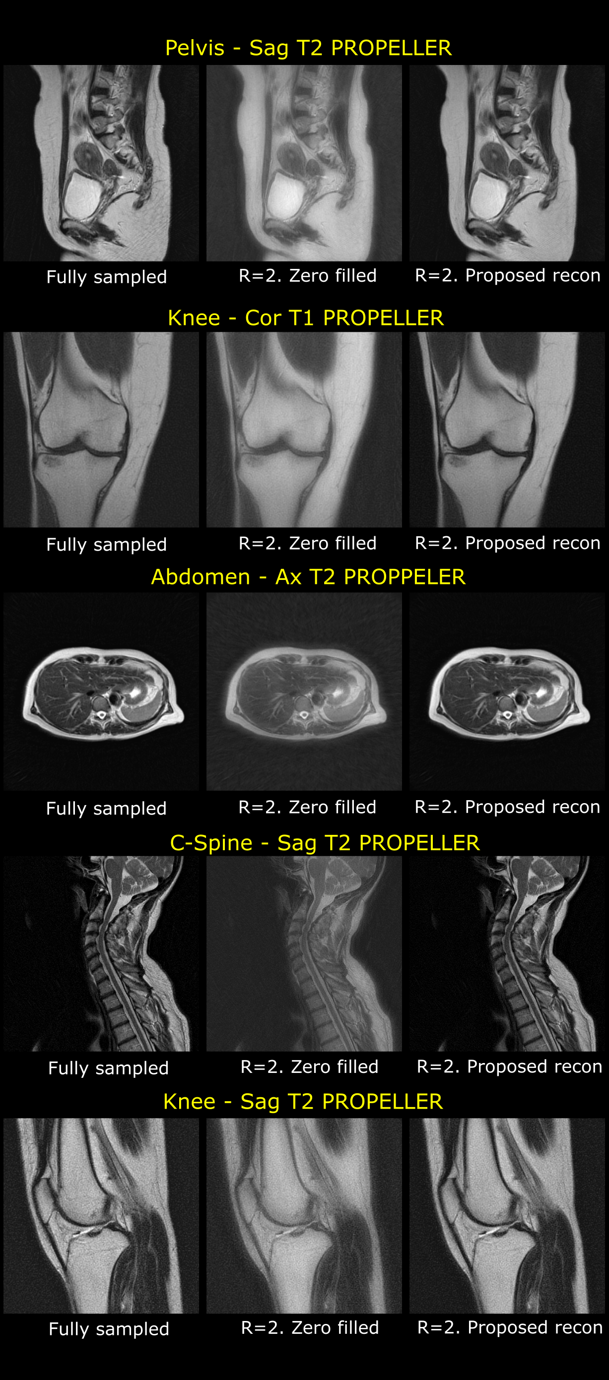
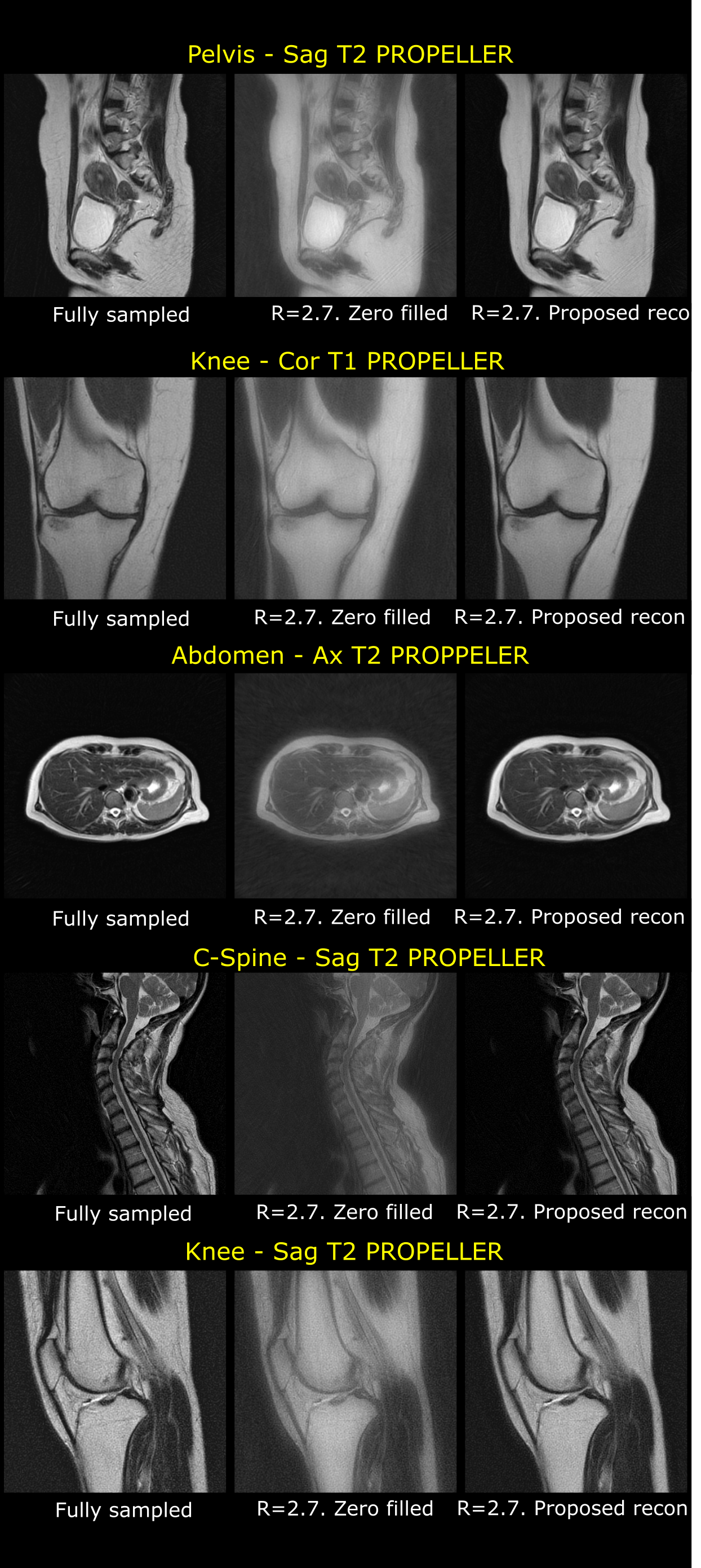
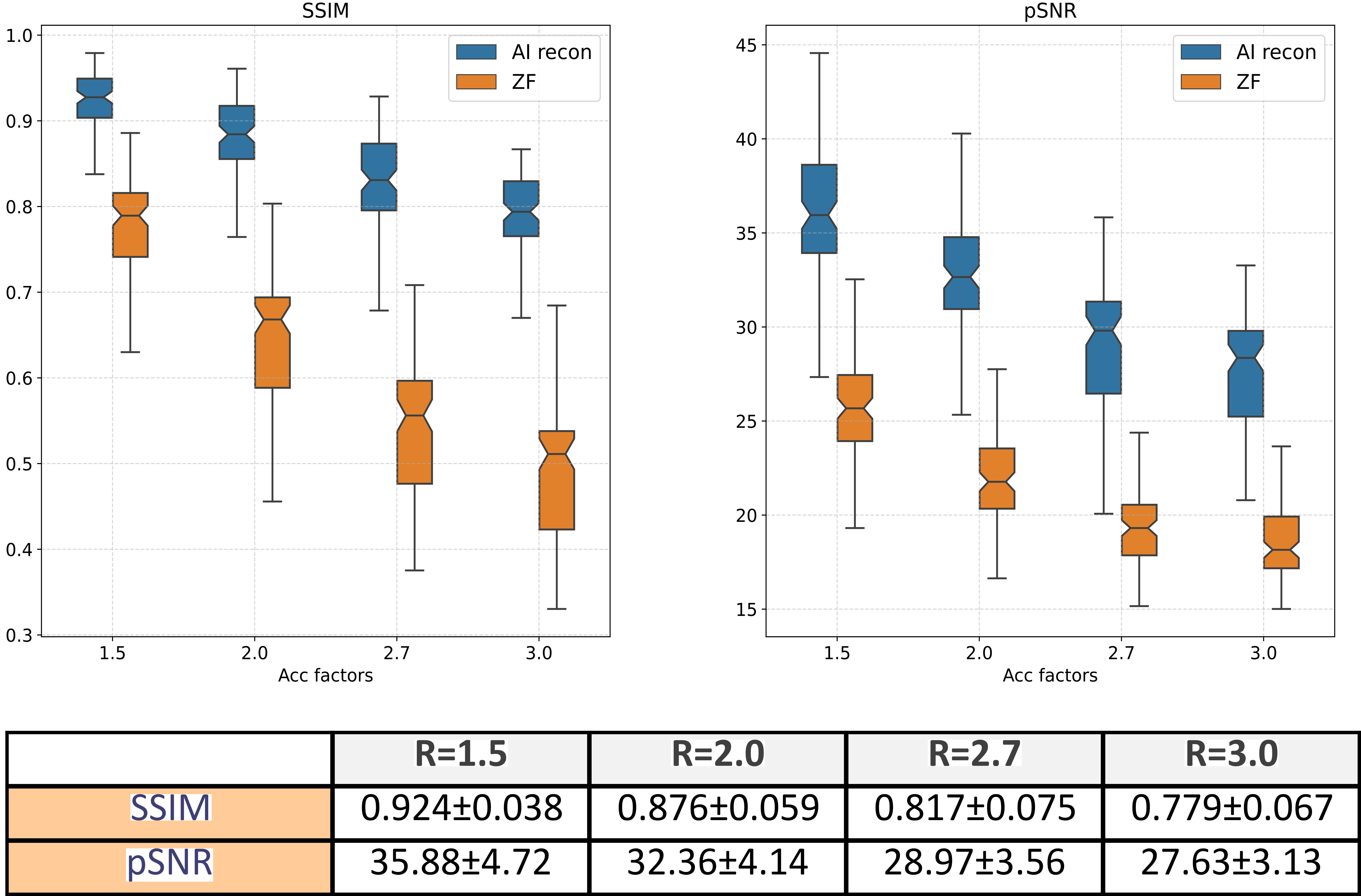
Datasets acquired using a single coil (volume coil) were used to evaluate the method’s performance. Fully sampled PROPELLER data were retrospectively undersampled using various levels of accelerations, $$$R=\{1.5,2.0,2.7,3.0\}$$$, to simulate undersampled PROPELLER MRI data. The same model was used to reconstruct retrospectively undersampled data for multiple anatomies and contrasts. Results using the proposed method are shown in Figure-2, Figure-3 and Figure-4. Structural similarity index (SSIM) and pSNR were computed between the fully sampled images and AI-reconstruction to evaluate performance of the proposed method. Metrics were computed over 101 2D slices which were reconstructed for multiple levels of acceleration. Results are shown in Figure-5.Discussion
In this work, we proposed a method to reduce scan times for PROPELLER MRI acquisitions. By virtue of this method, it is agnostic to number of blades present in the acquired data. The proposed method has been tested on several anatomies and contrasts. Since the method was evaluated on data acquired using body array coil (single coil), acceleration levels were limited to 3. However, this reconstruction method can be easily (and similarly) extended to undersampled multichannel PROPELLER MRI acquisitions. In future work, we shall evaluate the proposed method on prospectively acquired undersampled PROPELLER MRI data and extend the observations to accelerated multi-channel PROPELLER MRI data.Acknowledgements
No acknowledgement found.References
- Pipe, J.G., 1999. Motion correction with PROPELLER MRI: application to head motion and free‐breathing cardiac imaging. Magnetic Resonance in Medicine: An Official Journal of the International Society for Magnetic Resonance in Medicine, 42(5), pp.963-969
- Arfanakis, K., Tamhane, A.A., Pipe, J.G. and Anastasio, M.A., 2005. k‐space undersampling in PROPELLER imaging. Magnetic Resonance in Medicine: An Official Journal of the International Society for Magnetic Resonance in Medicine, 53(3), pp.675-683.
- Ramzi, Z., Starck, J.L. and Ciuciu, P., 2021, April. Density compensated unrolled networks for non-cartesian MRI reconstruction. In 2021 IEEE 18th International Symposium on Biomedical Imaging (ISBI) (pp. 1443-1447). IEEE.
- Ramzi, Z., Chaithya, G.R., Starck, J.L. and Ciuciu, P., 2022. NC-PDNet: A density-compensated unrolled network for 2D and 3D non-Cartesian MRI reconstruction. IEEE Transactions on Medical Imaging, 41(7), pp.1625-1638.
- Ravishankar, S. and Bresler, Y., 2010. MR image reconstruction from highly undersampled k-space data by dictionary learning. IEEE transactions on medical imaging, 30(5), pp.1028-1041.
- Schlemper, J., Caballero, J., Hajnal, J.V., Price, A.N. and Rueckert, D., 2017. A deep cascade of convolutional neural networks for dynamic MR image reconstruction. IEEE transactions on Medical Imaging, 37(2), pp.491-503.
- Aggarwal, H.K., Mani, M.P. and Jacob, M., 2018. MoDL: Model-based deep learning architecture for inverse problems. IEEE transactions on medical imaging, 38(2), pp.394-405.
- Zhang, Y., Li, K., Li, K., Wang, L., Zhong, B. and Fu, Y., 2018. Image super-resolution using very deep residual channel attention networks. In Proceedings of the European conference on computer vision (ECCV) (pp. 286-301).
Figures