4490
Volumetric Reconstruction Resolves Off-Resonance Artifacts in Static and Dynamic PROPELLER MRI1University of California, Berkeley, Berkeley, CA, United States, 2Stanford University, Stanford, CA, United States
Synopsis
Keywords: AI/ML Image Reconstruction, Motion Correction, Off-Resonance Correction, Video Reconstruction
Motivation: Off-resonance correction typically requires field map measurements or pretraining data, which are slow/difficult to collect.
Goal(s): We propose a physics-based strategy to address off-resonance artifacts using only PROPELLER measurements, by modeling an additional spectral dimension.
Approach: This strategy exploits an equivalence between measuring a PROPELLER blade at a certain angle, and viewing a relief sculpture at the same angle. In this equivalence, three-dimensional structures (fat) appear shifted along the blade/view direction relative to flatter structures (water).
Results: Our method resolves continuous chemical shift artifacts while allowing for video reconstruction of dynamic tissues. We provide preliminary results on synthetic static and dynamic data.
Impact: We use volumetric reconstruction to correct off-resonance artifacts and perform fat/water separation in PROPELLER MRI, without additional field map measurements or pretraining data. We hope our method opens the door to shorter scan times and higher temporal resolution imaging.
Introduction
Off-resonance artifacts in magnetic resonance imaging (MRI) are visual distortions that occur when the actual resonant frequencies of spins within the imaging volume differ from the expected frequencies used to encode spatial information [1-3]. We propose to resolve these artifacts by lifting the 2D MRI reconstruction problem to 3D, introducing an additional "spectral" dimension to model this off-resonance. Our approach is inspired by recent progress in modeling radiance fields [4,5], and is capable of reconstructing both static and dynamic MR images. We demonstrate our approach in the context of PROPELLER (Periodically Rotated Overlapping ParallEL Lines with Enhanced Reconstruction) [6] MRI acquisitions, which are popular for their robustness to motion artifacts. For these acquisitions, off-resonance artifacts appear as chemical shifts of fat relative to water, which can be interpreted as perspective effects in our volumetric model. Our method operates in a few minutes on a single GPU, and to our knowledge is the first to correct for chemical shift in raw gradient echo PROPELLER MRI reconstruction.Method
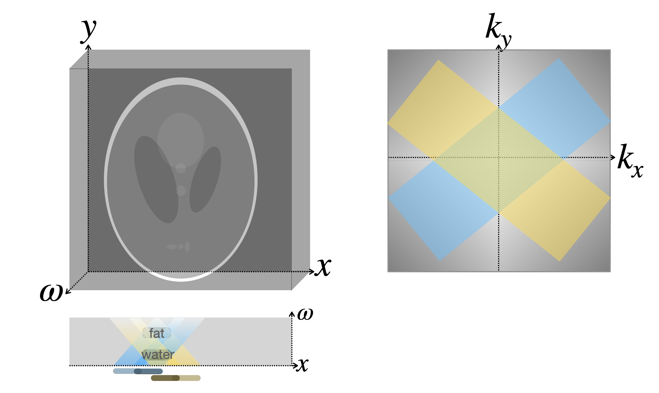
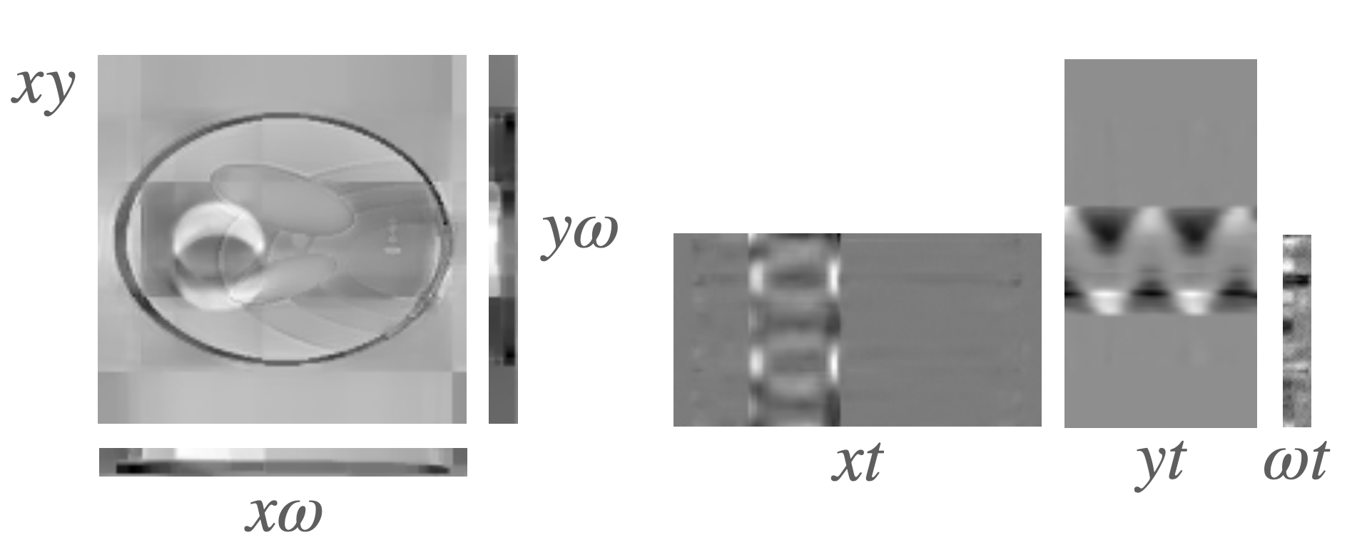
Our method is illustrated in Figure 1, and consists of: (1) a volumetric 3D or 4D model of the image to be reconstructed, including x and y spatial dimensions, a spectral dimension, and a time dimension for dynamic reconstructions, (2) a forward model mapping this volumetric model to the k-space measurements collected with each PROPELLER blade, and (3) gradient-based optimization through this forward model, to optimize the parameters of the volumetric model.Our volumetric model can be thought of as a relief sculpture in which water-based tissues lie near the back of the sculpture while fat-based tissues are more three-dimensional. For static reconstruction our volumetric model is a simple 3D grid in $$$xy\omega$$$ with complex-valued entries. For dynamic reconstruction we use a K-Planes [5] model that decomposes the 4D space into a set of six 2D grids representing $$$xy$$$, $$$x\omega$$$, $$$y\omega$$$, $$$xt$$$, $$$yt$$$, and $$$\omega t$$$.
Our forward model leverages the mathematical equivalence between sampling a PROPELLER blade along a certain angle in k-space, and viewing the volume (relief image) at the same angle, so that fatty tissues appear shifted in that direction relative to water-based tissues. We simulate a PROPELLER blade measurement by (1) projecting our volumetric model at the same angle, to produce an image with chemical shift artifacts, and (2) computing the discrete Fourier transform (DFT) of this image and masking out k-space coefficients outside the PROPELLER blade.
We optimize our volumetric model using the L2 loss function encouraging high fidelity to both the k-space and corresponding image space PROPELLER blade measurements. We also include spatial and spectral total variation and temporal smoothness regularizers. At each gradient step we consider a single PROPELLER blade, iterating among the blades randomly until convergence. We produce a corrected reconstruction without chemical shifts by projecting our optimized volumetric model along the spectral dimension, which corresponds to viewing the relief sculpture head-on instead of at an angle. For our dynamic reconstruction we project at each timestep to produce a corrected video, and denoise the final predictions using a nonlocal means filter.
Results
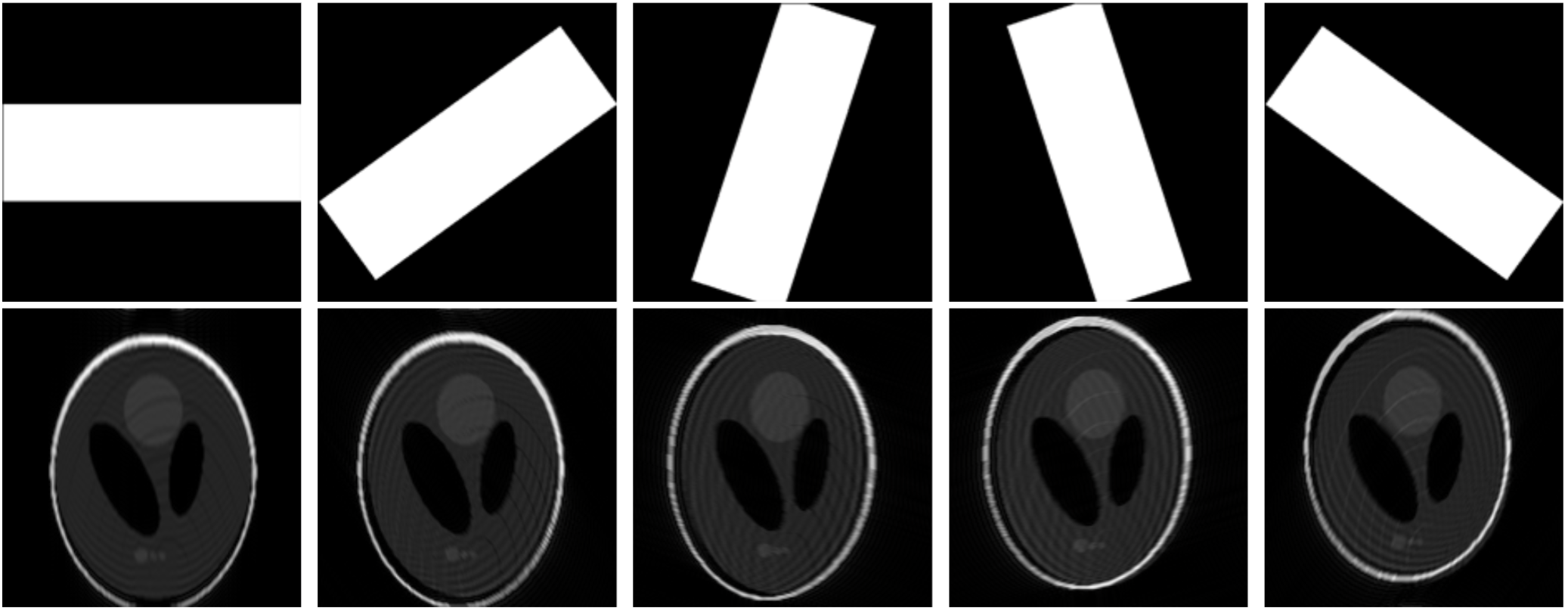
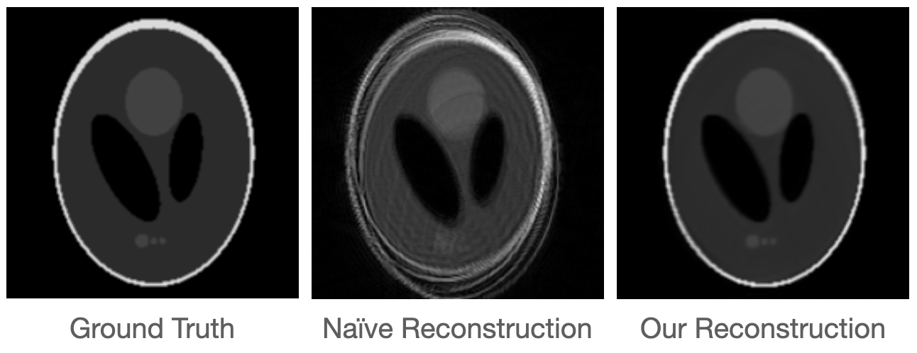
We evaluate our method on synthetic static and dynamic datasets based on the Shepp-Logan phantom separated into two components, with the bright outer ellipse simulating fat and its interior simulating water. We embed each of these layers in a static 3D slab following a shallow Gaussian contour, so that there is small, smooth spectral variation within each layer and a larger spectral jump between the water layer and the fat layer. This setup is designed to recapitulate the reality that fat exhibits larger chemical shift artifacts compared to water, but there are also smaller spectral differences between different tissues of each type.Our dynamic dataset is similar, except that the original Shepp-Logan phantom is modified so that one of the ellipses follows a sinusoidal trajectory left and right over time. We measure a single k-space blade at each timestep, cycling among the five blades shown in Figure 2 for a total of 67 timesteps and two periods of ellipse motion. We show training images and final corrected reconstructions for both datasets in Figures 2, 3, and 4, as well as the parameters from our K-Planes model in Figure 5, which shows recovery of both the spectral and dynamic aspects of the dataset.
Discussion
Our experiments are a preliminary but encouraging illustration of the potential for volumetric imaging to resolve off-resonance artifacts in MRI. Eventually, we hope that the proposed method enables faster imaging times with higher temporal resolution, without sacrificing image quality to off-resonance artifacts.Acknowledgements
We would like to thank Miki Lustig for encouraging us to pursue this research, as well as John Pauly, Efrat Shimron, and Suma Anand for helpful pointers. This material is based upon work supported by the National Science Foundation under award number 2303178 to SFK. Any opinions, findings, and conclusions or recommendations expressed in this material are those of the authors and do not necessarily reflect the views of the National Science Foundation.References
D. Y. Zeng, J. Shaikh, S. Holmes, R. L. Brunsing, J. M. Pauly, D. G. Nishimura, S. S. Vasanawala, and J. Y. Cheng, “Deep residual network for off-resonance artifact correction with application to pediatric body MRA with 3d cones,” Magnetic Resonance in Medicine, vol. 82, no. 4, pp. 1398–1411, 2019. [Online]. Available: https://onlinelibrary.wiley.com/doi/abs/10.1002/mrm.27825
Y. Lim, Y. Bliesener, S. Narayanan, and K. S. Nayak, “Deblurring for spiral real-time MRI using convolutional neural networks,” Magnetic Resonance in Medicine, vol. 84, no. 6, pp. 3438–3452, 2020. [Online]. Available:https://onlinelibrary.wiley.com/doi/abs/10.1002/mrm.28393
M. W. Haskell, A. A. Cao, D. C. Noll, and J. A. Fessler, “Deep learning field map estimation with model-based image reconstruction for off-resonance correction of brain images using a spiral acquisition,” ISMRM Workshop on Data Sampling and Image Reconstruction, 2020.
E. R. Chan, C. Z. Lin, M. A. Chan, K. Nagano, B. Pan, S. De Mello, O. Gallo, L. Guibas, J. Tremblay, S. Khamis, T. Karras, and G. Wetzstein, “Efficient geometry-aware 3D generative adversarial networks,” Computer Vision and Pattern Recognition, 2022.
S. Fridovich-Keil*, G. Meanti, F. R. Warburg, B. Recht, and A. Kanazawa, “K-Planes: explicit radiance fields in space, time, and appearance,” Computer Vision and Pattern Recognition, 2023.
K.P.N. Forbes, J. G. Pipe, C. R. Bird, and J. E. Heiserman, “PROPELLER MRI: clinical testing of a novel technique for quantification and compensation of head motion.” Journal of Magnetic Resonance Imaging, 14(3):215–222, 2001.
Figures