4488
Paired Conditional Generative Adversarial Network for Highly Accelerated Liver 4D MRI1Radiation Oncology, UCSF, San Francisco, CA, United States, 2Siemens Healthineers, Boston, MA, United States, 3Radiology, UCSF, San Francisco, CA, United States, 4Radiation Oncology, UCLA, Los Angeles, CA, United States
Synopsis
Keywords: AI/ML Image Reconstruction, Radiotherapy, 4D MRI
Motivation: Densely sampled k-space leads to high-quality MR but can be impractical due to lengthy scanning time. Accelerating MR acquisition by reducing sampling density can decrease image quality and/or increase reconstruction complexity and time.
Goal(s): This work aims to design an algorithm for efficient and high-quality reconstruction of highly accelerated radial-sampling liver 4D MR.
Approach: We proposed a novel Paired Conditional generative adversarial network term Re-Con-GAN, evaluated on a 4D liver MR dataset at 3x, 6x, and 10x acceleration ratios.
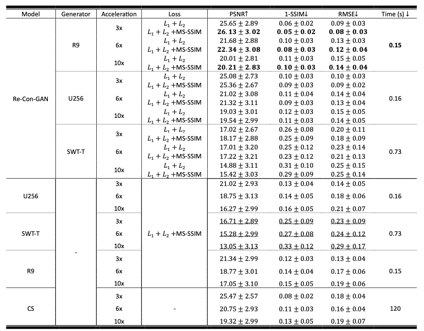
Results: Re-Con-GAN achieved better PSNR, SSIM, and RMSE with sub-second inference speed (0.15s) than compressed sensing (120s) and non-GAN deep learning methods (0.15s-0.73s).
Impact: A robust and efficient framework, Re-Con-GAN, is proposed in the current work with sub-second inference speed (0.15s) and promising reconstruction results demonstrated on an in-house curated 4D liver MRI dataset.
Introduction
4D MRI is a powerful tool to quantify tumor boundary and motion for image-guided liver radiation therapy (RT) owing to its superior soft tissue contrast1–6. In a free-breathing liver RT treatment planning workflow, the tumor contours are delineated at each respiratory phase of 4D MRI to form an internal target volume (ITV) for the ultimate target region of radiation. Full-liver coverage and high spatiotemporal resolutions are required for imaging to ensure accurate ITV definition. Continuous free-breathing scanning of 3D golden angle stack-of-star sequence with self-navigation is commonly used for acquiring 4D MRI data5. Though radial sampling is relatively robust to motion sensitivity7 and acceleration8, there are a few limitations of this technique, including long scanning time (8-10 min5), limited inter-slice resolution9, and under-sampling streaking artifacts that are evident with less regular breathing patterns.Parallel imaging10,11 and compressed sensing (CS)12–14 have been extensively studied. However, CS reconstruction tends to be slow and shows worsening performance at high under-sampling rates15–18. Recent deep learning (DL) advances have offered a data-driven approach for efficient 4D MRI reconstruction. Existing works have explored 4D MRI reconstruction with convolutional19 and recurrent20,21 neural networks as well as Transformers22. Promising results were reported using these models with explicitly defined loss functions, yet detail loss was evident at high acceleration ratios. Since balancing image fidelity, noise suppression, and detail retention in an explicitly defined loss function is difficult, we postulate that including implicit loss terms through Generative Adversarial Networks (GANs)23 training can circumvent the challenge. Though some previous studies have demonstrated superior MRI reconstruction using GANs24–27, none of them explored the capability of GANs in 4D MRI temporal profiling and reconstruction. The current study explored the feasibility of using GANs for 4D MRI reconstruction. We developed a novel architecture termed reconstruct conditional GAN (Re-Con-GAN).
Methods
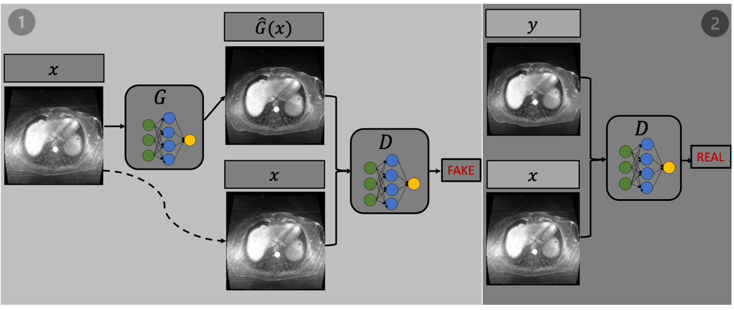
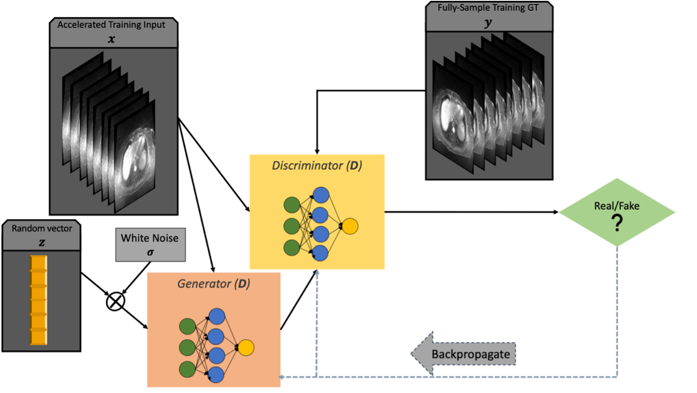
17 patients who underwent liver contrast-enhanced 4D MRI were included in the study. A prototype free-breathing T1-weighted volumetric golden angle stack-of-stars sequence was used for acquisition. The scanning parameters were - TE=1.5ms, TR=3 ms, matrix size=288 × 288 , FOV=374 mm × 374 mm, in-plane resolution=1.3 mm × 1.3 mm, slice thickness=3 mm, radial views (RV) per partition=3000, number of slices per partition=64-75, acquisition time=8-10 min. The pulse sequence ran continuously over multiple respiratory cycles. Retrospective under-sampling was performed by keeping the first 1000, 500, and 300 spokes from the total 3000 spokes (3×, 6× and 10×). For initial image reconstruction, data sorting based on self-gating signal was performed to divide the contiguously acquired k-space data into 8 respiratory phases. Nonuniform fast Fourier transform (nuFFT) was applied to reconstruct each phase. We organized the images in 2D+t. The images were resized to 256 × 256 and normalized using Z-score normalization. Data augmentation was employed, including random rotation, flipping, and cropping.Re-Con-GAN trains input (nuFFT reconstruction of under-sampled data) and output (fully sampled image series) in pairs. Re-Con-GAN is a versatile architecture with plug-and-play generator, discriminator, and loss objective, as shown in Figure 1 and 2. Three types of networks, including 3D ResNet928, UNet23, and reconstruction swin transformer (RST)22, were explored as generators. PatchGAN was selected as the discriminator. A novel loss objective fused with L1, L2, and multi-scale structure similarity indexed measurement was designed for network training.
Results
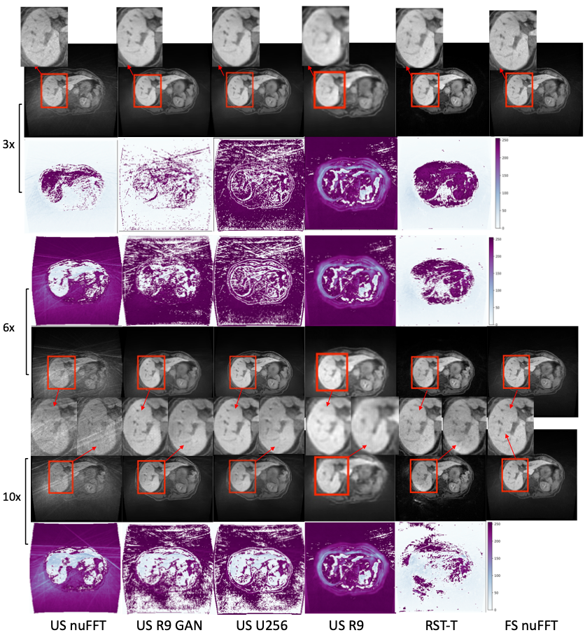
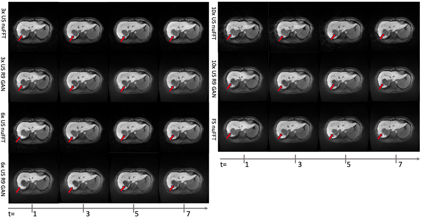
A conventional CS approach was included as the baseline. Ablation studies that solely tune generators are also conducted to underpin the improvement from Re-Con-GAN. Statistical results and visualization are reported in Table 1 Figure 3 and Figure 4. Re-Con-GAN based models is consistently superior to that of CS and ablated generators across all acceleration levels. Re-Con-GAN with 3D ResNet9 generator performs moderately better than other generators. The inference time of Re-Con-GAN with 3D ResNet9, 3D ResNet9 and CS are 0.15s, 0.15s and 120s.Discussion
RT provides effective tumor suppression for non-surgical hepatocellular carcinoma patients and the patients waiting for liver transplantation. The success of liver RT largely depends on the efficiency and quality of its imaging guidance. Therefore, we developed Re-Con-GAN for sub-second 4D reconstruction of highly accelerated MR sequences. The platform using implicit loss function is conducive to testing more aggressive acceleration ratios and future extension into raw coil signal reconstruction.Conclusion
A robust and efficient framework, Re-Con-GAN, is proposed in the current work with promising and efficient reconstruction results demonstrated on an in-house curated 4D liver MRI dataset. Re-Con-GAN has demonstrated superior performance than CS and non-GANs DL methods. Re-Con-GAN can potentially benefit the advance of online adaptive MR-guided liver RT.Acknowledgements
The study is supported in part by NIH R01CA259008 and DOD W81XWH2210044.References
1. Witt JS, Rosenberg SA, Bassetti MF. MRI-guided adaptive radiotherapy for liver tumours: visualising the future. Lancet Oncol. 2020;21(2):e74-e82. doi:10.1016/S1470-2045(20)30034-6
2. Hussain SM, Semelka RC. Hepatic imaging: comparison of modalities. Radiol Clin North Am. 2005;43(5):929-947, ix. doi:10.1016/j.rcl.2005.05.006
3. Voroney JP, Brock KK, Eccles C, Haider M, Dawson LA. Prospective comparison of computed tomography and magnetic resonance imaging for liver cancer delineation using deformable image registration. Int J Radiat Oncol Biol Phys. 2006;66(3):780-791. doi:10.1016/j.ijrobp.2006.05.035
4. Stemkens B, Paulson ES, Tijssen RHN. Nuts and bolts of 4D-MRI for radiotherapy. Phys Med Biol. 2018;63(21):21TR01. doi:10.1088/1361-6560/aae56d
5. Thomas HR, Miao X, Ferguson D, et al. Contrast-enhanced 4D-MRI for internal target volume generation in treatment planning for liver tumors. Radiother Oncol. 2022;173:69-76 doi:10.1016/j.radonc.2022.05.037
6. Paulson ES, Ahunbay E, Chen X, et al. 4D-MRI driven MR-guided online adaptive radiotherapy for abdominal stereotactic body radiation therapy on a high field MR-Linac: Implementation and initial clinical experience. Clin Transl Radiat Oncol. 2020;23:72-79. doi:10.1016/j.ctro.2020.05.002
7. Glover GH, Pauly JM. Projection reconstruction techniques for reduction of motion effects in MRI. Magn Reson Med. 1992;28(2):275-289. doi:10.1002/mrm.1910280209
8. Winkelmann S, Schaeffter T, Koehler T, Eggers H, Doessel O. An optimal radial profile order based on the Golden Ratio for time-resolved MRI. IEEE Trans Med Imaging. 2007;26(1):68-76. doi:10.1109/TMI.2006.885337
9. Li Z, Huang C, Tong A, Chandarana H, Feng L. Kz-accelerated variable-density stack-of-stars MRI. Magnetic Resonance Imaging. 2023;97:56-67. doi:10.1016/j.mri.2022.12.017
10. Pruessmann KP, Weiger M, Scheidegger MB, Boesiger P. SENSE: sensitivity encoding for fast MRI. Magn Reson Med. 1999;42(5):952-962.
11. Griswold MA, Jakob PM, Heidemann RM, et al. Generalized autocalibrating partially parallel acquisitions (GRAPPA). Magn Reson Med. 2002;47(6):1202-1210. doi:10.1002/mrm.10171
12. Feng L, Grimm R, Block KT, et al. Golden-angle radial sparse parallel MRI: combination of compressed sensing, parallel imaging, and golden-angle radial sampling for fast and flexible dynamic volumetric MRI. Magn Reson Med. 2014;72(3):707-717. doi:10.1002/mrm.24980
13. Feng L, Benkert T, Block KT, Sodickson DK, Otazo R, Chandarana H. Compressed sensing for body MRI: Compressed Sensing for Body MRI. J Magn Reson Imaging. 2017;45(4):966-987. doi:10.1002/jmri.25547
14. Lustig M, Donoho D, Pauly JM. Sparse MRI: The application of compressed sensing for rapid MR imaging. Magn Reson Med. 2007;58(6):1182-1195. doi:10.1002/mrm.21391
15. Gamper U, Boesiger P, Kozerke S. Compressed sensing in dynamic MRI. Magn Reson Med. 2008;59(2):365-373. doi:10.1002/mrm.21477
16. Chen L, Schabel MC, DiBella EVR. Reconstruction of dynamic contrast enhanced magnetic resonance imaging of the breast with temporal constraints. Magn Reson Imaging. 2010;28(5):637-645. doi:10.1016/j.mri.2010.03.001
17. Adluru G, McGann C, Speier P, Kholmovski EG, Shaaban A, Dibella EVR. Acquisition and reconstruction of undersampled radial data for myocardial perfusion magnetic resonance imaging. J Magn Reson Imaging. 2009;29(2):466-473. doi:10.1002/jmri.21585
18. Lingala SG, Hu Y, DiBella E, Jacob M. Accelerated dynamic MRI exploiting sparsity and low-rank structure: k-t SLR. IEEE Trans Med Imaging. 2011;30(5):1042-1054. doi:10.1109/TMI.2010.2100850
19. Schlemper J, Caballero J, Hajnal JV, Price AN, Rueckert D. A Deep Cascade of Convolutional Neural Networks for Dynamic MR Image Reconstruction. IEEE Trans Med Imaging. 2018;37(2):491-503. doi:10.1109/TMI.2017.2760978
20. Qin C, Schlemper J, Caballero J, Price AN, Hajnal JV, Rueckert D. Convolutional Recurrent Neural Networks for Dynamic MR Image Reconstruction. IEEE Trans Med Imaging. 2019;38(1):280-290. doi:10.1109/TMI.2018.2863670
21. Huang Q, Xian Y, Yang D, et al. Dynamic MRI reconstruction with end-to-end motion-guided network. Medical Image Analysis. 2021;68:101901. doi:10.1016/j.media.2020.101901
22. Xu D, Liu H, Ruan D, Sheng K. Learning Dynamic MRI Reconstruction with Convolutional Network Assisted Reconstruction Swin Transformer. Published online September 18, 2023. Accessed September 19, 2023. http://arxiv.org/abs/2309.10227
23. Isola P, Zhu JY, Zhou T, Efros AA. Image-to-Image Translation with Conditional Adversarial Networks. Published online 2016. doi:10.48550/ARXIV.1611.07004
24. Yang G, Yu S, Dong H, et al. DAGAN: Deep De-Aliasing Generative Adversarial Networks for Fast Compressed Sensing MRI Reconstruction. IEEE Trans Med Imaging. 2018;37(6):1310-1321. doi:10.1109/TMI.2017.2785879
25. Mardani M, Gong E, Cheng JY, et al. Deep Generative Adversarial Neural Networks for Compressive Sensing MRI. IEEE Trans Med Imaging. 2019;38(1):167-179. doi:10.1109/TMI.2018.2858752
26. Liu F, Samsonov A, Chen L, Kijowski R, Feng L. SANTIS: Sampling‐Augmented Neural neTwork with Incoherent Structure for MR image reconstruction. Magnetic Resonance in Med. 2019;82(5):1890-1904. doi:10.1002/mrm.27827
27. Gao C, Ghodrati V, Shih SF, et al. Undersampling artifact reduction for free-breathing 3D stack-of-radial MRI based on a deep adversarial learning network. Magnetic Resonance Imaging. 2023;95:70-79. doi:10.1016/j.mri.2022.10.010
28. Johnson J, Alahi A, Fei-Fei L. Perceptual Losses for Real-Time Style Transfer and Super-Resolution. Published online 2016. doi:10.48550/ARXIV.1603.08155
Figures