4485
Improving Image Quality of Dynamic Contrast Enhanced Abdominal MRI Using a Novel Deep Learning Reconstruction1GE HealthCare, Boston, MA, United States, 2Department of Radiology, Massachusetts General Hospital, Boston, MA, United States, 3GE HealthCare, Houston, TX, United States
Synopsis
Keywords: AI/ML Image Reconstruction, DSC & DCE Perfusion, DISCO-Star, DL Stack-of-stars
Motivation: Free breathing DCE imaging utilizes stack-of-stars sampling, which can lead to streak artifacts and noise reduction when too few spokes are used.
Goal(s): Our goal was to validate application of deep learning to 3D DISCO-Star DCE imaging in the abdomen via image quality assessment and noise characterization.
Approach: DL and conventionally reconstructed images were assessed by two radiologists across different IQ attributes. Noise characteristics were evaluated by calculation of total variation. AUC was also calculated.
Results: The radiologists preferred DL across many of the IQ attributes, with noticeably lowered noise and decreased streaks in DL images. AUC was similar between the two reconstructions.
Impact: The application of DL to DISCO-Star DCE imaging provides enhanced diagnostic quality, with reduced streaking, higher SNR, and better in-plane resolution. This has the potential to improve care for abdominal patients who have trouble holding their breath.
Introduction
Abdominal MRI protocols routinely utilize dynamic contrast-enhanced (DCE) imaging in the assessment of abdominal pathology1,2. Cartesian acquisitions with breath-holds, such as LAVA-Flex and DISCO, have been among the most popular techniques for DCE-MRI, although extended breath-holds (>10s) can be demanding for many patients2–4. One recently developed technique is DISCO-Star, which incorporates a stack-of-stars (radial in x and y directions; cartesian in z direction) trajectory, and results in a free breathing, respiratory and motion robust sequence with suitable temporal resolution2,5,6. However, increasing spatial and temporal resolution with radial trajectories results in streaking artifacts and decreased image quality5. In this study, we apply a novel deep learning (DL) reconstruction aimed at denoising and destreaking DISCO-Star acquisitions. We perform comparisons between DL and non-DL reconstructions via qualitative and quantitative analyses.Methods
Fourteen patients underwent free-breathing dynamic contrast-enhanced MRI (DISCO-Star) on a GE Signa Premier XT 3T MRI system (GE HealthCare, Waukesha, WI, USA) as part of a routine liver protocol exam. The scanning parameters were as follows: axial, FOV=38x38cm2, matrix size=256x256, slice thickness/resolution=3mm/50%, # of slices=80, TR/TE=2.9/1.3ms, FA=12 degrees, phase/slice acceleration=1.5/2, 13 total phases, 8 wash-in phases (temporal resolution of 8.3s), and 5 delayed phases (temporal resolution of 37s). The resulting raw data was reconstructed conventionally (non-DL) and with a DL algorithm based on a Convolutional Neural Network trained to reduce noise, Gibbs ringing, and streaks7–9.Two radiologists independently assessed the image quality of the DL and non-DL reconstructed images in the arterial, portal venous, and transitional phases, using a 5-point Likert scale. The following IQ characteristics were assessed by the readers: Pulsation Artifact/Respiratory Motion, Streak Artifact, Liver Edge sharpness, Image quality, Diagnosability, and Scan time appropriateness.
For both DL and non-DL reconstructions, the signal-to-noise ratio (SNR) and Total Variation (TV) were calculated. TV was chosen as a representation of effective image denoising and destreaking from application of DL. The TV was determined by calculating the gradient across a representative slice containing the liver (imgradient function in MATLAB 2022b, MathWorks Inc., Natick, MA) and summing across the 13 timepoints for that slice. Additionally, the area under the curve (AUC) analysis was performed for each voxel in the representative slice, and the mean AUC was calculated across the whole slice, and an ROI within the liver/kidney. Separate ordinal logistic regression models were generated to group differences in reader evaluations; models were summarized using odds ratios and their 95% confidence intervals. Unweighted Fleiss’ Kappa were also used for reader evaluations; paired t-test for quantitative measures with a p-value ≤ 0.05 considered statistically significant.
Results
Application of DL was associated with improved overall image quality, with noticeably reduced noise and reduced streaking as scored by the radiologists (Table 1). This allowed for improved visual conspicuity within the liver and generally across the abdomen. Figures 1A and 2A show the timepoints from a representative slice of the liver from two patients. There was a statistically significant reduction in TV and increased SNR in the representative slice after application of DL (p < 0.001). Figures 1B and 2B show the gradient images from the four timepoints in Figures 1A and 2A. Zoomed-in images of the liver show reduced streak artifacts after application of DL. The AUC within ROIs of the liver/kidney was not significantly different between the DL and non-DL reconstruction. However, the AUC across the whole representative slice was greater with DL in most cases with a difference in mean of 1.5% (p<0.005). Figures 3 and 4 show AUC maps and dynamic curves from an ROI in the liver/kidney, respectively.Discussion and Conclusion
The study demonstrated that our novel DL reconstruction can be an effective technique to improve the image quality of free-breathing dynamic contrast-enhanced MRI sequences without compromising quantitative measurements. The readers preferred the DL reconstructed images in most of the IQ categories. Although the AUC across the slice was significantly different, this could be due to reduction of noise and streak artifacts reduction, causing a modest increase in AUC. The findings have the potential to improve the diagnostic accuracy and optimize resource utilization in patients who may have difficulty holding their breath during contrast-enhanced acquisitions.Acknowledgements
No acknowledgement found.References
1. Kaltenbach B, Roman A, Polkowski C, et al. Free-breathing dynamic liver examination using a radial 3D T1-weighted gradient echo sequence with moderate undersampling for patients with limited breath-holding capacity. European Journal of Radiology. 2017;86:26-32.
2. Zins M, Legou F. In pursuit of fast and consistent free-breathing abdominal MR exams. Signa Pulse of MR. 2020.
3. Saranathan M, Rettmann DW, Hargreaves BA, Clarke E, Vasanawala SS. DIfferential Subsampling with Cartesian Ordering (DISCO): a High Spatio-temporal Resolution Dixon Imaging Sequence for Multiphasic Contrast Enhanced Abdominal Imaging. Journal of Magnetic Resonance Imaging. 2012;35(6):1484-1492.
4. Li et al. XH. Abdominal MRI at 3.0 T: LAVA‐flex compared with conventional fat suppression. Journal of Magnetic Resonance Imaging. 2014;40:58-66.
5. Ichikawa S, Motosugi U, Wakayama T, et al. An Intra-individual Comparison between Free-breathing Dynamic MR Imaging of the Liver Using Stack-of-stars Acquisition and the Breath-holding Method Using Cartesian Sampling or View-sharing. Magnetic Resonance in Medical Sciences. 2023;22(2).
6. Ichikawa S, Motosugi U, Kromrey ML, et al. Utility of Stack-of-stars Acquisition for Hepatobiliary Phase Imaging without Breath-holding. Magnetic Resonance in Medical Sciences. 2020;19(2).
7. Wang X, Lee Y, Lee J, et al. Convolutional Neural Network based Stack-of-Star Imaging with Noise and Artifacts Removal. In: Proceedings of the 31st Annual Meeting of the International Society for Magnetic Resonance in Medicine (ISMRM). Toronto, Ontario, Canada; 2023.
8. Park EJ, Lee Y, Lee HJ, et al. De-streaking effect Deep Learning Reconstruction in free-breathing dynamic contrast enhanced Liver MRI. In: Proceedings of the 31st Annual Meeting of the International Society for Magnetic Resonance in Medicine (ISMRM). Toronto, Ontario, Canada; 2023.
9. Lebel RM. Performance characterization of a novel deep learning-based MR image reconstruction pipeline. August 2020. doi:10.48550/arXiv.2008.06559
Figures
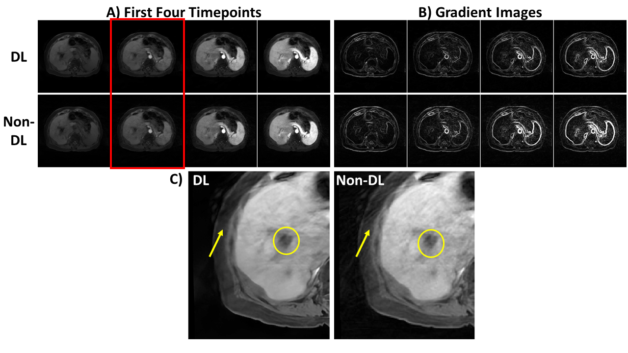
Figure 1: Example DL and non-DL images from the first four timepoints (A) along with the respective gradient images (B). The DL images show higher SNR and fewer streaks, which is apparent on the gradient images. This can be seen particularly in the liver (yellow circle) and the area between the liver and outer edge of the patient’s contour (yellow arrow) of the zoomed images from timepoint 2 (red box; rescaled to better depict streaking).
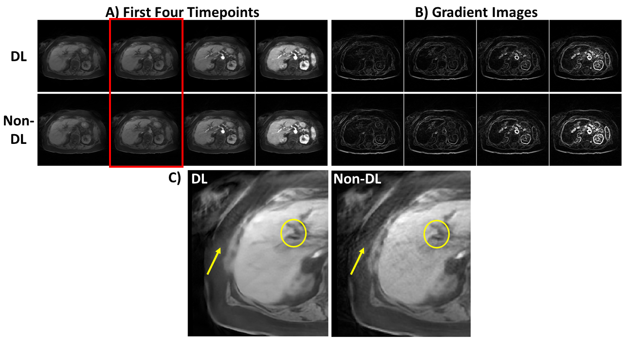
Figure 2: Example DL and non-DL images from the first four timepoints (A) along with the respective gradient images (B) from another patient. The denoising and destreaking is also apparent here, similar to Figure 1.
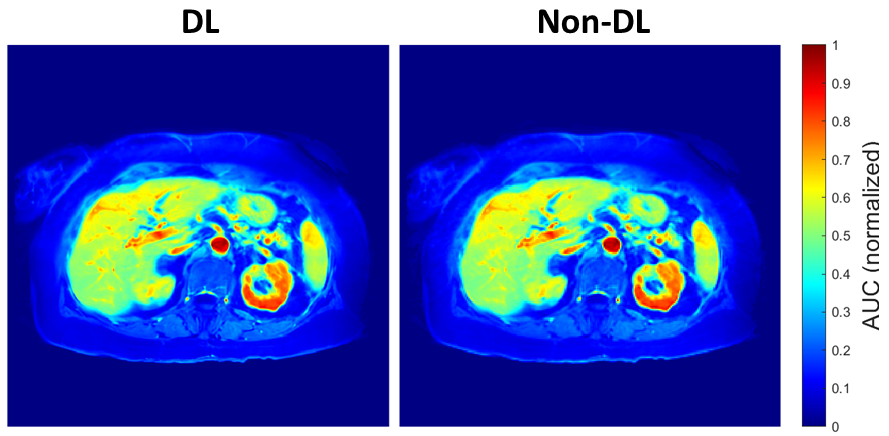
Figure 3: Example DL and non-DL AUC maps from the patient in Figure 2. Visually, the AUC maps look very similar to each other, particularly in the liver and kidneys. The mean difference between the AUC across the whole images was 1.5%.
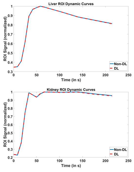
Figure 4: Example DL and non-DL dynamic curves from an ROI in the liver and kidneys of the patient in Figure 2. The curves match across all timepoints, indicating application of DL does not distort quantitative analysis of DSICO-Star data.
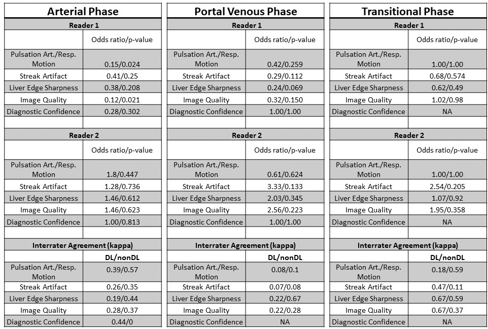
Table 1: Statistical analysis results for the radiologists’ reads. These values may be interpreted as the increase in odds when comparing nDL (reference) and DL. For example, if the OR is > 1, then we can conclude that DL measures tended to be higher than nDL measures. If the OR < 1, then we can conclude that nDL measures were higher than DL. Reader 2 appeared to prefer the DL more often than Reader 1 across the IQ characteristics and three different phases. The DL showed the best results in the transitional phase across the two readers.