4478
Neural Bloch-McConnell fitting (NBMF): unsupervised test-time learning of clinical semisolid MT/CEST MRF reconstruction1Department of Biomedical Engineering, Tel Aviv University, Tel-Aviv, Israel, 2Institute of Neuroradiology, University Hospital Erlangen, Friedrich-Alexander-Universität Erlangen-Nürnberg (FAU), Erlangen, Germany, 3Magnetic Resonance Center, Max-Planck-Institute for Biological Cybernetics, Tübingen, Germany, 4Department of Artificial Intelligence in Biomedical Engineering, Friedrich-Alexander-Universität Erlangen-Nürnberg (FAU), Erlangen, Germany, 5Sagol School of Neuroscience, Tel Aviv University, Tel-Aviv, Israel
Synopsis
Keywords: CEST / APT / NOE, Molecular Imaging, AI, Deep Learning, Unsupervised Learning, Bloch-McConnell, Differentiable Physics
Motivation: MRF-based quantification of semi-solid MT/CEST proton-exchange requires a computationally demanding dictionary synthesis/matching. Recently reported unsupervised learning alternatives were incompatible with pulsed clinical CEST and multi-pool imaging.
Goal(s): To develop a training-set-free MRF reconstruction method, learning directly from the acquired data via pulsed-saturation-compatible physical modeling.
Approach: A differentiable multi-pool Bloch-McConnel simulator was designed and embedded within a test-time learning framework. Validation was performed using L-arginine phantoms and a human subject at 3T.
Results: The method enabled quantitative MT/CEST reconstruction in ~1 minute. The resulting maps were highly correlated with ground-truth in-vitro (Pearson’s r>0.95). In-vivo, semi-solid volume fractions were in agreement with MRF-based maps (r~0.8).
Impact: A one-stop-shop for semisolid MT and CEST MRF reconstruction was developed, enabling a training-set-free rapid quantification of exchange parameters on clinical scanners. This accessible approach could help a variety of Bloch-fitting applications to benefit from deep learning through differentiable spin-physics.
Introduction
Magnetic resonance fingerprinting (MRF) is a powerful quantitative imaging strategy, capable of jointly extracting multiple tissue parameter maps.1 MRF was recently expanded to quantify the proton exchange parameters of mobile proteins, lipids, and metabolites, via the semisolid MT and chemical exchange saturation transfer (CEST) mechanisms2,3. Such molecular imaging scenarios, involving multiple proton pools, impose technical challenges for MRF. The dictionary size scales exponentially with the number of tissue parameters, rapidly elevating the compute/memory demands of parameter matching or the training of a reconstruction neural network (NN) 2,4,5. A recently proposed alternative6 introduces unsupervised learning of convolutional NN on real subject scans, but comes short of clinical applicability. One of its limitations is a reliance on limited-diversity training data, a disadvantage w.r.t fully-sampled dictionary training. Another challenge lies in simulating spin physics within the computational graph, for clinically relevant multi-pool and pulsed-scanner settings. We address the latter by developing a generic differentiable multi-pool simulation of any pulsed saturation protocol. To address the data issue, we drop the train-test paradigm and embed the physical model into a test-time- (sometimes referred to as internal- or zero-shot-)7,8,9,10 learning procedure. This results in a deep-learning-infused Bloch-fitting of single scan’s voxels, hence dubbed NBMF - Neural Bloch-McConnell Fitting, which quantifies semisolid MT/CEST parameters on the fly.Methods
NBMF Pipeline ArchitectureA test-time learning framework (Fig. 1b) was designed, with its two basic blocks being the:
1. Decoder (Fig. 1c) - a voxel-wise multi-layer perceptron (MLP) NN, aligned to standard deep reconstruction3,4,5.
2. Simulator (Fig. 1d) - a differentiable multi-pool spin physics solver, accommodating standard clinical (SAR-limited) pulsed-saturation acquisition protocols in two variations (Fig. 1d):
(i) The isar2 approximate analytical solution11 of regularly-pulsed two-pool saturation.
(ii) Multi-pool solution by chaining Bloch-McConnell (BM) matrix exponentiation across each pulse.
The simulators were implemented using the open-source JAX framework12 for auto-differentiation and GPU-acceleration.
Pseudo-Random Non-Steady-State Semisolid MT/CEST Data Acquisition
An in-vitro phantom was assembled, comprised of 7 vials with 25-100 mM L-arginine at pH 4-6 and scanned at room temperature. Human imaging was performed under approval by the local ethics/IRB committee. A healthy volunteer, having given a written informed consent, was scanned using a 3D whole-brain MRF protocol with a single-shot EPI readout13 (implemented using the pulseq-CEST framework14). The semisolid MT/CEST acquisition protocol varied the saturation pulse power and frequency offsets across 30 iterations15. B0, T1 and T2 maps were acquired using the same 3D snapshot EPI readout16 via WASABI17, saturation recovery, and multi-echo sequences, respectively. Imaging was performed using a clinical 3T scanner (Prisma, Siemens, Germany).
Scan Processing and Reference Data Analysis
In-vivo images were motion-corrected and registered using elastix. Quantitative MRF reference maps were obtained using a fully connected deep reconstruction network, trained using large simulated single-voxel signal dictionaries, as described in ref13,15, see Fig. 1a for an overview). NBMF-based reconstruction was performed via test-time training of a MLP on-the-fly, as described in Fig. 1b. B0, T1, and T2 maps served as additional inputs. A GeForce RTX 3060 GPU was used for neural network training and inference.
Results
In-Vitro studyA representative result for exchange parameter quantification in phantoms is shown in Fig. 2 and Fig. 3. A good agreement was obtained between the NBMF-estimated L-arginine concentrations and the known concentrations (Pearson’s r=0.95), and between the estimated proton exchange rates and the reference QUESP calculated values (Pearson’s r=0.97).
Human study
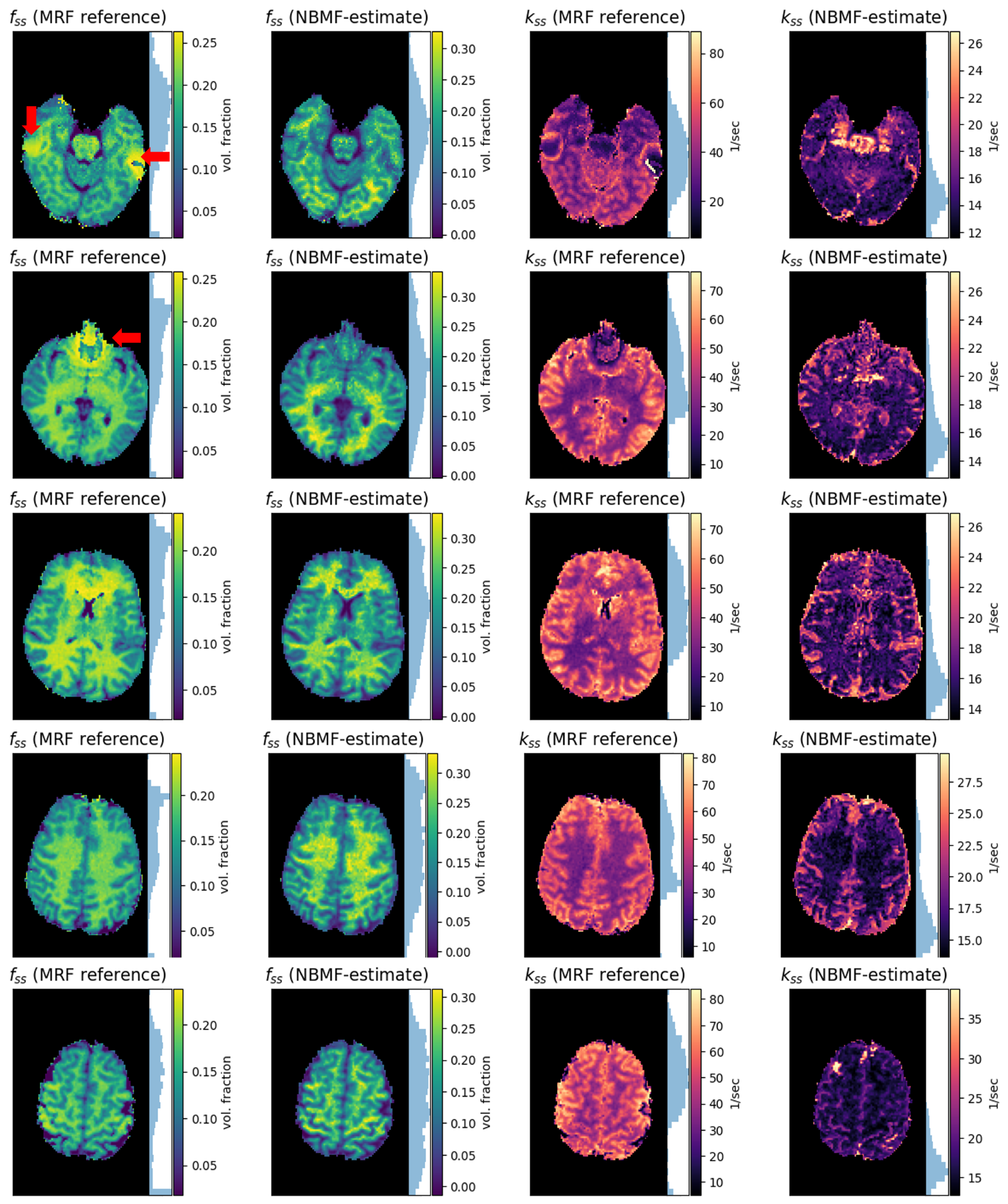
A good agreement was obtained between the NBMF-based and MRF-based fss (Pearson’s r=0.8), see Fig. 4. The kssw was generally lower in NBMF compared to MRF, yet comparable to previous literature18. Interestingly, NBMF was able to mitigate B0-related artifacts that were visible in MRF-based maps (Fig. 4, red arrows).
Discussion
The NBMF pipeline demonstrated a rapid convergence and reconstruction of quantitative tissue parameter maps in-vivo (~1 min for two-pool isar2, <10 min for 3-pool matrix-exponentiation for a ~1M-voxel 3D whole brain data). An attractive byproduct of NBMF is a trained reconstruction network, directly reusable for a real-time (<10 sec) reconstruction of parameter maps from unseen new subjects.
Conclusions
A dictionary-free, pulsed-acquisition-compatible, and rapid means for semisolid MT/CEST quantification was developed. NBMF may serve as a one-stop-shop for reconstructing quantitative molecular MRI data, circumventing the lengthy and error-prone computational efforts required as new CEST protocols are being investigated. The neural solver could accelerate/improve traditional Bloch fitting in a variety of other imaging applications, and facilitate protocol optimization.Acknowledgements
This project received funding from the European Research Council under the Horizon Europe program (grant agreement no. 101115639), the Ministry of Innovation, Science and Technology, Israel, and a grant from the Tel Aviv University Center for AI and Data Science (TAD).References
Ma, D., Gulani, V., Seiberlich, N., Liu, K., Sunshine, J.L., Duerk, J.L. and Griswold, M.A., 2013. Magnetic resonance fingerprinting. Nature, 495(7440), pp.187-192.
Cohen, O., Huang, S., McMahon, M.T., Rosen, M.S. and Farrar, C.T., 2018. Rapid and quantitative chemical exchange saturation transfer (CEST) imaging with magnetic resonance fingerprinting (MRF). Magnetic resonance in medicine, 80(6), pp.2449-2463.
Kim, B., Schär, M., Park, H. and Heo, H.Y., 2020. A deep learning approach for magnetization transfer contrast MR fingerprinting and chemical exchange saturation transfer imaging. Neuroimage, 221, p.117165.
Cohen, Ouri, Bo Zhu, and Matthew S. Rosen. "MR fingerprinting deep reconstruction network (DRONE)." Magnetic resonance in medicine 80, no. 3 (2018)
Cohen, Ouri, Victoria Y. Yu, Kathryn R. Tringale, Robert J. Young, Or Perlman, Christian T. Farrar, and Ricardo Otazo. CEST MR fingerprinting (CEST‐MRF) for brain tumor quantification using EPI readout and deep learning reconstruction. Magnetic Resonance in Medicine 89, no. 1 (2023)
Kang, B, Kim, B, Schär, M, Park, H, Heo, H-Y. Unsupervised learning for magnetization transfer contrast MR fingerprinting: Application to CEST and nuclear Overhauser enhancement imaging. Magn Reson Med. 2021; 85: 2040–2054https://onlinelibrary.wiley.com/doi/10.1002/mrm.28573
V. Lempitsky, A. Vedaldi and D. Ulyanov, 2018. Deep Image Prior. 2018 IEEE/CVF Conference on Computer Vision and Pattern Recognition, doi: 10.1109/CVPR.2018.00984.
Shocher, A., Cohen, N., & Irani, M. (2018). “zero-shot” super-resolution using deep internal learning. In Proceedings of the IEEE conference on computer vision and pattern recognition
Sun, Y., Wang, X., Liu, Z., Miller, J., Efros, A. and Hardt, M., 2020. Test-time training with self-supervision for generalization under distribution shifts. In International conference on machine learning
Yaman, B., Hosseini, S.A.H. and Akçakaya, M., 2021. Zero-shot self-supervised learning for MRI reconstruction. arXiv preprint arXiv:2102.07737.
Roeloffs, Volkert & Meyer, Christian & Bachert, Peter & Zaiss, Moritz. (2014). Towards quantification of pulsed spinlock and CEST at clinical MR scanners: An analytical interleaved saturation-relaxation (ISAR) approach. NMR in Biomedicine. 28.10.1002/nbm.3192.
J.Bradbury, R.Frostig, P.Hawkins, M.J.Johnson, C.Leary, D.Maclaurin, G.Necula, A.Paszke, J.Vander, S.Wanderman-Milne, Q.Zhang, 2018. JAX: composable transformations of Python+NumPy programs, https://github.com/google/jax
Weigand‐Whittier, Jonah, Maria Sedykh, Kai Herz, Jaume Coll‐Font, Anna N. Foster, Elizabeth R. Gerstner, Christopher Nguyen, Moritz Zaiss, Christian T. Farrar, and Or Perlman. Accelerated and quantitative three‐dimensional molecular MRI using a generative adversarial network. Magnetic Resonance in Medicine 89, no. 5 (2023)
Herz, K., Mueller, S., Perlman, O., Zaitsev, M., Knutsson, L., Sun, P.Z., Zhou, J., van Zijl, P., Heinecke, K., Schuenke, P. and Farrar, C.T., 2021. Pulseq‐CEST: Towards multi‐site multi‐vendor compatibility and reproducibility of CEST experiments using an open‐source sequence standard. Magnetic resonance in medicine
Perlman, O., Ito, H., Herz, K., Shono, N., Nakashima, H., Zaiss, M., ... & Farrar, C. T. (2022). Quantitative imaging of apoptosis following oncolytic virotherapy by magnetic resonance fingerprinting aided by deep learning. Nature biomedical engineering, 6(5), 648-657.
Mueller S, Stirnberg R, Akbey S, et al. Whole brain snapshot CEST at 3T using 3D-EPI: aiming for speed, volume, and homogeneity. Magn Reson Med. 2020; 84: 2469-2483.
Schuenke P, Windschuh J, Roeloffs V, Ladd ME, Bachert P, Zaiss M. Simultaneous mapping of water shift and B1 (WASABI)—application to field-inhomogeneity correction of CEST MRI data. Magn Reson Med. 2017; 77:
Samsonov, A. et al. Quantitative MR imaging of two-pool magnetization transfer model parameters in myelin mutant shaking pup. Neuroimage 62, 1390–1398 (2012).
Figures
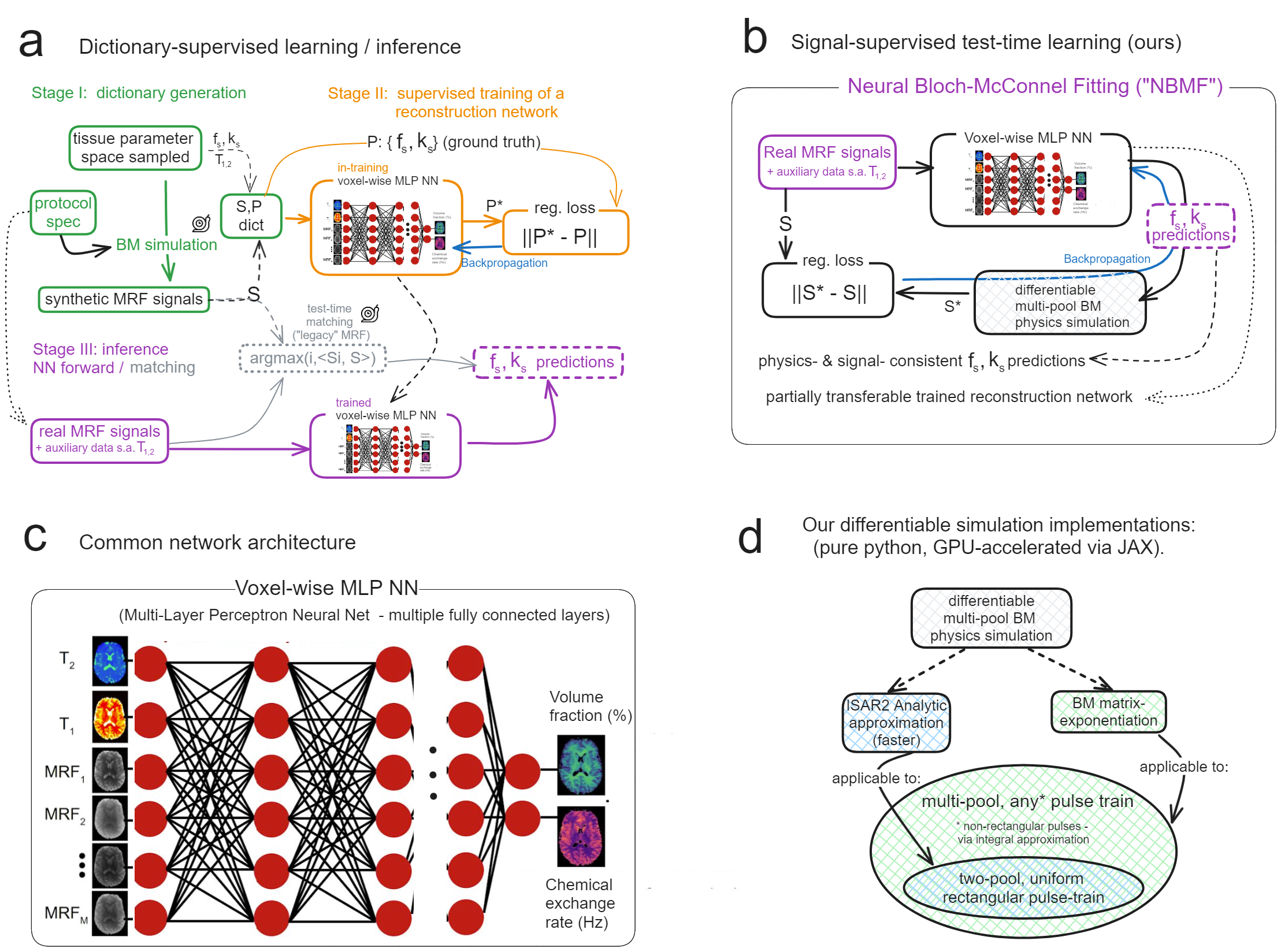
Fig. 1: Overview of explored methods. (a.) MRF pipeline (reference method). Raw non-steady-state MT/CEST images, as well as any auxiliary (T1,2, B0,1) maps serve as inputs to an MLP (c), trained on a large dictionary (synthesized over hours/days) to reconstruct MT/CEST parameters. (b.) NBMF pipeline (ours). A test-subject MRF data serves as both the input for an MLP decoder (c) and as the regression target for the decoder-simulator circuit. Convergence yields a reconstruction for the test subject, and an inference-ready neural decoder. (d.) Solvers' features/limitations.
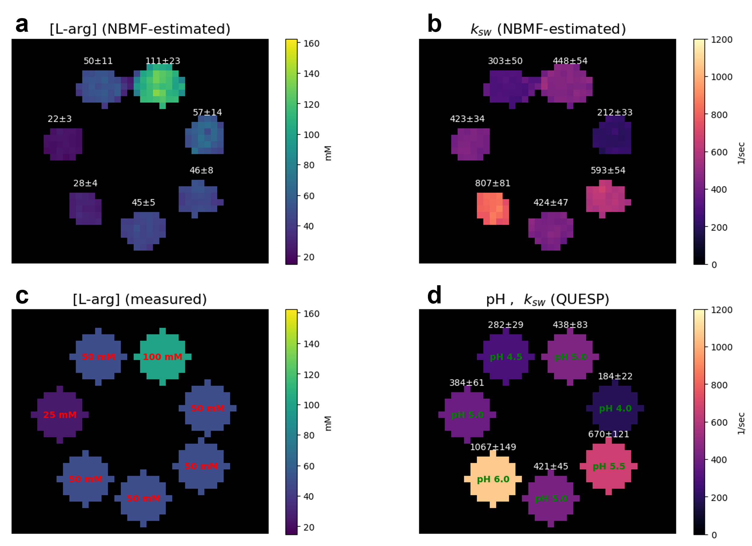
Fig. 2: Typical in-vitro scan reconstruction. (a, b.) L-arginine concentration and amine proton exchange rate (ksw) maps generated using NBMF. (c.) Ground truth concentrations (d.) Ground truth pH and QUESP-estimated exchange rates. An excellent agreement was obtained between NBMF and ground truth: r(ksw)=0.97, r([L-arg])=0.95 (r - Pearson's correlation across all voxels).
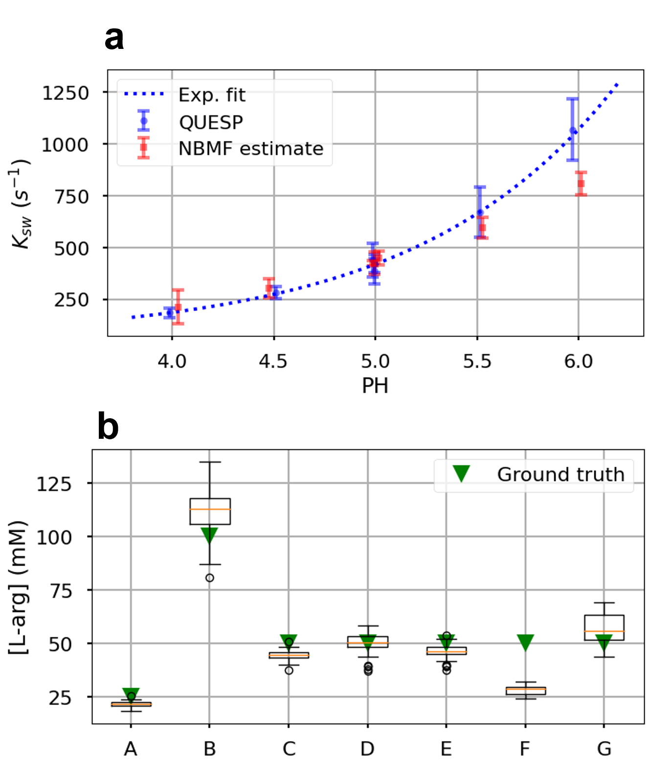
Fig. 3: Statistical analysis of the in-vitro study. (a.) NBMF-based exchange rates compared to QUESP and a base-catalyzed proton exchange model. (b.) L-arginine concentrations estimated across the different vials (A-G). The green triangles represent the ground truth.