4476
Accelerating CEST MRI using Data-Driven Z-Spectral Compressed Sensing1Department of Electronic Science, Fujian Provincial Key Laboratory of Plasma and Magnetic Resonance, School of Electronic Science and Engineering, National Model Microelectronics College, Xiamen University, Xiamen, China, 2Departments of Radiology, Shandong Provincial Hospital Affiliated to Shandong First Medical University, Jinan, Shandong, China
Synopsis
Keywords: CEST / APT / NOE, CEST & MT
Motivation: CEST MRI requires the collection of multiple saturated images with different saturation offsets, resulting in prolonged scan times, which hinders its clinical applications.
Goal(s): we aim to reduce the scan time of CEST MRI by recovering the undersampled Z-spectrum to full-sampling counterpart using data-driven Z-spectral compressed sensing method.
Approach: The modified U-Net was employed for Z-spectral recovery. Training data were generated using Bloch equation. Numerical simulations and in vivo experiments on rat brains were conducted to validate the proposed method.
Results: The results demonstrate that our method outperformed conventional interpolation methods, and threefold undersampling rate can be achieved without discernible degradation in quantification.
Impact: The proposed method can efficiently reduce the scan time of CEST MRI, potentially facilitating its clinical applications.
Introduction
CEST MRI is a versatile technique that provides valuable diagnostic insights for various diseases, including tumors, neurodegeneration diseases, and ischemic stroke. Conventional CEST experiments acquire multiple saturated images with different saturation offsets, leading to prolonged scan time, which hinders its clinical applications. Furthermore, the long scan time also makes CEST MRI vulnerable to motion artifacts and degrades quantification reliability.Various methods have been developed to accelerate CEST imaging, including fast readout1, parallel imaging2, and compressed sensing3 (CS) in the k-space domain. Recently, exploiting sparsity in the Z-spectrum domain has attracted significant attention in accelerating CEST MRI4. In this study, we proposed a data-driven deep learning method, named Z-spectral CS for short, to fully exploit the sparsity in the Z-spectrum domain and recover the undersampled Z-spectrum to its full-sampling counterpart. The proposed method was validated through numerical simulations and in vivo rat brain experiments.
Methods
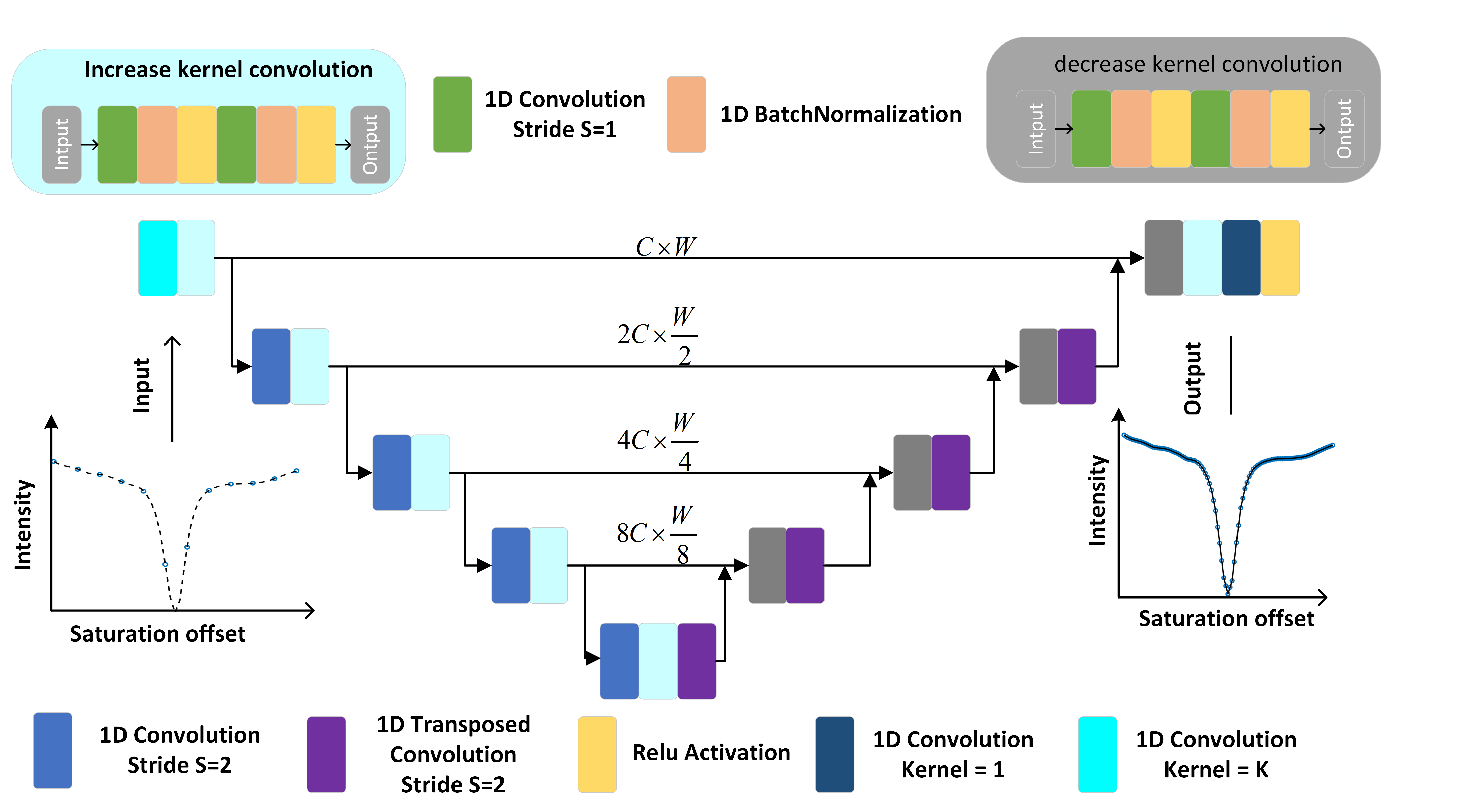
The neural network used in this study is a modified U-Net, consisting of an encoder-decoder network with multi-scale skip connections and 1D convolution, as shown in Fig. 1. The input and output are undersampled Z-spectrum and fully sampled Z-spectrum, both with the same dimension size of 1 × W, where W refers to the number of frequency offsets. For the input, missing data were set to zero. We adopted the modified U-Net with four scales. Each scale features a skip connection between the downsampling and upsampling operations. The channel numbers for the four scales are C, 2C, 4C, and 8C, respectively, corresponding to feature maps with dimensions of C × W, 2C × W/2, 4C × W/4, and 8C × W/8. In this study, we adopted C=64 and W=51.The training data were generated using the five-pool Bloch-McConnell equation (i.e. bulk water, NOE, Amide, MT, and creatine) with frequency offsets ranging from -5 to 5 ppm. Various combinations of different experimental parameters, including concentration, exchange rate, T1, and T2, saturation power and duration, and different magnetic field strength, were considered. The in vivo rat experiments were performed on a 9.4T Bruker scanner. The linear and pchip interpolation methods, accomplished by Matlab's built-in functions, were performed for comparison.
Results and Discussion
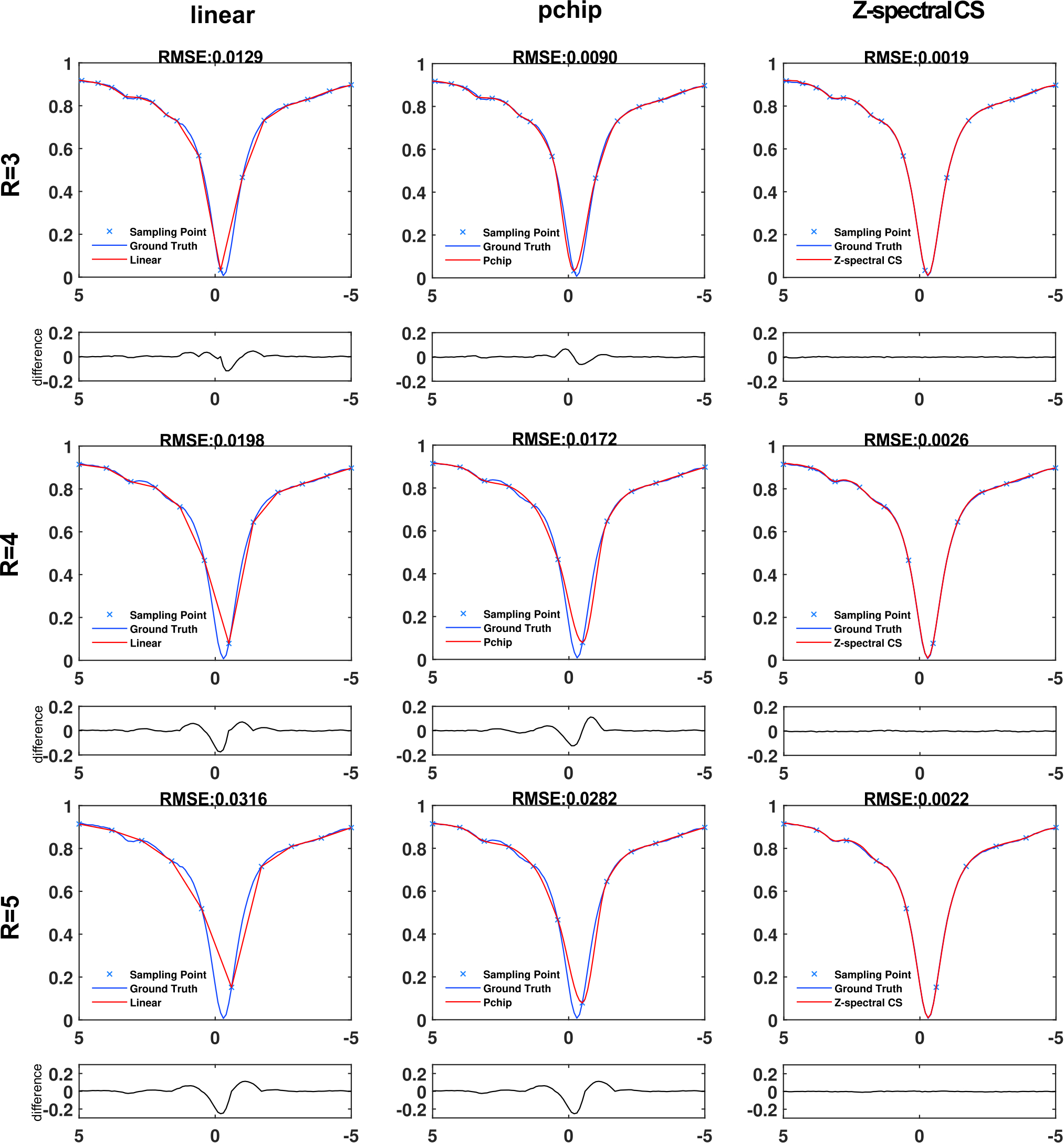
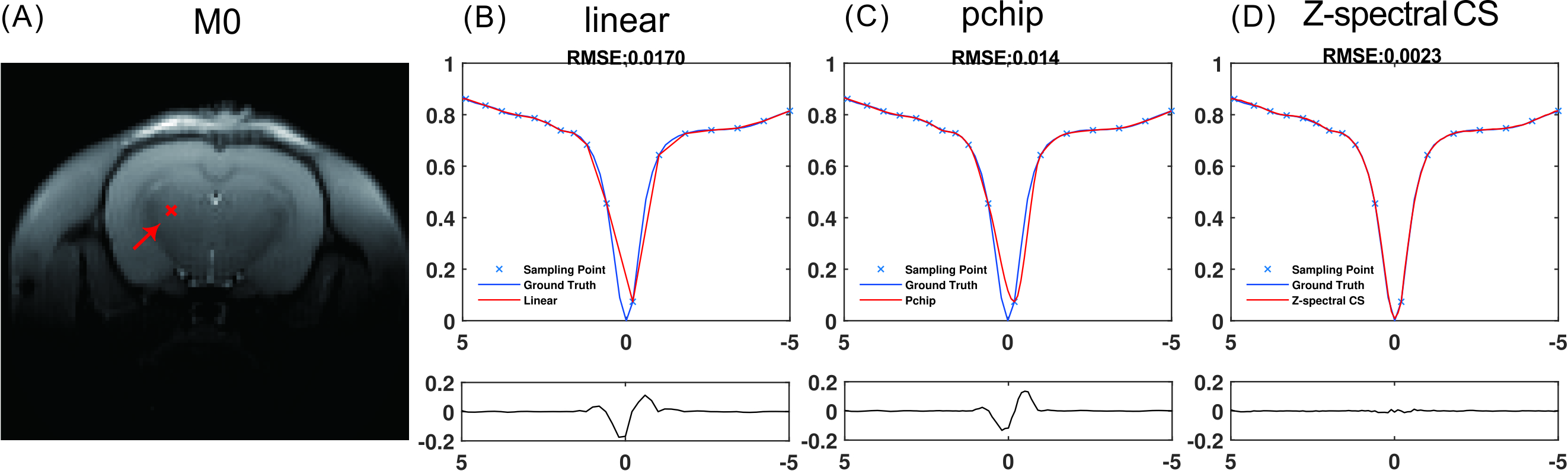
In neural network training, the Adam optimizer is employed to minimize the mean squared error (MSE) loss function, the neural network was converged after 80 epochs.The performance of the proposed method with different acceleration factors was demonstrated in Fig. 2. From the results, Z-CS outperformed the other two comparison methods in both root mean square error (RMSE) and difference evaluation. The interpolation methods show inferior performance around 0 ppm due to the significant signal variation in this region. This is because these methods rely on the local smoothness assumption, which becomes invalid around 0 ppm. In contrast, Z-CS recovered the full Z-spectrum with better fidelity even around 0 ppm, indicating that the neural network successfully learned the prior information embedded in the Z-spectrum. Similar results were also observed in the in vivo rat brain experiment with an acceleration factor of R=3, as shown in Fig. 3.
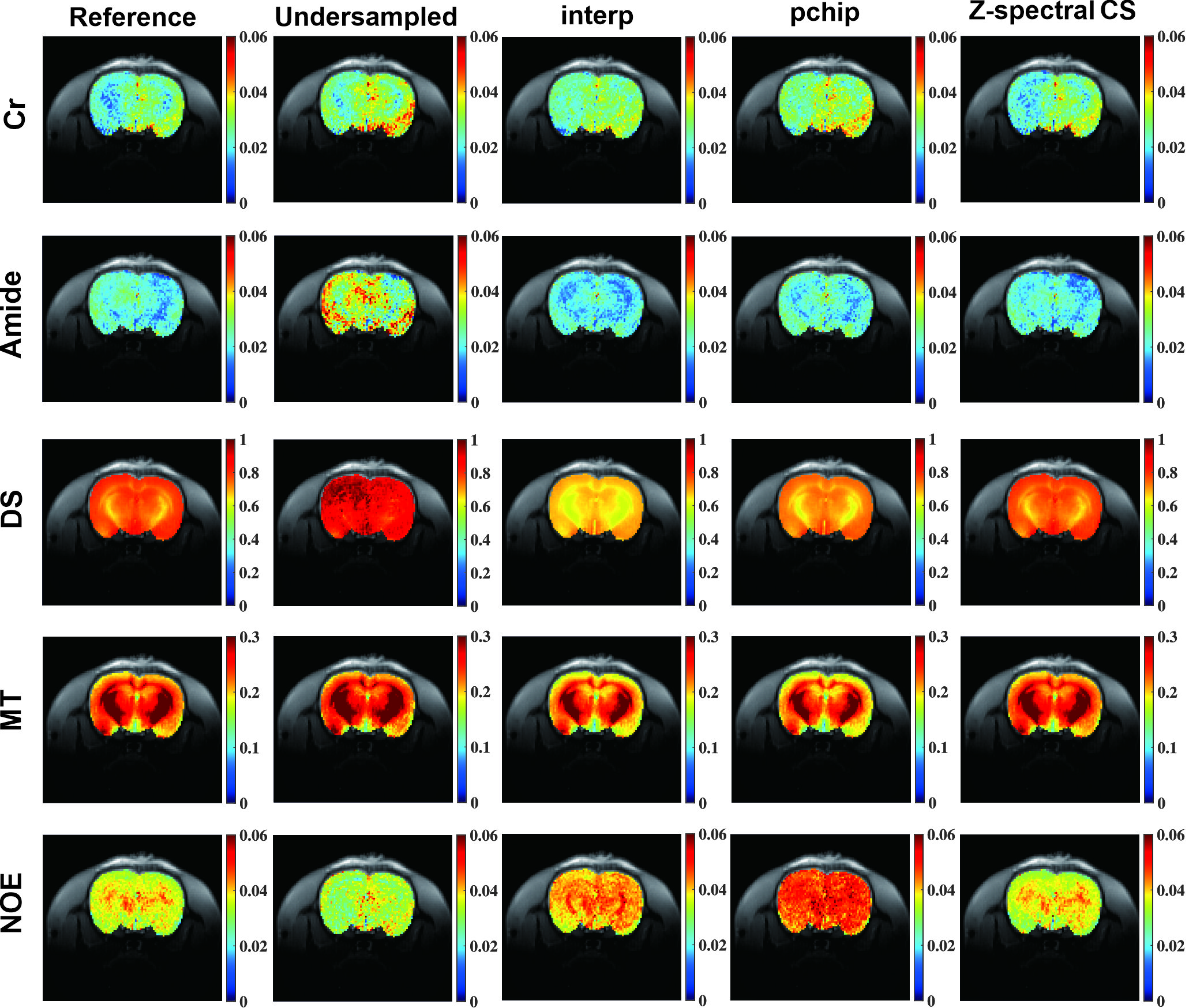
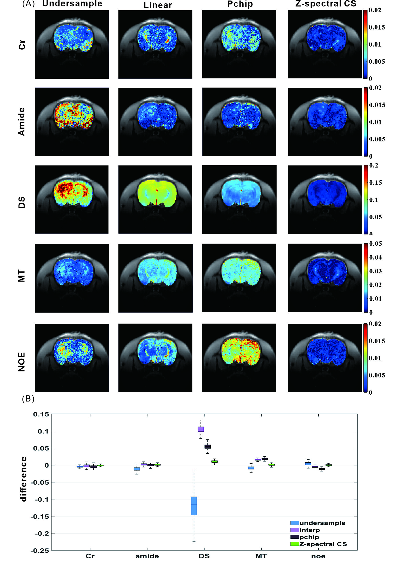
The five-pool Lorentzian fitting was performed to quantify the Z-spectrum recovered by different methods, and the results are shown in Fig. 4. The difference maps, along with statistical analyses, are presented in Fig. 5. The results indicate that our method yields satisfactory quantification accuracy with an acceleration factor of R=3. The modified U-Net is known for its ability to recover missing data by leveraging features extracted from different levels, making it well-suited for Z-spectral compressed sensing, as demonstrated in this study. As a data-driven method, the accuracy of the proposed method highly depends on the training data. In this study, the five-pool Bloch-McConnell equation was adopted to generate training data, which is sufficient for the conditions of this study. In more complex situations, the use of the Bloch-McConnell equation with additional exchangeable pools is recommended.
Conclusion
In this study, a data-driven Z-Spectral compressed sensing based on a modified U-Net was proposed to accelerate CEST MRI without discernible compromise in quantification accuracy, potentially facilitating the clinical applications of CEST MRI.Acknowledgements
This work is supported by the National Natural Science Foundation of China, Grant/Award Number:82302151; Shenzhen Science and Technology Program, Grant/Award Number: JCYJ20220818101213029; Fujian Province Science and Technology Project, Grant/Award Number: 2022J05013; Xiamen University Nanqiang Outstanding Talents Program.References
1.Zhu, H., et al., Fast 3D Chemical Exchange Saturation Transfer (CEST) Imaging of the Human Brain. MAGNETIC RESONANCE IN MEDICINE, 2010. 64(3): p. 638-644.
2.Zhang, Y., et al., Fast 3D chemical exchange saturation transfer imaging with variably-accelerated sensitivity encoding (vSENSE). MAGNETIC RESONANCE IN MEDICINE, 2019. 82(6): p. 2046-2061. 3.Lustig, M., D. Donoho, and J.M. Pauly, Sparse MRI: The application of compressed sensing for rapid MR imaging. MAGNETIC RESONANCE IN MEDICINE, 2007. 58(6): p. 1182-1195.
4.Kwiatkowski, G. and S. Kozerke, Accelerating CEST MRI in the mouse brain at 9.4 T by exploiting sparsity in the Z -spectrum domain. NMR IN BIOMEDICINE, 2020. 33(9).
Figures