4468
Investigating the feasibility of reverse cross polarization for the development of a new myelin detection technique in MRI1Physics and Astronomy, University of British Columbia, Vancouver, BC, Canada, 2International Collaboration on Repair Discoveries (ICORD), University of British Columbia, Vancouver, BC, Canada, 3Radiology, University of British Columbia, Vancouver, BC, Canada, 4UBC MRI Research Centre, University of British Columbia, Vancouver, BC, Canada, 5Pathology and Laboratory Medicine, University of British Columbia, Vancouver, BC, Canada
Synopsis
Keywords: Non-Proton, Non-Proton, myelin, spectroscopy, phosphorus, hydrogen, spinal cord, cross polarization, NMR, magnetization transfer, brain
Motivation: The potential of phosphorous MRI to enhance in vivo myelin detection and improve neurodegenerative disease diagnosis inspired our project. However, the challenge lies in transferring the solid phosphorus signal to aqueous protons for MRI measurement.
Goal(s): To demonstrate the feasibility of detecting an aqueous proton signal originating from myelin phosphorous, providing a proof of principle result.
Approach: We employed gradients in solid-state NMR experiments to investigate signal transfer between aqueous proton signal originating from myelin phosphorous, incorporating encoding and decoding gradients.
Results: While individual transfer steps were successful, the complete transfer experiment yielded an unexpected negative result, indicating that further investigation is needed.
Impact: The successful transfer of signal from phosphorous in myelin to aqueous hydrogen would lead to a new method for direct myelin detection. This could potentially offer earlier and more direct measurements of demyelination, benefiting those with neurodegenerative diseases.
Introduction
Various MRI techniques are used to study white matter1-6. Our prior work demonstrated the potential of solid-state NMR techniques like cross polarization (CP)7 to probe myelin via phosphorous (31P) nuclei in the myelin sheath’s phospholipid bilayers8,9. The broad chemical shift anisotropy of the 31P nuclei in the phospholipid head group make detection with an MRI scanner difficult. We propose using CP to transfer magnetization between semi-solid 31P (31Ps) and nearby semi-solid hydrogen (1Hs). Then using magnetization transfer (MT), move the magnetization from 1Hs to aqueous hydrogen (1Haq).While we have shown the feasibility of transferring magnetization from 1Haq through 1Hs to 31Ps via Goldman-Shen MT-CP10, the reverse remains to be seen. Detecting a small 1Haq signal in the presence of background water is a significant challenge, requiring selective water suppression, as our desired signal also resides in water.
Our objective was to investigate the use of gradient pulses to encode and decode the transfer of magnetization from 31Ps to 1Haq, while simultaneously suppressing any extraneous water signal. Success could lead to a new MRI technique capable of more directly detecting myelin in-vivo.
Methods
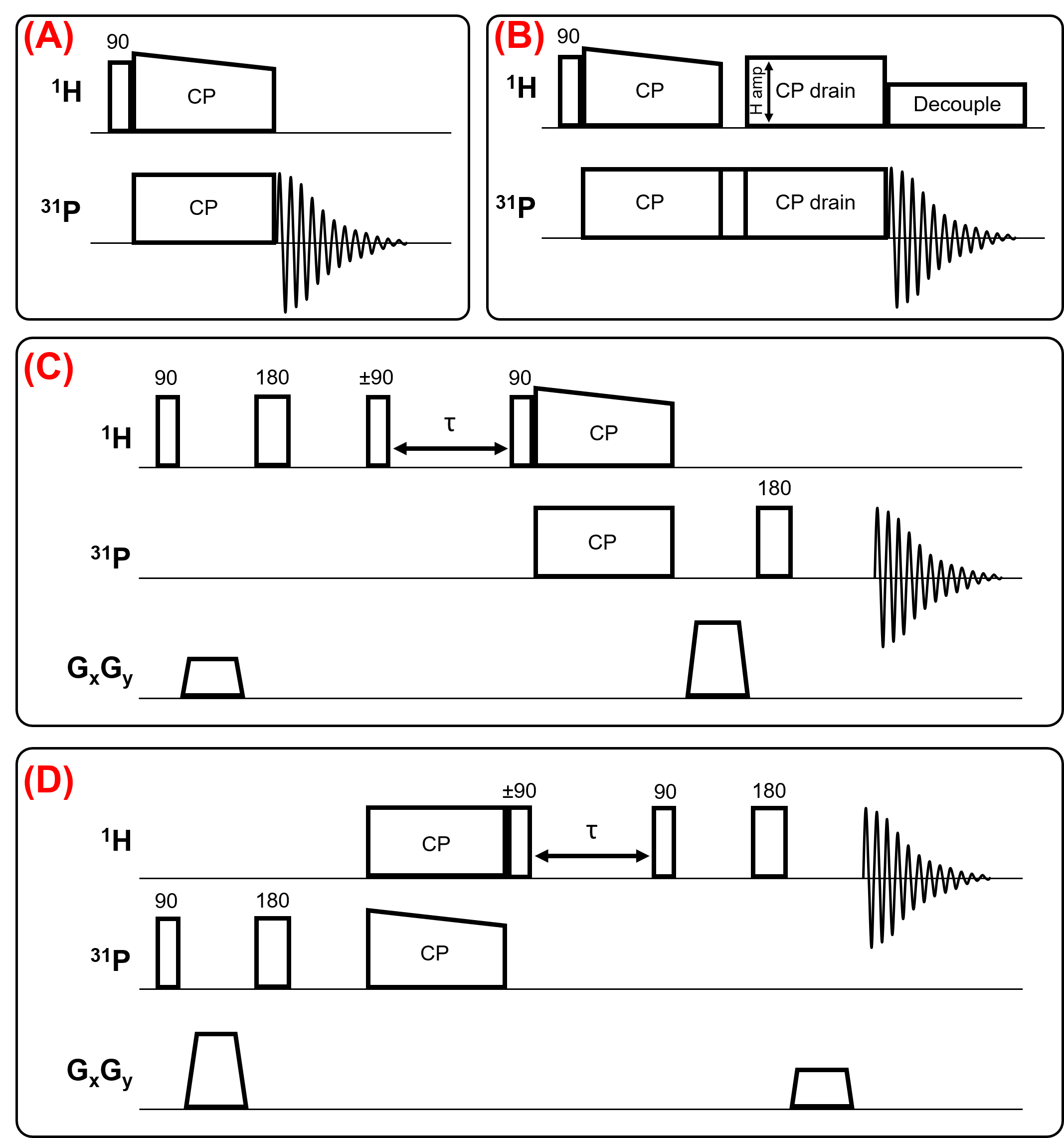
Experiments on porcine spinal cord white matter were conducted using a horizontal coil double-resonant probe on a lab-built 8.4T NMR spectrometer11, with a 4cm bore Bruker (MIC2.5 model) gradient set driven by a BGU-II gradient unit with BAFPA-40 gradient amplifiers.Pulse sequences included ramped-amplitude CP12 (Fig.1A), locally designed HPH CP drain (Fig.1B), gradient modified Goldman-Shen MT-CP with 31P-detect (Fig.1C), and 1H-detect (Fig.1D). The probe was rotated to align the coil 45° to the x and y-gradients to maximize the gradient amplitude (trapezoidal gradients, slew rate=15.4T/m/ms rising to amplitude=0.77T/m for 200µs). The gradient amplitude applied to 1H signal was reduced by the ratio of 1H and 31P gyromagnetic ratios (~2.47).
Results
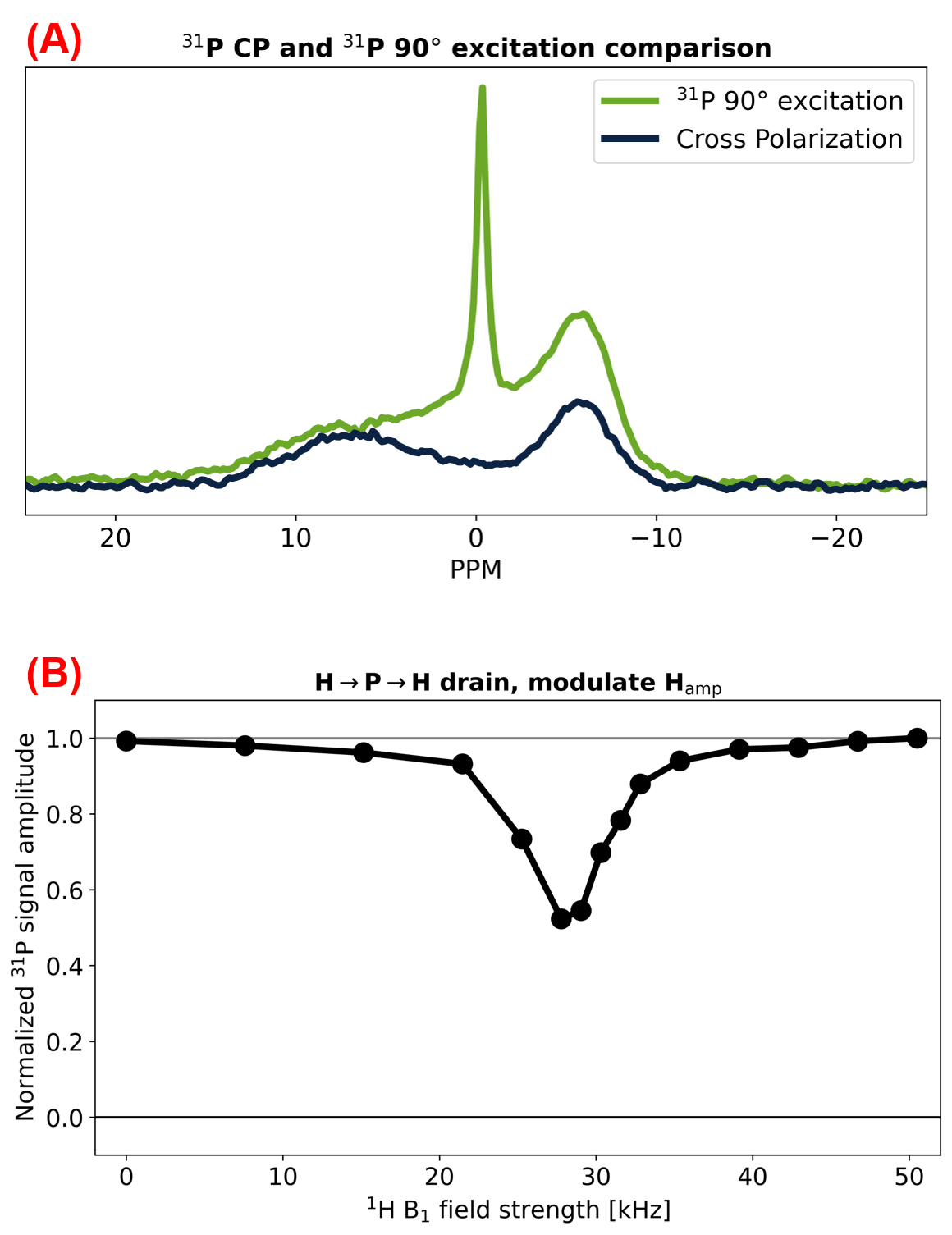
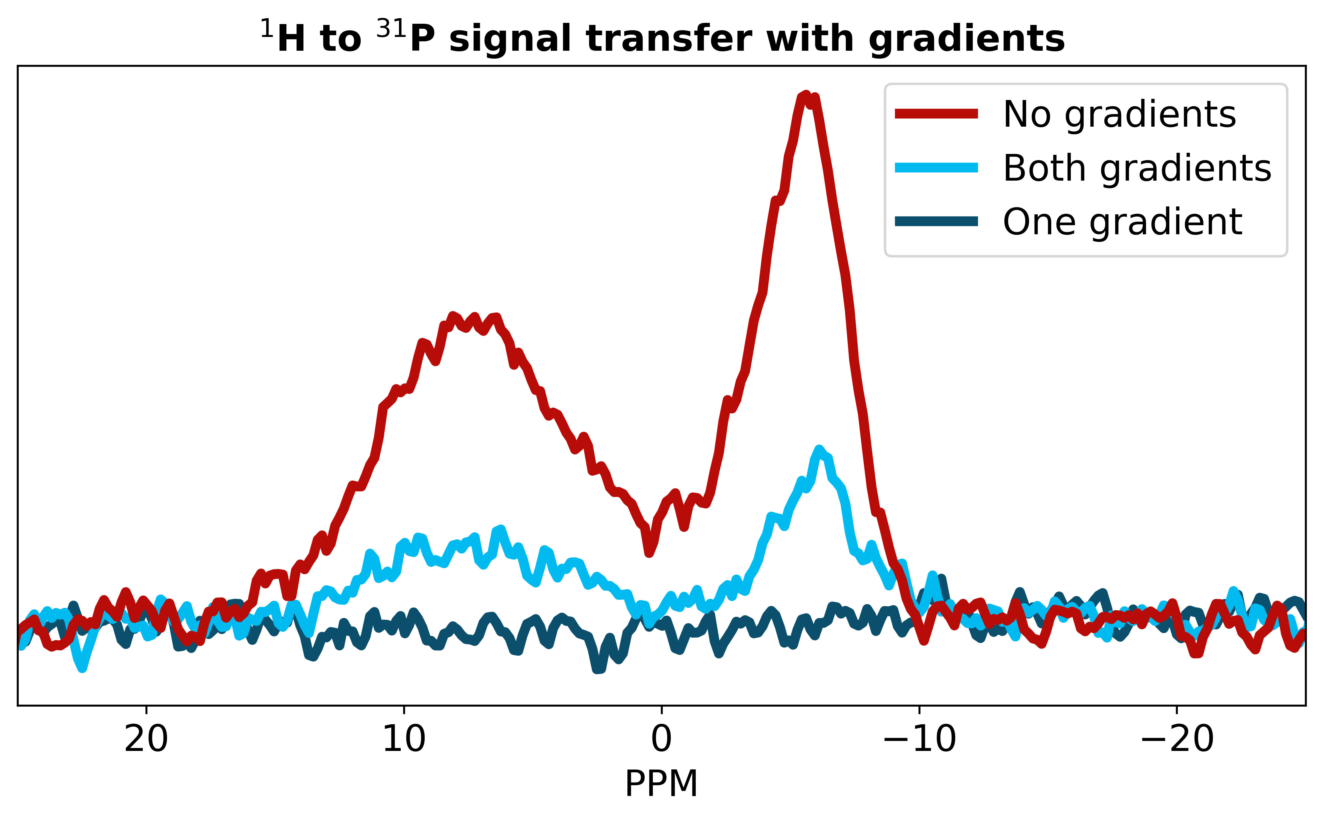
Fig.2A compares acquisition via ramped-CP and via 90° excitation pulse with detection on 31P. Fig.2B from previous work9, demonstrates the transfer of magnetization from 31Ps to 1Hs.Fig.3 plots the spectra of experiments using the pulse sequence in Fig.1C, comparing three gradient configurations. With both gradients off, we see the largest signal. When both gradients are on, the signal has a reduced amplitude as the combination of gradients produce a stimulated echo that is reduced in amplitude by 2x compared to a full echo. With only the decode gradient on, we see no signal, indicating this gradient removes any signal that does not originate from the initial 1H excitation pulse encoded by the first gradient pulse.
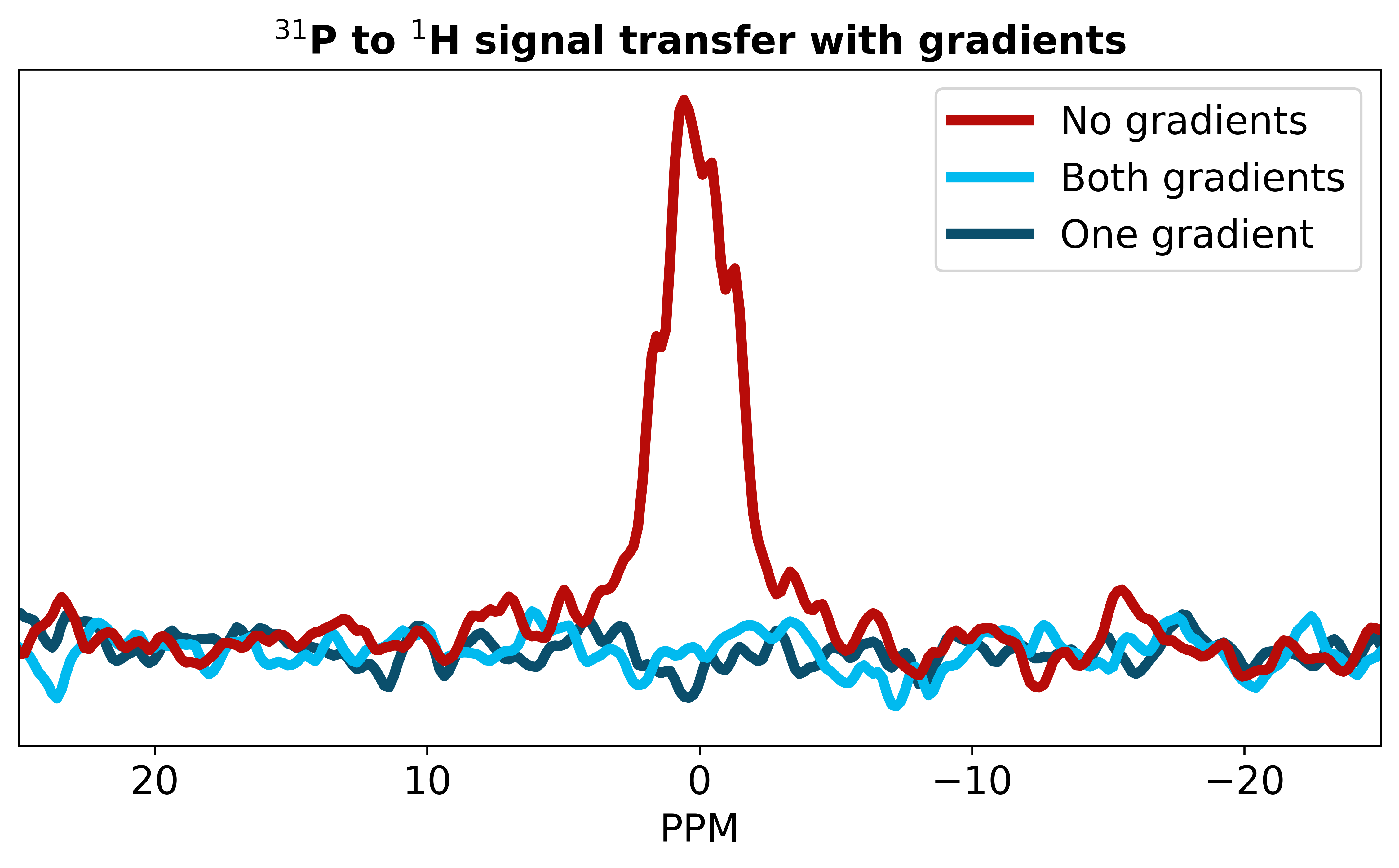
Fig.4 plots the spectra of the pulse sequence in Fig.1D with 1H-detect, in the same gradient configuration as in Fig.3. The spectra show a signal with the gradients off and show no signal with the gradients on.
Discussion
We have successfully demonstrated each part of the reverse transfer: signal transfer from 1H to 31P (Fig.2A), from 31P to 1H (Fig.2B), and from 1Haq through 1Hs to 31Ps (previous work and Fig.3). Additionally, Fig.3 demonstrates this 3-pool transfer with encode and decode gradients.However, the reverse transfer experiment, with 1H detect, led to an unexpected negative result (Fig.4). We see (spurious) signal when both gradients are off resulting from the accidental excitation of 1Haq during the later RF pulses. Phase cycling alone is insufficient to remove this unwanted signal. The encode and decode gradients address this: the first gradient encodes the 31Ps signal, and the second gradient decodes the desired 1Haq signal while dephasing the unwanted 1Haq signal. This works as expected when detecting on 31Ps in Fig.3, however no signal is detected when detecting on 1Haq with both gradients on in Fig.4.
The discrepancy between our expectations and experimental results warrants further investigation. One theory is that rapid T1ρ relaxation of some subset of the 1Hs pool drains away the transferred magnetization. However, variable contact-time CP drain experiments9 contradict this theory. Efforts are ongoing to understand these unexpected findings to achieve the desired transfer. This will be a significant step forward in making this technique applicable to MRI scanners.
Conclusion
This work provides crucial evidence for the transfer of polarization between 1Haq, 1Hs, and 31Ps in white matter. We’ve successfully demonstrated the transfer from 1Haq to 31Ps using encode and decode gradient pulses. However, the reverse transfer from 31Ps to 1Haq remains elusive, despite expectations from successful experiments investigating each step individually, suggesting reverse transfer is feasible. This unexpected outcome presents a compelling challenge for future research. Once we achieve reverse transfer, a novel method for directly detecting myelin can be implemented on an MRI scanner, paving the way for in vivo quantification of myelin.Acknowledgements
AE extends appreciation to MS Canada for a Doctoral Studentship Award. CL, PK, AM and CM gratefully acknowledge funding from the NSERC Discovery grants program. This work was conducted on the traditional, ancestral, and unceded territories of Coast Salish Peoples, including the territories of the xwməθkwəy̓əm (Musqueam), Skwxwú7mesh (Squamish), Stó:lō and Səl̓ílwətaʔ/Selilwitulh (Tsleil- Waututh) Nations.
References
1. Stanisz, G.J., Kecojevic, A., Bronskill M.J., Henkelman RM. Characterizing White Matter With Magnetization Transfer and T2. Magn Reson Med. 1999;42:1128-1136. doi:10.2307/2316017
2. Henkelman RM, Stanisz GJ, Graham SJ. Magnetization transfer in MRI: A review. NMR Biomed. 2001;14(2):57-64. doi:10.1002/nbm.683
3. Mackay A, Whittall K, Adler J, Li D, Paty D, Graeb D. In vivo visualization of myelin water in brain by magnetic resonance. Magn Reson Med. 1994;31(6):673-677. doi:10.1002/mrm.1910310614
4. Whittall KP, MacKay AL, Graeb DA, Nugent RA, Li DKB, Paty DW. In vivo measurement of T2 distributions and water contents in normal human brain. Magn Reson Med. 1997;37(1):34-43. doi:10.1002/mrm.1910370107
5. Le Bihan D, Mangin JF, Poupon C, et al. Diffusion tensor imaging: Concepts and applications. J Magn Reson Imaging. 2001;13(4):534-546. doi:10.1002/jmri.1076
6. Manning AP, Chang KL, MacKay AL, Michal CA. The physical mechanism of “inhomogeneous” magnetization transfer MRI. J Magn Reson. 2017;274:125-136. doi:10.1016/j.jmr.2016.11.013
7. Pines A, Gibby MG, Waugh JS. Proton-enhanced nuclear induction spectroscopy. a method for high resolution nmr of dilute spins in solids. J Chem Phys. 1972;56(4):1776-1777. doi:10.1063/1.1677439
8. Ensworth A, Knight C-A, Kozlowski P, Laule C, MacKay AL, Michal CA. 1H-31P Cross-polarization: A new frontier to study myelin in white matter. ISMRM. 2022.
9. Ensworth A, Knight C-A, Kozlowski P, Laule C, MacKay AL, Michal CA. Solid state NMR in white matter: Unconventional 31P→1H cross polarization interrogates the proton pool. ISMRM. 2023.
10. Goldman M, Shen L. Spin-spin Relaxation in LaF3. Phys Rev. 1966;144(1):321-331. doi:10.1103/physrev.144.321
11. Michal CA, Broughton K, Hansen E. A high performance digital receiver for home-built nuclear magnetic resonance spectrometers. Rev Sci Instrum. 2002;73(2 I):453. doi:10.1063/1.1433950
12. Metz G., X.L. Wu, Smith S.O. Ramped-Amplitude Cross Polarization in Magic-Angle-Spinning NMR. J Magn Reson, Ser A. 1994;110:219-227. doi: https://doi.org/10.1006/jmra.1994.1208
Figures