4452
Renal Metabolic CEST-MRI Based on Golden-Angle Radial Sampling Under Free Breathing1Department of Rehabilitation Medicine, Zhujiang Hospital, Southern Medical University, Guangzhou, China, 2Guangdong Provincial Key Laboratory of Medical Image Processing, Southern Medical University, Guangzhou, China, 3Guangdong Provincial Engineering Laboratory for Medical Imaging and Diagnostic Technology, Guangzhou, China, 4Medical Imaging Center, Nanfang Hospital, Southern Medical University, Guangzhou, China, 5Philips Healthcare, Shenzhen, China, 6Philips Healthcare, Guangzhou, China, 7School of Biomedical Engineering, Southern Medical University, Guangzhou, China, 8Department of Radiology, Zhujiang Hospital, Southern Medical University, Guangzhou, China
Synopsis
Keywords: CEST / APT / NOE, Kidney
Motivation: Respiration motion may induce artifact in CEST image and the in-accurate quantization of CEST signal.
Goal(s): We aimed to develop clinical motion-insensitive CEST imaging of kidney.
Approach: Here, we used turbo filed echo (TFE) based on golden-angle radial sampling to readout renal CEST image under free breathing of three normal volunteers and also evaluated its effectiveness for motion artifact suppression.
Results: The renal Z-spectrum is float and there is no motion artifact in CEST images. This motion in-sensitive sequence showed high repeatability for renal CEST imaging.
Impact: It may provide a motion in-sensitive CEST imaging sequence for renal imaging in clinical nephropathy patients under free breathing, and improve the accuracy of injury detection.
Introduction
The metabolic CEST technique has been approved to be sensitive to kidney disease[1]. However, the respiratory motion is the major obstacle in clinical renal imaging of CEST technique, which may lead the in-correction of CEST signal quantification. Breath hold is the common method to avoid the influence of respiration motion, however, this will hinder the clinical usage of CEST imaging. Some non-Cartesian acquisition methods that are insensitive to respiratory movement are used for CEST imaging of brain[2] and liver[3]. Here, we aimed to develop a motion in-sensitive CEST sequence readout by TFE based on golden-angle radial sampling.Method
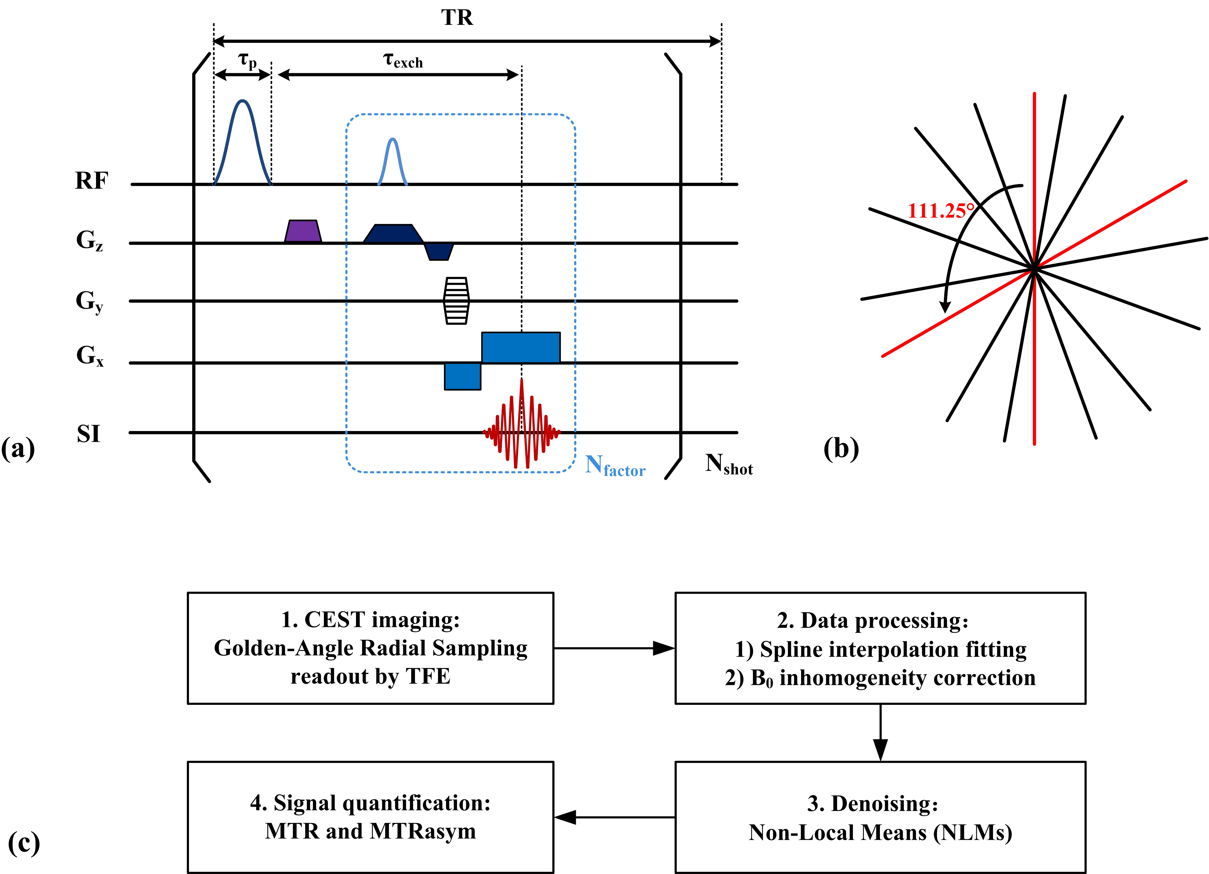
Sequence design: Each CEST module contains a 30 ms Gaussian-shape saturation pulse, followed by a spoiler gradient and three TFE readout, and the k-space was filled by golden-angle radial (Fig. 1a and Fig. 1b).MRI: The experiment were approved by local Institutional Review Board. Written informed consent was obtained for all participating subjects. MRI data were acquired in three (n = 3) healthy volunteers under free breathing on a Philips 3.0T scanner (Ingenia, Philips Healthcare, Best, The Netherlands) equipped with 32 channels torso coil. The CEST parameters were FOV = 384×384 mm2, matrix size = 256×256, repetition time/echo time/saturation time/(TR/TE/ST) = 50/4.4/30 ms, saturation power = 1 μT. The frequency offsets were from -6 to 6 ppm with 0.25 ppm interval (Mz), and the other image at 200 ppm was acquired as reference (M0).
Data Analysis: All data were analyzed using MATLAB (MathWorks, Inc., Natick, MA, USA). The CEST data was interpolated to a 0.01 ppm interval using spline interpolation, and followed by B0 inhomogeneity correction. The non-local means (NLMs) was used to image denoising, and the MTRasym, MTR at -3.5 ppm and 3.5 ppm were calculate. The CEST signal were showed as mean ± std. The imaging scheme was showed in Fig. 1c. The total acquisition time is 6 min and 48 s.
Results
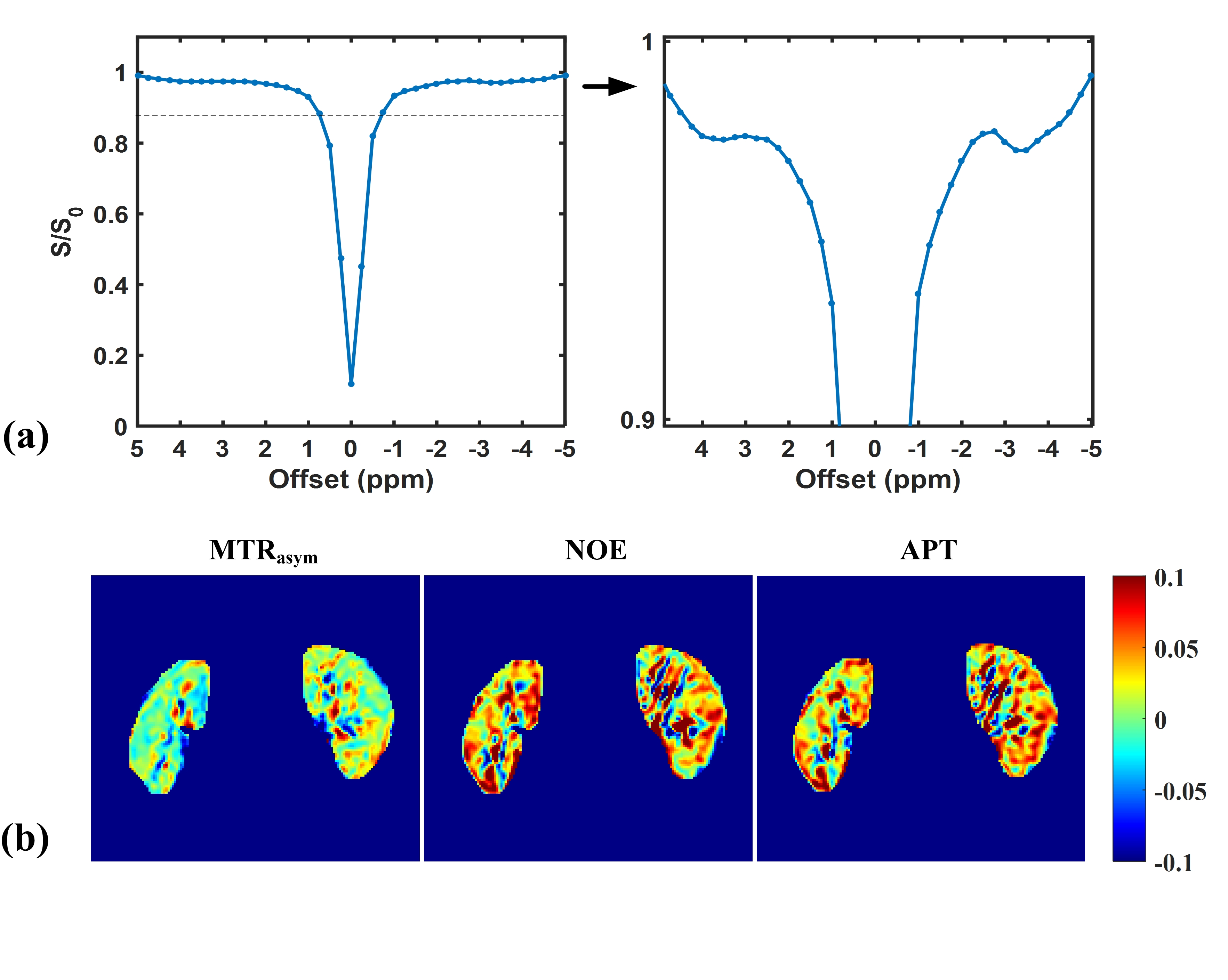
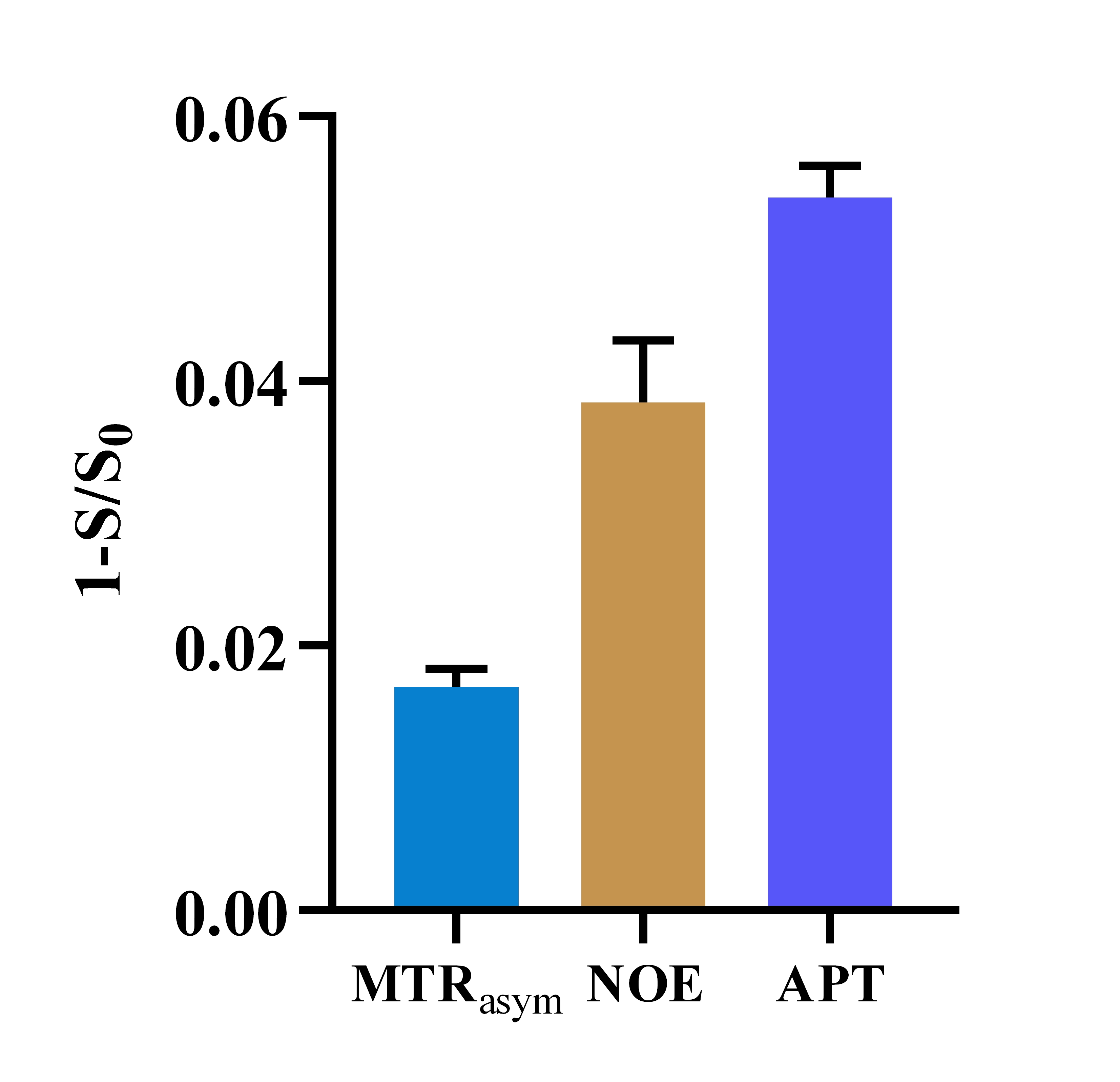
Fig 2a shows the cortical Z-spectrum of kidney in a volunteer under free breathing, there is no data float induced by respiratory motion, and also shows two obvious CEST signal at 3.5 ppm and -3.5 ppm, which were assumed to be amide proto transfer (APT) and nuclear Overhauser effect (NOE) signal. The pseudo-color images of MTRasym, NOE, and APT were showed in Fig. 2b, the renal cortex and medulla showed clear boundary and morphology. The value of MTRasym, NOE, and APT signal were 0.017 ± 0.01, 0.044 ± 0.01, and 0.054 ± 0.02 (n = 3).Discussion
Respiration motion is the main hamper in the clinical transformation of CEST technique, here, we designed the CEST sequence readout by TFE base on golden-angle radial sampling, the results showed it is effective in suppressing motion artifact. However, the small number of volunteers and long acquisition time are the limitations of this study, more appropriate renal imaging protocol of this sequence should be investigated.Conclusion
The CEST-TFE sequence based on golden-angle radial sampling provides a motion in-sensitive technique for renal CEST imaging, and may with the potential to clinical imaging for patients with kidney diseases.Acknowledgements
This study was supported by National Natural Science Foundation of China (U21A6005), Key-Area Research and Development Program of Guangdong Province (2018B030340001, 2018B030333001).References
1. Van Zijl P C M, Yadav N N. Chemical exchange saturation transfer (CEST): what is in a name and what isn't?[J]. Magnetic resonance in medicine, 2011, 65(4): 927-948.
2. Sui R, Chen L, Li Y, et al. Whole‐brain amide CEST imaging at 3T with a steady‐state radial MRI acquisition[J]. Magnetic resonance in medicine, 2021, 86(2): 893-906.
3. Han P, Cheema K, Cao T, et al. Free‐breathing 3D CEST MRI of human liver at 3.0 T[J]. Magnetic Resonance in Medicine, 2023, 89(2): 738-745.
Figures