4451
Mutli-modal MRI confirms HIV-1-linked neurometabolomic impairments in humanized mice1Department of Radiology, University of Nebraska Medical Center, Omaha, NE, United States, 2Department of Pharmacology and Experimental Neuroscience, University of Nebraska Medical Center, Omaha, NE, United States
Synopsis
Keywords: CEST / APT / NOE, CEST & MT, HIV
Motivation: While anti-retroviral therapy (ART) is essential for combating the type-one human immunodeficiency virus (HIV-1), patients with undetectable viral load still experience HIV-associated neurocognitive disorders.
Goal(s): We aimed to use the chemical exchange saturation transfer (CEST) effects of brain metabolites with MRI to elucidate HIV-associated neurocognitive outcomes on ART patients.
Approach: Humanized mice were infected with HIV-1, then given daily treatment of ART or vehicle. CEST-MRI and MRS were used to evaluate brain metabolites at four key timepoints.
Results: Untreated mice showed declining 2ppm and 3ppm CEST signal in key brain regions, alongside increasing NOE signal. This was partially validated with MRS.
Impact: The metabolic insights that CEST-MRI offers to HIV immunological care may aid in the development of increasingly effective ART drugs, such as long-acting injectables. Increased efficacy of ART will allow for increased quality of life for people living with HIV.
Background
Though anti-retroviral therapy (ART) has transformed the human immunodeficiency virus (HIV) from the deadliest modern epidemic pathogen into a manageable chronic illness, this treatment plan is incomplete. Even when people living with HIV can access care and adhere to treatment, HIV-associated neurocognitive disorders (HAND) still impact their quality of life[1]. As such, elucidation of HIV-associated long-term neurocognitive outcomes on ART patients is imperative for the refinement of HIV immunological care. In this study, we utilized chemical exchange saturation transfer (CEST) effects[2-5] of metabolites to measure virus-associated metabolic alterations linked to ART using magnetic resonance imaging (MRI).Methods
Humanized mice[6] (n=14) were scanned on a 7T Bruker scanner (PharmaScan), using CEST-MRI and MRS. CEST was done using a RARE sequence with frequency range [-5ppm, 5ppm]. Nonuniform offsets with were used with step = 0.2ppm in ranges [5, -4] and [4, 5] and with step = 0.1 ppm in [-4, 4]. MRS was carried out using SemiLASER (TR/TE = 4000/40ms) in a single hippocampus voxel (4.86mm x 1mm x 1mm). Immediately after baseline scans, the mice were infected with HIV-1 and monitored for viral load. After infection was confirmed by plasma viral load (six weeks post-infection), the surviving mice were scanned again, then given daily oral gavage of either ART (n=5) or vehicle (n=5) for 28 days. The chosen treatment was TLD, a combination of tenofovir disoproxil (123mg/kg), lamivudine (123mg/kg), and dolutegravir (20.5mg/kg). After viral loads were undetectable, the scans were reproduced a final time. CEST data was analyzed using 5-pool Lorentzian fitting, with CEST contrasts at 2, 3 and -3.5ppm evaluated on the cortex (CTX), hippocampus (HIP), hypothalamus (HY), piriform cortex (PIR), and thalamus (TH). MRS data was preprocessed using in-house MATLAB software and quantified using LCModel. Metabolite concentrations were calculated relative to water concentration in the voxel.Results
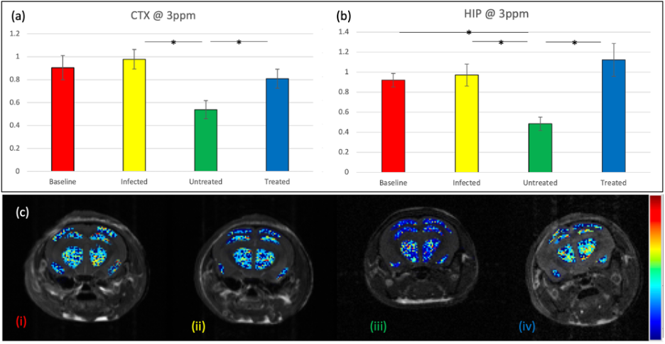
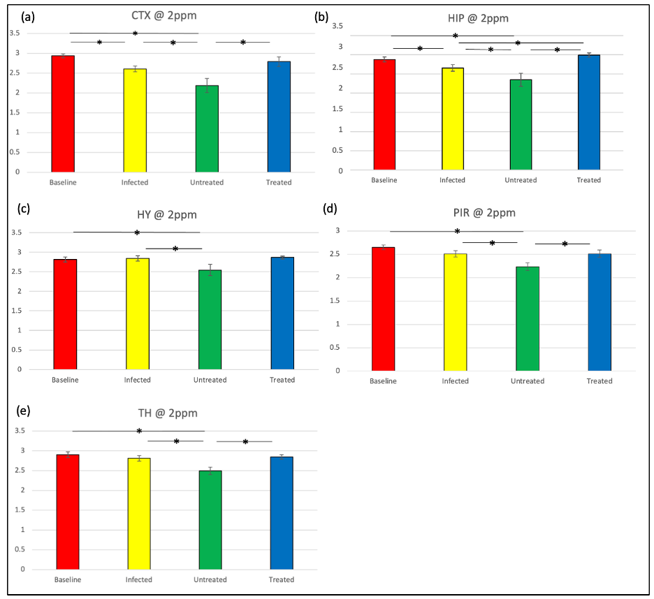
Figure 1 shows significant differences in 3ppm CEST in the CTX and HIP. In the CTX, untreated mice demonstrated decreased signal relative to treated mice (p=0.036) and early-infection mice (p=0.017). In the HIP, untreated mice showed decreased signal relative to treated (p=0.014), early-infection (p=0.03), and baseline mice (p=0.032).The 2ppm CEST effect in untreated mice decreased in all regions compared to all other groups (Figure 2). Relative to early-infection mice, there was a signal decrease in the CTX (p<0.01), HIP (p=0.049), HY (p=0.018), PIR (p=0.02), and TH (p=0.013). When compared to treated mice, there was a decrease in the CTX (p=0.02), HIP (p<0.01), PIR (p=0.02), and TH (p<0.01). Relative to the baseline, the untreated mice saw a signal decrease in all regions (p<0.01).
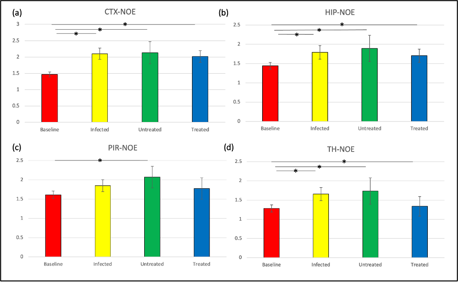
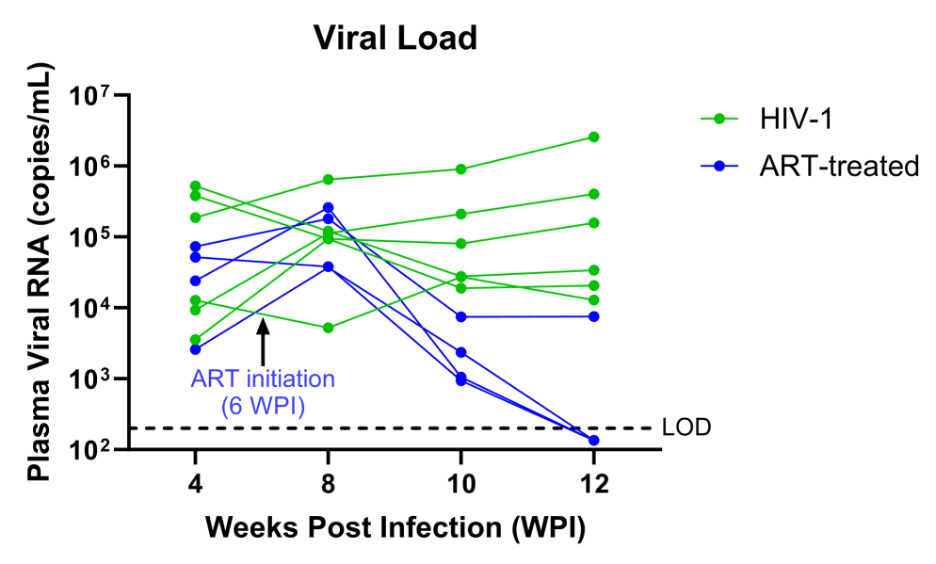
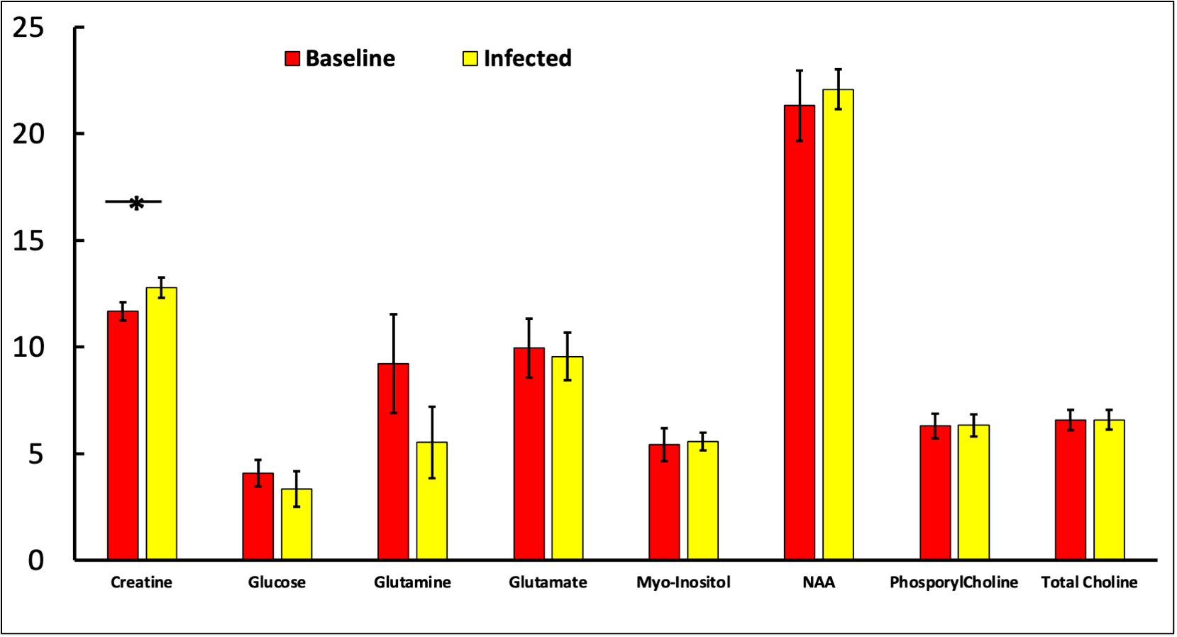
The Nuclear Overhauser Effect (NOE) signal (Figure 3), was lower in untreated mice compared to baseline in the CTX and TH (p<0.01), as well as on the HIP and PIR (p=0.03). Early-infection mice showed reduced NOE signal in the CTX and HIP compared to the baseline, while the untreated group showed a decrease in the HYP compared to all others (p<0.03). Progressive viral load is displayed in Figure 4. This was monitored via biweekly cheek-bleeding, verifying ART’s impact on infection severity. Within untreated mice, viral RNA count trends upward. Within treated mice, however, viral load decreases steadily after 4 weeks post-infection (WPI), typically reaching the limit of detection (LOD) around week 12. MRS data, provided in Figure 5, suggests a significant increase in post-infection creatine, with no trend in other metabolites of interest. This table only contains the baseline and early-infection data, as the remaining data is still being processed.
Discussion
It is believed that the 3ppm CEST contrast results from glutamate, a primary excitatory neurotransmitter[7]. Long-term infection (~10 weeks) caused decreasing glutamate in all regions, indicating impaired neuronal function. While glutamate MRS showed no significant change caused by short-term infection (6 weeks), the precursor of glutamate–glutamine was decreased. However, glutamate was restored during ART treatment, evidenced via increased 3ppm CEST contrasts. The 2ppm CEST contrast reflects creatine level[8-9], which facilitates cellular metabolism. Long-term infection resulted in declining creatine, suggesting bioenergetic shifts in brain cells. ART treatment restored creatine levels in all regions. CEST results showed no significant changes in creatine by short-term infection. MRS, however, showed increased creatine levels. This disparity may be due to different sensitivity between these methods or potential tissue contamination in the single-voxel MRS. The increased NOE of long-term infected mouse membrane lipids indicates damage to brain cellular integrity[9], which was restored by ART treatment. In summary, our CEST MRI study showed that long-term HIV infection is linked to neuronal impairments, altered brain cellular energy consumption, and damage to cellular integrity. This was partially validated by MRS.Acknowledgements
This study was partially supported by NIH R21MH128123, R21MH134678, P20GM130447, Nebraska Research Initiative, UNMC Graduate Fellowship. The authors thank the Small Animal MRI core facility at UNMC for technical support.References
- Saylor D, Dickens AM, Sacktor N, Haughey N, Slusher B, Pletnikov M, et al. HIV-associated neurocognitive disorder--pathogenesis and prospects for treatment. Nat Rev Neurol 2016; 12(4):234-248.
- Ward KM, Aletras AH, Balaban RS. A New Class of Contrast Agents for MRI Based on Proton Chemical Exchange Dependent Saturation Transfer (CEST). Journal of Magnetic Resonance 2000; 143(1):79-87.
- van Zijl PC, Yadav NN. Chemical exchange saturation transfer (CEST): what is in a name and what isn't? Magnetic resonance in medicine 2011; 65(4):927-948.
- Liu G, Song X, Chan KW, McMahon MT. Nuts and bolts of chemical exchange saturation transfer MRI. NMR Biomed. 2013 Jul;26(7):810-28. doi: 10.1002/nbm.2899. Epub 2013 Jan 10. PMID: 23303716; PMCID: PMC4144273.
- Wu B, Warnock G, Zaiss M, Lin C, Chen M, Zhou Z, et al. An overview of CEST MRI for non-MR physicists. EJNMMI physics 2016; 3(1):19.
- Mathews, S., Branch Woods, A., Katano, I. et al. Human Interleukin-34 facilitates microglia-like cell differentiation and persistent HIV-1 infection in humanized mice. Mol Neurodegeneration 14, 12 (2019). https://doi.org/10.1186/s13024-019-0311-y
- Hadar, PN, Kini, LG, Nanga, RPR, et al. Volumetric glutamate imaging (GluCEST) using 7T MRI can lateralize nonlesional temporal lobe epilepsy: A preliminary study. Brain Behav. 2021; 11:e02134. https://doi.org/10.1002/brb3.2134
- Cai, K., Singh, A., Poptani, H., Li, W., Yang, S., Lu, Y., Hariharan, H., Zhou, X. J., and Reddy, R. (2014) CEST signal at 2 ppm (CEST@2ppm) from Z-spectral fitting correlates with creatine distribution in brain tumor, NMR Biomed., 28, 1–8, doi: 10.1002/nbm.3216.
- Chen, L, Zeng, H, Xu, X, et al. Investigation of the contribution of total creatine to the CEST Z-spectrum of brain using a knockout mouse model. NMR in Biomedicine. 2017; 30:e3834. https://doi.org/10.1002/nbm.3834
- Yu Zhao, Casey Sun, Zhongliang Zu. Assignment of molecular origins of NOE signal at −3.5 ppm in the brain. bioRxiv: 2023.02.03.526979; doi: https://doi.org/10.1101/2023.02.03.526979
Figures