4449
APTw/CEST-MRI for body oncology with SPIR fat suppression using improved Z-spectral modeling for olefinic fat components1Philips Research, Hamburg, Germany, 2Philips Healthcare, Gainesville, FL, United States, 3Advanced Imaging Research Center, University of Texas Southwestern Medical Center, Dallas, TX, United States, 4Department of Radiology, University of Texas Southwestern Medical Center, Dallas, TX, United States
Synopsis
Keywords: CEST / APT / NOE, CEST & MT, Z-spectral fitting, olephinic fat, aliphatic fat
Motivation: Fat-artifacts are challenging in CEST-MRI for body applications as they overlap spectrally with clinically relevant signals like amide or hydroxyl.
Goal(s): Improve the quality of in vivo fat correction based on previously explored Z-spectral fitting of data obtained with SPIR fat suppression.
Approach: Olefinic fat is modeled separately while scaling and inverting aliphatic components. Fitting was tested on Z-spectral data from breast ROIs with variable fat content.
Results: The new model is accurate for a large range of fat fractions. The opposite phase of aliphatic and olefinic fat components provides a better explanation for the observed Z-spectra in vivo.
Impact: Understanding and accurate modeling of fat signals in fat-suppressed APT/CEST-MRI will allow unbiased assessment of CEST effects in body-oncology (breast, kidney cancer).
INTRODUCTION
Fat signal correction remains a challenge in APT/CEST-MRI for body-oncology1,2,3,5. Signal models including water saturation (DWS), MT-effect, and modified fat spectra were previously shown4 to fit Z-spectra acquired with SPIR fat suppression. SPIR pulses potentially improve body CEST-MRI by lowering the initial fat contribution. Here, application in breast imaging reveals fat components close to water, identified as olefinic fat components not touched by the SPIR pulse. The signal model is extended with relative weighting and phases imposed by SPIR and tested on volunteer breast imaging data with different fat fractions.METHODS
Examinations were performed at 3T (Ingenia, Philips) with alternating dual-channel RF-transmission (100% duty-cycle) using a 16-channel breast coil on a female volunteer (IRB approved, informed consent obtained).40×50ms saturation pulses were used (Tsat=2s and B1,rms=2μT) in a single-shot 2D fast-spin echo sequence: SPIR fat-suppression, FOV=(160mm)2, voxel size 1.2×1.2×8mm3, TR/TE=5700/7.0ms, 90° flip angle (FA), 120° refocusing, centric k-space ordering, and N=17 offsets: {-1560(S0),±7.8,±6.3,±5.1,±3.9,±2.9,±1.8, ±0.9,±0.3} ppm (relative to water), taking Tacq=2min. A 3-point multi-acquisition gradient-echo Dixon sequence, same geometry and TE, was used for B0 mapping with TR=14 ms, DTE=0.6 ms, FA=35°, 8 averages, Tacq=45 s. Z-spectra were interpolated to 100 points including B0-correction.
The updated model (Eq.1) includes Lorentzian (DWS), Gaussian (MT) and a fat spectral model with SPIR fat suppression, using Nl1 (olefinic) spectral fat components not touched by the SPIR pulse (+sign) and further Nl2 components (-sign) reduced by the SPIR effect s (s≈0.06, empirically established).
$$ S_{model} (f)=\left| S_0- \left( A_L \frac{w_L^2}{f^2+ w_L^2 }+A_G e^{\large{-\frac{1}{2}(\frac{f}{w_G})^2}}+A_F\left\{ \sum_{l=1}^{N_{l1}}a_l\frac{w_F^2}{(f-f_l )^2+ w_F^2 }-\sum_{l=N_{l1}+1}^{N_{l1}+N_{l2}}a_l\frac{s * w_F^2}{(f-f_l )^2+ w_F^2 }\right\} \right) \right| \ \ \ [1] $$
5 parameters are essential for the fitting: AL, AG, AF (amplitudes) and wL, wG (Lorentzian/Gaussian width). The frequency center was fitted to confirm the quality of B0 correction. The SPIR effect s was fitted to confirm the model validity. As Z-spectral magnitude levels close to zero can show a positive noise bias, a noise level estimation was included in the fit. APTw signals were obtained as fit residuals (APT#), omitting [1.7…5.1]ppm around 3.5ppm from the fit (weighting shown in Fig. 2B).
Fixed fat linewidths (wF=1.1 ppm) and fat frequencies fl (Nl1 +Nl2 =7 lines) were used, olefinic +0.6 ppm (relative to water), aliphatic {-3.7, -3.4, -3.0, -2.6, -2.3, -1.8} ppm. The individual fat amplitudes al were normalized such that
$$ \sum_{l=1}^{N_{l1}}a_l+\sum_{l=N_{l1}+1}^{N_{l1}+N_{l2}}s*a_l = 1.0$$
Initial conditions and boundaries: AG =0.2(>10-5), AL =0.6(>10-5), AL + AG = S0, wG =78 ppm (10-7..390), wL =2.3 ppm (>10-7), AF =0.4 (10-5..5.0), s=0.01 (10-3..0.09), noise=0.08 (<0.6).
RESULTS
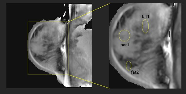
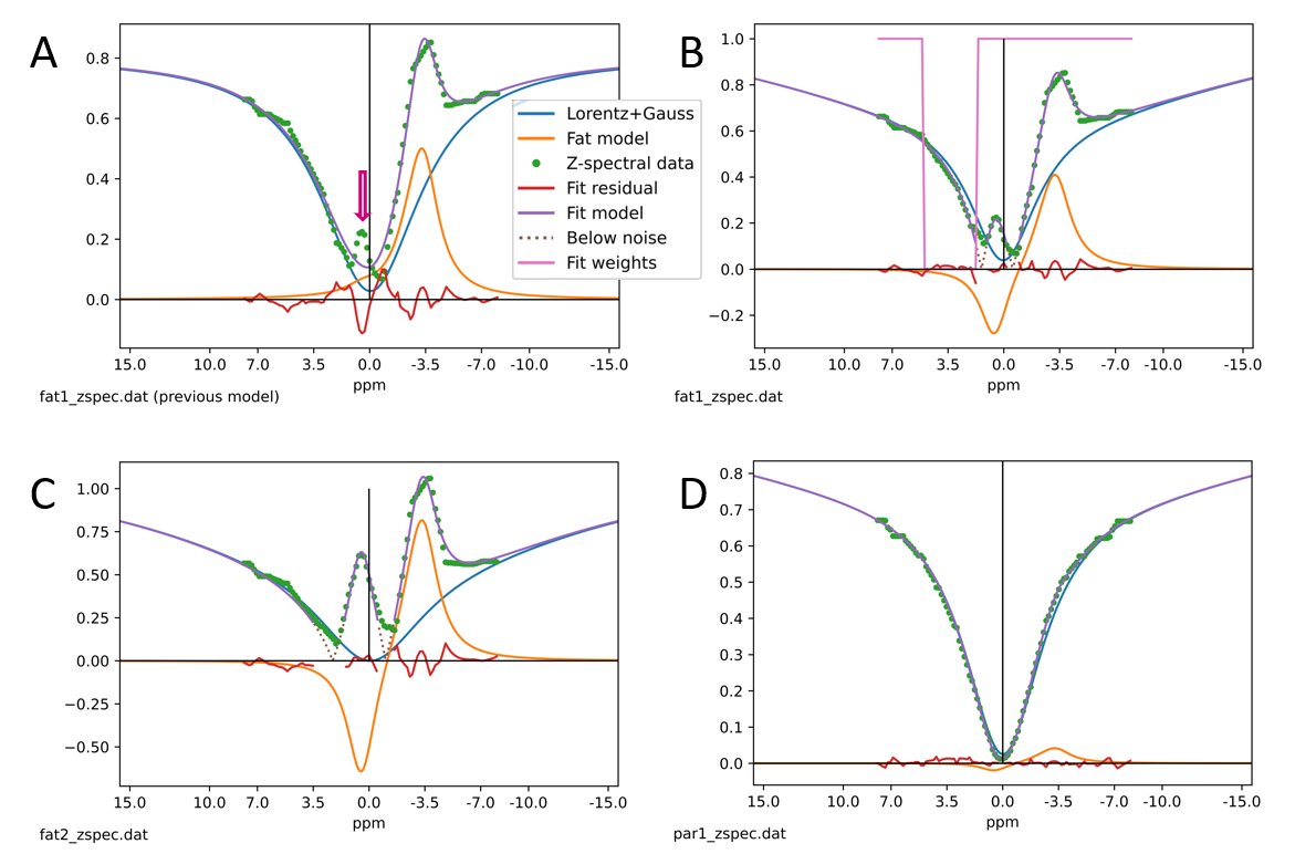
Fig.2 shows example Z-spectra (green dots) and results from 3 different ROIs (fat1, fat2, par1, see Fig. 1). Lorentz-Gauss components (blue), the fat model (orange), the combined fat model (violet) and the fit residual (red) are shown separately (legend in Fig. 2A). Values below the estimated noise level are shown as dotted line. A previous fitting model4 (Fig. 2A) misses the olefinic fat signal at +0.6ppm (arrow). For the extended model (Fig.2B/C/D), the quality of the fit is good (R>0.98). Table 1 lists obtained example fitting parameter results for the cases shown.DISCUSSION
Inclusion of olefinic components without SPIR suppression improves Z-spectral fitting. Actual “zero crossing” of the model function is not visible in the data because of the positive noise bias, which increases with fat content. Small positive APT# values are found in ROIs with significant non-fat tissue content (fat1, par1), as expected. Apparently, residual olefinic fat signal with SPIR suppression may influence the assessment of APT# for moderate or high fat levels (Fig.2B/C). Even at low fat levels (Fig.2D), remaining fat contributes to data asymmetry at ±3.5ppm and alters the spectral shape. Fitting of s appears less reliable in low-fat compartments. For a given MR-protocol, the SPIR effect is similar throughout different compartments and could be fixed.While previously attributed to saturation recovery4, the opposite phase of aliphatic and olefinic fat components provides a better explanation of the observed Z-spectra. Without RF saturation, aliphatic and olefinic components (partly) cancel. When saturating aliphatic components near -3.4ppm, olefinic components remain with elevated signal (positive phase). Vice versa, when saturating at the olefinic frequencies, aliphatic components remain, albeit strongly reduced by SPIR. At low signal level, the inverted phase of aliphatic components leads to negative signal amplitudes, appearing mirrored in magnitude Z-spectra (elevated signal). Minimum Z-spectral requirements for the improved fitting model and robustness are currently under investigation in body-oncology applications.
CONCLUSION
Better understanding of RF saturation and SPIR effects lead to an improved model to obtain fat-signal corrected APTw signals in body-oncology APT/CEST-MRI.Acknowledgements
This work was supported by NIH-1R01CA252281-01A1.References
1. Gao T, Zou C, Li Y, Jiang Z, Tang X, Song X. A Brief History and Future Prospects of CEST MRI in Clinical Non-Brain Tumor Imaging. Int J Mol Sci. 2021 Oct 26;22(21):11559.
2. Vinogradov E, Keupp J, Dimitrov IE, Seiler S, Pedrosa I. CEST-MRI for body oncologic imaging: are we there yet? NMR Biomed. 2023 Jun;36(6):e4906.
3. Zhang S, Keupp J, Wang X, Dimitrov I, Madhuranthakam AJ, Lenkinski RE, Vinogradov E. Z-spectrum appearance and interpretation in the presence of fat: Influence of acquisition parameters. Magn Reson Med. 2018 May;79(5):2731-2737.
4. Keupp J, Dimitrov I, Eggers H, and Vinogradov E. Z-spectral fitting for fat-correction of APT/CEST-MRI including fat-suppression pulses to improve body applications. Proc. Intl. Soc. Mag. Reson. Med. 31 (2023) 2989
5. Zimmermann F, Korzowski A, Breitling J, Meissner JE, Schuenke P, Loi L, Zaiss M, Bickelhaupt S, Schott S, Schlemmer HP, Paech D, Ladd ME, Bachert P, Goerke S. A novel normalization for amide proton transfer CEST MRI to correct for fat signal-induced artifacts: application to human breast cancer imaging. Magn Reson Med. 2020 Mar;83(3):920-934.
Figures