4448
The impact of deep learning based image reconstruction on CEST MRI for distinguishing inactive from active thyroid-associated ophthalmopathy1Graduate School of Bengbu Medical College,, Bengbu, China, 2Department of Radiology, Second Affiliated Hospital of Naval Medical University, Shanghai, China, 3MR Research, GE Healthcare, Beijing, China
Synopsis
Keywords: CEST / APT / NOE, CEST & MT, thyroid-associated ophthalmopathy, diffusion weighted imaging, deep learning reconstruction
Motivation: Thyroid-associated ophthalmopathy (TAO) is characterized by accumulation of collagen in extraocular muscle. CEST-MRI can evaluate the collagen content by focusing on amide compound. However, CEST effect is small and sensitive to low image SNR. A vendor-provided deep learning reconstruction (DLR) algorithm can dramatically increase image SNR.
Goal(s): Investigate if CEST-MRI can distinguish inactive from active TAO and the impact of DLR on its diagnostic performance.
Approach: 11 active and 12 inactive TAO were enrolled. CEST imaging was reconstructed with DLR and conventional reconstruction.
Results: DLR can significantly increase SNR of CEST imaging and improved the diagnostic performance for discriminating inactive from active TAO.
Impact: The treatment of TAO depends on the disease phase. DLR image reconstruction improved the performance of CEST in differentiation between inactive and active TAO. It would help in the evaluation and management of TAO patients.
Introduction
Thyroid-associated ophthalmopathy (TAO) is an autoimmune disorder which begins with an initial active phase of progression and follows by subsequent static inactive phase[1-2]. Active and inactive TAO were treated with hormonal/anti-inflammatory drugs and surgical decompression, respectively[1-2]. The diagnosis of TAO phase bases on symptoms or signs in clinic[1-2]. It remains challenging for clinicians to evaluate and management TAO[2].One pathological characteristic of TAO is an accumulation of collagen in extraocular muscle (EOM)[2]. In inactive phase, the long-lasting and increased production of collagen leads ultimately to atrophy, fibrosis, and sclerosis of EOM and subsequently to restrictive strabismus[2]. Thus, the detection of collagen may be a way to evaluate and management of TAO.
Chemical exchange saturation transfer (CEST) imaging, as a promising magnetic resonance (MR) contrast mechanism, relies on proton exchange between specific chemical compounds and bulk water pool[3]. Amide protons transfer weighted (APTw) imaging is one type of CEST which focuses on the exchange between amide protons and bulk water[3-4]. It has been applied in diagnosing tumors and predicting tumor treatment output[4-6].
Taking pathology into consideration, we assumed that APTw might hold the potential in diagnosing TAO phase. However, CEST effect is usually small[3-4] and may be sensitive to signal with low SNR. A vendor provided deep learning reconstruction algorithm (DLR; AIRTM ReconDL, GE Healthcare) has recently been proposed to dramatically increase the imaging SNR[7]. It had been applied for CEST in brain glioma and resulted with improved lesion conspicuity[8]. Thus, this study aimed at investigating if APTw can be used to diagnose TAO phase and the impact of DLR on the diagnostic performance.
Material and Methods
Patients23 patients were enrolled. According to clinical active score, 11 were in active phase and the rest 12 were in inactive phase.
MRI Acquisition
All MRI examinations were scanned at a 3.0T scanner (SIGNA Premier; GE Healthcare, USA) with 21ch head-and-neck combined coil. A single-shot fast-spin-echo based sequence was applied for CEST imaging. 29 frequencies ranging from 7ppm to -7ppm with an increment of 0.5ppm were applied with the RF saturation power and duration of 2μT and 2000ms. With WASSR method[4], additional 11 frequencies from 1.875 to -1.875ppm were measured with the saturation power and duration of 0.5μT and 2000ms for B0 inhomogeneity correction. Other scan parameters were TR/TE=3000ms/minimal, FOV=24×24cm2, matrix=132×132, slice thickness=3mm. The acquired CEST imaging was reconstructed in conventional manner (ConR) and DLR, separately.
Data Analysis
The signal-to-noise ratio (SNR) of EOM was defined as: SNR=S(muscle)/SD(background), where S(muscle) is the average signal of extraocular musculature and SD(background) is the standard derivation of background signal.
CEST data reconstructed with ConR (CEST_ConR) and DLR (CEST_DLR) were analyzed with custom-written MATLAB (2018b) scripts. Magnetic-transfer-ratio-asymmetry, representing CEST effect at 3.5ppm, was calculated as MTRasym=[S(-3.5ppm)-S(3.5ppm)]/S0, where S0 represents the signal without saturation. S(-3.5ppm) and S(3.5ppm) are the signals obtained at ±3.5ppm away from water pool.
Statistical Analysis
SNR and MTRasym of EOM in all patients were separately compared between CEST_ConR and CEST_DLR. Mann-Whitney-U test was used to assess the differences of MTRasym between active and inactive TAO. Subsequently, receiver operating characteristics curve (ROC) analysis was employed to evaluate the diagnostic performance of MTRasym in differentiating inactive from active TAO patients. P<0.05 was considered statistical significance.
Results
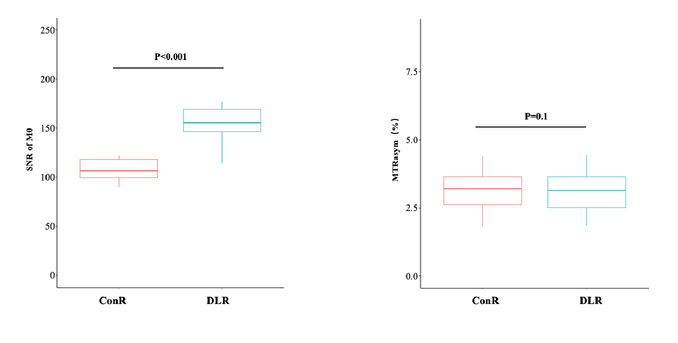
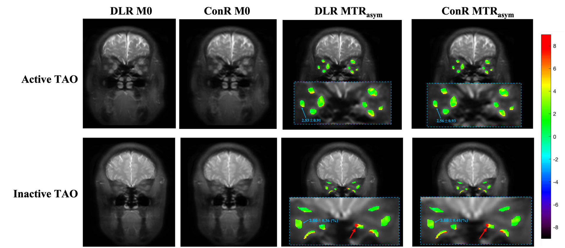
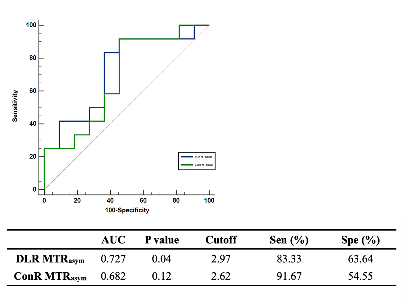
Figure 1 showed DLR, relative to ConR, significantly improved the SNR of CSET images (163±30 vs. 108±14; P<0.001). No significant difference of EOM MTRasym was observed between CEST_DLR and CEST_ConR (3.07%±0.70% vs. 3.14%±0.67%; P>0.05).The EOM MTRasym calculated from CEST_DLR was significantly lower in active than in inactive TAO (Table 1 and Figure 2). Similar trend was also observed in CEST_ConR, but the difference was not significant (P=0.075). The CEST_DLR showed improved diagnostic performance for distinguishing inactive from active TAO patients (Figure 3).
Discussion and Conclusion
This study investigated the role of CEST in diagnosing TAO phase and the impact of DLR on the diagnostic performance. Our results showed DLR can significantly increase the SNR of CEST images and had no significantly impact on the EOM MTRasym. Moreover, the EOM MTRasym derived from CEST_DLR was significantly lower in active than in inactive TAO and may be used as an effecive index to discriminate disease phase. More importantly, probably due to higher SNR, CEST_DLR with dervied MTRasym was proved with more robust diagnostic efficacy, relative to CEST_ConR, in differentiating active from inactive TAO.In conclusion, DLR image reconstruction can improve the diagnostic performance of CEST for distinguishing active and inactive TAO patients. It would be beneficial for TAO evaluation and management.
Acknowledgements
No acknowledgement found.References
[1] Henry B Burch, Petros Perros, Tomasz Bednarczuk, et al. Management of thyroid eye disease: a Consensus Statement by the American Thyroid Association and the European Thyroid Association. Eur Thyroid J. 2022; 11(6): e220189.
[2] Jesús Barrio-Barrio, Alfonso L Sabater, Elvira Bonet-Farriol, et al. Graves’ Ophthalmopathy: VISA versus EUGOGO Classification, Assessment, and Management. J Ophthalmol. 2015; 2015:249125.
[3] Van Zijl, P. C., Yadav, N. N. Chemical exchange saturation transfer (CEST): What is in a name and what isn’t? Magn Reson Med, 2011, 65(4): 927-948.
[4] Jinyuan Zhou, Moritz Zaiss, Linda Knutsson, et al. Review and consensus recommendations on clinical APT-weighted imaging approaches at 3T: Application to brain tumors. Magn Reson Med. 2022; 88(2): 546-574.
[5] Mancini, L., Casagranda, S., Gautier, G., et al, CEST MRI provides amide/amine surrogate biomarkers for treatment-naïve glioma sub-typing. Eur J of Nucl Med and Mol Imaging, 2022, 49(7): 2377-2391.
[6] Chan, K. W., Jiang, L., Cheng, M., et al. CEST-MRI detects metabolite levels altered by breast cancer cell aggressiveness and chemotherapy response. NMR in biomedicine, 2016, 29(6): 806-816.
[7] Lebel RM. Performance characterization of a novel deep learning-based MR image reconstruction pipeline. arxiv.org/abs/2008.06559.[8] Shu Zhang, Xinzeng Wang, F.Wolliam Schuler et al. Deep Learning-based image reconstruction improves CEST MRI. ISMRM.2021;1457.
Figures