4447
Altered amide in substantia nigra is concordant with motor asymmetry in Parkinson's disease: a multipool CEST study1Department of Radiology, Department of Radiology, Beijing Hospital, National Center of Gerontology, Institute of Geriatric Medicine, Chinese Academy of Medical Sciences, Beijing, China, 2Graduate School of Peking Union Medical College, Chinese Academy of Medical Sciences, Beijing, China, 3Clinical & Technique Support, Philips Healthcare, Beijing, China
Synopsis
Keywords: CEST / APT / NOE, CEST & MT, Parkinson's Disease
Motivation: The pathophysiological changes associated with motor asymmetry within midbrain nucleus in Parkinson’s Disease (PD) remains unclear.
Goal(s): We aimed to explore the mesencephalic change by using four-pool model-based chemical exchange saturation transfer (CEST) analysis in asymmetrical PD.
Approach: The difference of four-pool model-based CEST parameters in more and less affected side of mesencephalic region were compared, and its associations with motor asymmetry were estimated.
Results: Our data revealed the inherent asymmetry on nigral amide and relayed nuclear Overhauser effect (rNOE) in asymmetric PD patients, and the substantial consistency between imaging laterality of nigral amide and motor laterality in significant asymmetrical PD.
Impact: Our findings could benefit a better understanding of the mechanisms contributing to the asymmetry of PD and provide promising no-invasive neuroimaging biomarkers related to lateralization in PD.
Introduction
PD is a strikingly asymmetric condition with typical motor asymmetry related to the dopaminergic deficit and aggregation of misfold proteins. CEST imaging can assess alterations in endogenous proteins and peptides caused by these pathophysiological abnormalities, which has been indicated by two-pool CEST studies [1-3], but not yet took asymmetric involvement into consideration. Comparing with two-pool CEST analysis, multi-pool CEST analysis could separate certain confounding effects and provide additional value to PD. Furthermore, a potential asymmetric nigral amide signal in hemiparkinsonism patients was revealed using two-pool CEST model [2]. Therefore, we hypothesized that this change exists in PD patients with bilaterally asymmetrical motor symptoms, and we posited a correlation between alterations in CEST parameters derived from four-pool analysis and the motor asymmetry observed in PD patients.Methods
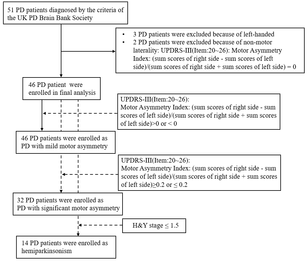
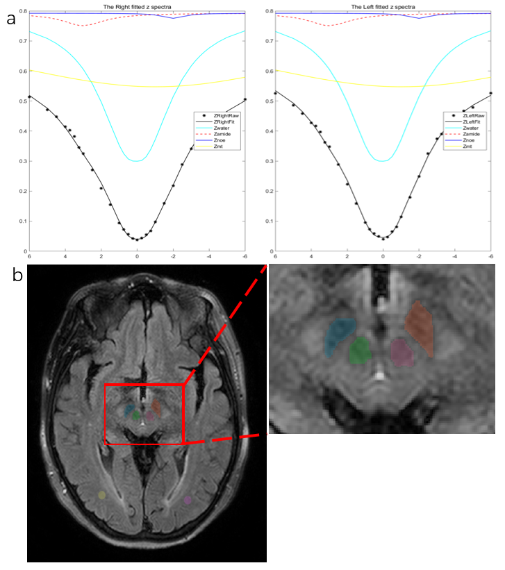
Forty-six PD patients and twenty-three normal controls (NC) were enrolled in this study. According to the motor asymmetry index, we classified the participants into three motor asymmetry subgroups: PD with mild motor asymmetry (PD_MMA), PD with significant motor asymmetry (PD_SMA), and hemiparkinsonism (PD_Hemi), as depicted in Figure 1. Additionally, we confirmed the motor laterality in these subgroups.All MRI examinations were performed on 3.0 T MRI scanner (Achieva, Philips Healthcare). As previously stated[1], the CEST imaging was performed with 31 offsets based on an off-resonance continuous-wave RF saturation pulse with four block, each with a duration of 200ms and a saturation power level of 2.0 μT. Four CEST related parameters, including amide, rNOE, direct water saturation (DS), and magnetization transfer contrast (MTC), were calculated using four-pool Lorentzian fitting model in bilateral SN, red nucleus (RN) and normal-appearing occipital white matter (OWM) based on FLAIR[4]. The regions contralateral to the motor laterality were defined as more affected side, otherwise, the less affected side. The representative 4-pool Lorentzian fit z spectra and anatomical segmentations were shown in Figure 2. Imaging asymmetry index was calculated and imaging laterality was confirmed.
Results
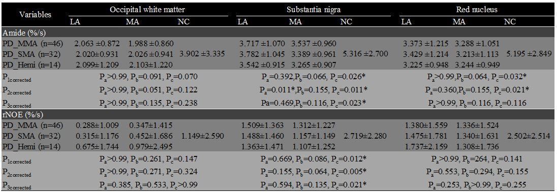
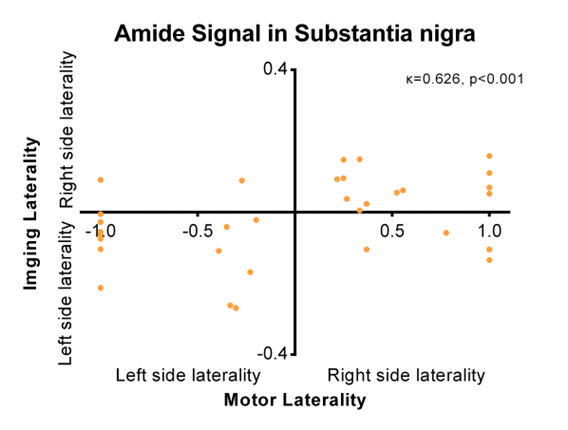
Our findings indicated that there were no significant differences in the comparison between DS and MTC across all regions and among various groups. In all three PD subgroups, amide and rNOE were decreased in both more affected side and less affected side in SN compared with NC, while the statistical differences only illustrated between NC and PD more affected side (all corrected P <0.05). Besides, significantly decreased amide was observed in more affected side of RN in PD_MMA and PD_SMA compared with NC (corrected p = 0.032 and 0.021, respectively). As for the paired comparison of amide and rNOE between PD more and less affected side in all regions and all subgroups, only nigral amide differed significantly in PD_SMA subgroup, with a statistical decrease in PD more affected side compared with less affected side (corrected p =0.011). Table 1. demonstrated the comparison details about amide and rNOE signals. The laterality of nigral amide exhibits a substantial consistency with the motor laterality in the PD_SMA subgroup (κ=0.629, p<0.001, as shown in Figure 3). However, there is no significant correlation between the imaging asymmetry index of any CEST parameters and the motor asymmetry index scores in the PD_MMA and PD_SMA subgroups across all regions.Discussion
Our findings revealed an inherent lateralized pattern of amide and rNOE in PD between the more and less affected sides of SN, since significant lower amide and rNOE exist in more affected side of SN, but not in less affected side, regardless of the extent of motor asymmetry. These could be partially explained by asymmetric neuronal degeneration in SN. However, the significant difference between the more and less affected side only existed in PD_SMA subgroup, but not in hemiparkinsonism (the earlier stage of PD). One possible explanation is the selected vulnerability of neuron in unilateral SN[5]. Another explanation could be the converge of differential trajectories in asymmetric motor symptoms over time[6], which may narrow the difference and prevent them from reaching the vulnerable point of amide and rNOE decrease. Additionally, the underlying asymmetric distribution of amide in RN in both PD_MMA and PD_SMA indicated the asymmetry of extrapyramidal system in asymmetrical PD. Furthermore, the substantial consistency between motor laterality and imaging laterality of nigral amide in PD_SMA indicated the potential of nigral amide as neuroimaging biomarkers for PD lateralization.Conclusion
Our findings revealed the inherent asymmetry of nigral amide, rNOE and amide within RN, along with the concordance of imaging laterality of nigral amide and motor laterality in asymmetrical PD patients. It enhances our comprehension of the mechanisms underlying PD's asymmetry and provides promising neuroimaging biomarkers associated with PD lateralization.Acknowledgements
This work was supported by the National Natural Science Foundation of China (No. 82071891 and No. 82272107).References
[1] Li C, Peng S, Wang R, Chen H, Su W, Zhao X, Zhou J, Chen M. Chemical exchange saturation transfer MR imaging of Parkinson's disease at 3 Tesla. Eur Radiol 2014;24(10):2631-9.
[2] Li C, Chen M, Zhao X, Wang R, Chen H, Su W, Li S, Lou B, Song G, Zhang S, Zhang J, Zhou J. Chemical Exchange Saturation Transfer MRI Signal Loss of the Substantia Nigra as an Imaging Biomarker to Evaluate the Diagnosis and Severity of Parkinson's Disease. Front Neurosci 2017;11:489
[3] Tian Y, Li X, Wang X, Su W, Li S, Wang W, Zhang Y, Li C, Chen M. CEST 2022-three-dimensional amide proton transfer (APT) imaging can identify the changes of cerebral cortex in Parkinson's disease. Magn Reson Imaging 2023;102:235-41.
[4] Mennecke A, Khakzar KM, German A, Herz K, Fabian MS, Liebert A, Blumcke I, Kasper BS, Nagel AM, Laun FB, Schmidt M, Winkler J, Dorfler A, Zaiss M. 7 tricks for 7 T CEST: Improving the reproducibility of multipool evaluation provides insights into the effects of age and the early stages of Parkinson's disease. NMR Biomed 2023;36(6):e4717.
[5] Surmeier DJ. Determinants of dopaminergic neuron loss in Parkinson's disease. FEBS J 2018;285(19):3657-68.
[6] Miller-Patterson C, Buesa R, McLaughlin N, Jones R, Akbar U, Friedman JH. Motor asymmetry over time in Parkinson's disease. J Neurol Sci 2018;393:14-7.
Figures