4437
MR Elastography Image Reconstruction using Spatio-Temporal Neural Networks-based Regularization1Physikalisch-Technische Bundesanstalt (PTB), Braunschweig and Berlin, Germany, 2Charité - Universitätsmedizin Berlin, Berlin, Germany
Synopsis
Keywords: Quantitative Imaging, Elastography
Motivation: MR Elastography (MRE) is a quantitative, noninvasive method to map viscoelastic properties in tissue. We propose a method of advanced image reconstruction to increase resolution and accuracy for fast MRE.
Goal(s): We aim to reduce scan time and, eventually, enable real-time MRE.
Approach: To overcome artifacts resulting from the accelerated data acquisition, we employ a data-driven regularization method. Our approach utilizes a physics-informed convolutional neural network (CNN) that exploits spatio-temporal correlation among the images.
Results: We show that the employed spatio-temporal approach can improve the image reconstruction performance and further outperforms iterative SENSE reconstructions and standard 2D U-Net approaches.
Impact: Our approach allows for the accurate estimation of elastograms from strongly undersampled data, thus allowing a highly reduced scan time. These improvements will eventually benefit clinical practice, making MRE an even more powerful imaging tool.
Introduction
The mechanical properties of tissue can alter when it is affected by disease. For instance, fibrosis can increase tissue stiffness of the liver1. Magnetic Resonance Elastography (MRE)2 is a non-invasive tool to assess pathological changes by transmitting mechanical waves through the tissue. The induced motion is evaluated with motion-encoding gradients and encoded in the phase of the obtained MR images.Subsequently, shear wave speed (SWS) maps can be estimated by applying inversion methods3 to the complex-valued wavenumber $$$k^*$$$. Currently, most of the MRE research is related to the development of these inversion methods. However, they are constrained by the quality of the images used, limiting their applicability to real-time MRE, since this requires acquiring undersampled data. Consequently, the obtained images suffer from reduced quality, resulting in a lack of details within the SWS maps. We propose utilizing unrolled CNN-based iterative reconstruction schemes to fill the gap between data acquisition and elastography inversion algorithms.
Methods
In MRE, the data acquisition can be described by$$ \mathbf{y} = \mathbf{A}\mathbf{q}(\mathbf{p})+\mathbf{e} $$ where $$$\mathbf{e}$$$ is Gaussian noise and $$$\mathbf{q}$$$ is a nonlinear process describing the interaction of the different quantitative parameters, e.g. SWS and wave penetration rate. In this work, since we apply the k-MDEV inversion4, we consider the problem $$ \begin{equation} \begin{cases} p = \text{k-MDEV}(\mathbf{x}^{\ast}),\\ \text{ where } \mathbf{x}^{\ast}:= \underset{\mathbf{x}}{{\arg\min}}\frac{1}{2}\| \mathbf{A}\mathbf{x} - \mathbf{y}\|_2^2 + \frac{\lambda}{2} \| \mathbf{x} - \mathbf{x}_{\mathrm{NN}}\|_2^2 \end{cases}\end{equation}$$ Thereby, $$$\mathbf{x}^{\ast}$$$ denotes the complex-valued MR images which are obtained by solving the CNN-regularized image reconstruction problem and $$$ \mathbf{y}=[\mathbf{y}^1 \ldots\mathbf{y}^{N_C}]^T $$$ is undersampled and noisy k-space data of multiple coils acquired on spiral trajectories. The regularizer $$$ {\mathbf{x}}_{\mathrm{NN}} $$$ is in our case the output of a CNN aiming to construct spatio-temporal slices mapped to the corresponding ground truth slices, which in the following will be referred to as the XT,YT network5. The output of the CNN is subsequently used as a starting point for solving the minimization problem above, which is achieved by solving a linear system with a conjugate gradient method6. This results in a physics-informed neural network that can be trained end-to-end7.
We simulated an undersampled spiral data acquisition by constructing the MRI operator $$$ \mathbf{A} $$$ using representative spiral trajectories containing few k-space points and retrospectively applying it to the data, comprising of brain MRE data of 13 subjects acquired with standard Cartesian sampling. Three test cases were examined to explore the effect of k-space points in total and the number of spirals - single, two and three shot spiral imaging with 800 k-space points per spiral were considered.
Each dataset is split into three motion encoding gradient (MEG) directions, four frequencies, and eight timesteps. The image size is given as $$$126 \times 126 \times 40$$$. The dataset was subsequently split into 4/1/8 subjcets for training, validation, and testing.
The proposed method using the 2D U-Net and the XT,YT Net trained for 15 epochs using the ADAM optimizer with an initial learning rate of 10-3. Both were constructed using a 2D U-Net with an initial number of 32 filters, three encoding stages, and two convolutions per encoding stage. Thereby, the number of trainable parameters is equal among the two methods, making them directly comparable in terms of 8. The network architecture is shown in Figure 1. Since the phase images are crucial for elastograms, a particular symmetric loss function in the form of $$$\perp$$$-loss9 was applied. All methods are evaluated in terms of PSNR, NRMSE, and SSIM.
Results
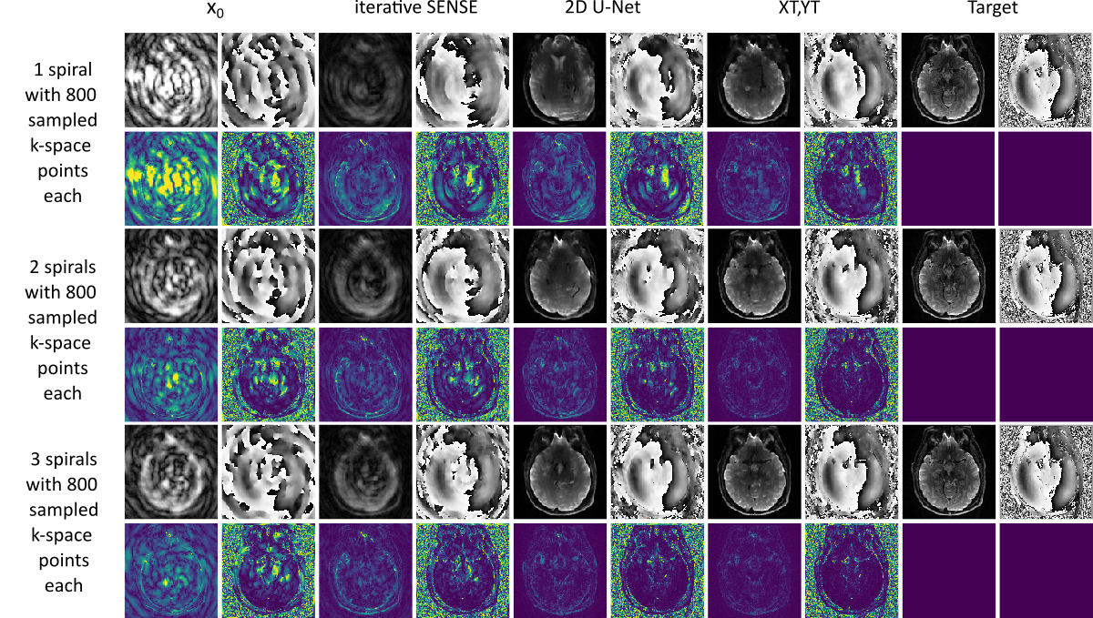
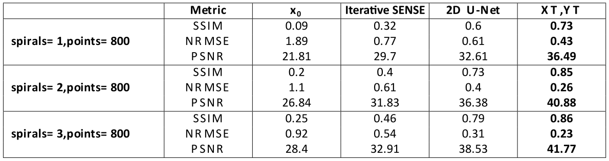
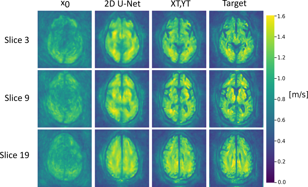
Both networks improve the magnitude and phase image quality, as shown in Figure 2, and confirmed by the SSIM, NRMSE, and PSNR metrics in Table 1. However, the XT,YT network showed better results and outperformed the 2D U-Net.Figure 3 confirms that the presented approach yields images that are suitable to obtain elastograms by further processing using the k-MDEV method. They were obtained from the BIOQIC-Apps website.4,10
Discussion
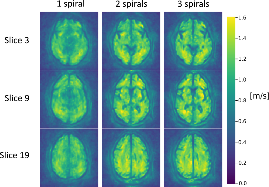
We have demonstrated that the use of CNN-regularized image reconstruction can enable the acceleration of the data-acquisition process, allowing an accurate reconstruction of MRE k-space data acquired along spirals. Even for single shot acquisition, the reconstruction is close to the target. The use of the XT,YT method surpasses the standard 2D U-Net due to the spatial-temporal correlation in the data.Currently, the k-MDEV inversion and the image reconstruction parts are treated differently. Integrating the latter as a trainable module to allow for a task-specific end-to-end11 could further enhance the results and improve the SWS maps from single-shot spiral acquisition, which presently still lack detail, see Figure 4. As for now, real-time MRE would be possible with 2D single-shot sequences12.
Acknowledgements
Funding from the German Research Foundation acknowledged (GRK2260, BIOQIC).References
1. Kennedy, P., Wagner, M., Castéra, L., Hong, C. W., Johnson, C. L., Sirlin, C. B., & Taouli, B. (2018). Quantitative elastography methods in liver disease: current evidence and future directions. Radiology, 286(3), 738-763.
2. Muthupillai, R., & Ehman, R.L. (1996). "Magnetic resonance elastography." Nature medicine 2.5: 601-603.
3. Sack, I. (2023). "Magnetic resonance elastography from fundamental soft-tissue mechanics to diagnostic imaging." Nature Reviews Physics 5.1: 25-42.
4. Meyer, T., Marticorena Garcia, S., Tzschätzsch, H., Herthum, H., Shahryari, M., Stencel, L., Braun, J., Kalra, P., Kolipaka, A, & Sack, I. (2022). Comparison of inversion methods in MR elastography: An open‐access pipeline for processing multifrequency shear‐wave data and demonstration in a phantom, human kidneys, and brain. Magnetic Resonance in Medicine, 88(4), 1840-1850.
5. Kofler, A., Dewey, M., Schaeffter, T., Wald, C., & Kolbitsch, C. (2019). Spatio-temporal deep learning-based undersampling artefact reduction for 2D radial cine MRI with limited training data. IEEE transactions on medical imaging, 39(3), 703-717.
6. Pruessmann, K. P., Weiger, M., Börnert, P., & Boesiger, P. (2001). Advances in sensitivity encoding with arbitrary k‐space trajectories. Magnetic Resonance in Medicine: An Official Journal of the International Society for Magnetic Resonance in Medicine, 46(4), 638-651.
7. Kofler, A., Haltmeier, M., Schaeffter, T., & Kolbitsch, C. (2021). An end‐to‐end‐trainable iterative network architecture for accelerated radial multi‐coil 2D cine MR image reconstruction. Medical Physics, 48(5), 2412-2425.
8. Kofler, A., Schaeffter, T., & Kolbitsch, C. (2022). The more the merrier?—On the number of trainable parameters in iterative neural networks for image reconstruction. In Proceedings of the Joint Annual meeting of ISMRM-ESMRMB and SMRT 31st Annual Meeting, London.
9. Terpstra, M. L., Maspero, M., Sbrizzi, A., & van den Berg, C. A. (2022). ⊥-loss: A symmetric loss function for magnetic resonance imaging reconstruction and image registration with deep learning. Medical Image Analysis, 80, 102509.
10. Sack, I. "Forschungsförderung–BIOQIC–neue generation von bildgebungsspezialisten." RöFo-Fortschritte auf dem Gebiet der Röntgenstrahlen und der bildgebenden Verfahren. Vol. 189. No. 01. © Georg Thieme Verlag KG, 2017.
11. Zimmermann, F. F., Kolbitsch, C., Schuenke, P., & Kofler, A. (2023). PINQI: An End-to-End Physics-Informed Approach to Learned Quantitative MRI Reconstruction. arXiv preprint arXiv:2306.11023.
12. Herthum, H., Shahryari, M., Tzschätzsch, H., Schrank, F., Warmuth, C., Görner, S., Hetzer, S., Neubauer, H., Pfeuffer, J., Braun, J., & Sack, I. (2021). Real-time multifrequency MR elastography of the human brain reveals rapid changes in viscoelasticity in response to the Valsalva Maneuver. Frontiers in Bioengineering and Biotechnology, 9, 666456.
Figures