4435
Distortion-free Diffusion Imaging Using BUDA-gSlider on the Connectome 2.0 System1Radiology, Athinoula A. Martinos Center for Biomedical Imaging, Charlestown, MA, United States, 2Harvard Medical School, Boston, MA, United States, 3Pediatric Imaging Research Center, Massachusetts General Hospital, Boston, MA, United States, 4Brigham and Women's Hospital, Boston, MA, United States, 5Zhejiang University, Hangzhou, China, 6Hongik University, Seoul, Korea, Republic of, 7Institute of Medical Physics and Radiation Protection, Mittelhessen University of Applied Sciences, Giessen, Germany, 8Department of Diagnostic and Interventional Radiology, University Hospital Marburg, Philipps University of Marburg, Marburg, Germany, 9Department of Radiology, Massachusetts General Hospital, Boston, MA, United States, 10Harvard/MIT Health Sciences and Technology, Cambridge, MA, United States
Synopsis
Keywords: Data Acquisition, Diffusion/other diffusion imaging techniques
Motivation: High-performance gradients of the Connectome 2.0 system drastically improve diffusion and imaging encoding to enable submillimeter diffusion imaging with high geometric fidelity.
Goal(s): To enable high-resolution, distortion-free diffusion imaging with adequate SNR.
Approach: Distortion-free BUDA-gSlider acquisition was deployed on the Connectome 2.0 system, and was compared against acquisitions using gradients derated to the level of clinical systems.
Results: The Connectome 2.0 system provides improved diffusion-weighted images with higher SNR and geometric fidelity, particularly at submillimeter resolutions.
Impact: High-performance Connectome 2.0 gradients enable high-resolution, distortion-free diffusion-weighted imaging with improved SNR at submillimeter resolutions.
Introduction
Connectome 2.0 is a next-generation human 3T scanner equipped with an ultra-high gradient strength of 500 mT/m and a slew rate of 600 T/m/s, optimized for the study of neural tissue microstructure and connectional anatomy across multiple length scales1. Its asymmetric head gradient coil is designed to minimize peripheral nerve stimulation (PNS). Diffusion MRI (dMRI) provides detailed information on white matter microstructure within the central nervous system2,3. Echo planar imaging (EPI) is typically used for dMRI because of its fast encoding per imaging slice. EPI with diffusion-encoding gradients utilizes strong gradients and their maximum slew rate to increase SNR by minimizing echo time (TE), but this significantly increases peripheral nerve stimulation (PNS). Despite using maximum gradient slew rate, long echo spacing (ESP) time between adjacent readout gradients induces geometrical distortions, T2/T2*-related voxel blurring, and voxel pile-ups. In this study, we performed high-resolution, distortion-free dMRI using BUDA-gSlider4 on the high-performance Connectome 2.0 system. The stronger gradients significantly increase SNR by reducing TE and mitigate EPI-related artifacts by reducing ESP, while minimizing PNS.Methods
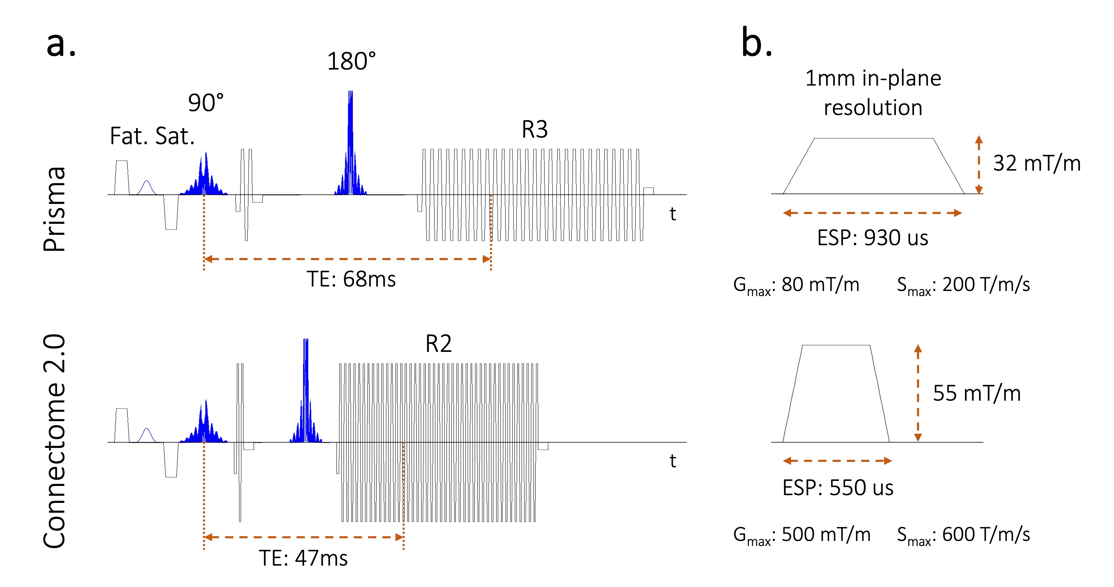
dMRI scans were performed on the Connectome 2.0 (MAGNETOM Connectome. X, Siemens Healthineers) equipped with a custom-built 72-channel in vivo head coil. We conducted in vivo experiments using BUDA-gSlider acquisitions4 with LORAKS constraints5, which acquires blip-up and blip-down multi-shot EPI data and reconstructs distortion-free multi-shot EPI images with low-rank constraints. We compared the images acquired with Connectome 2.0's powerful gradient against those obtained using the state-of-the-art Prisma research scanner (MAGNETOM Prisma, Siemens Healthineers) by derating the Connectome 2.0’s gradients down to the gradient strength of 80 mT/m and slew rate of 200 T/m/s. Simultaneous multislice (SMS) acceleration was retrospectively performed6.Fig. 1 shows the sequence diagram at 1mm isotropic resolution. The imaging parameters are;
Prisma-level gradients: TE=68ms, TR=3000ms, ESP=930us, Rinplane=3, Rsms=2, BW=1235Hz/px;
Connectome 2.0 gradients: TE=47ms, TR=3000ms, ESP=550us, Rinplane=2, Rsms=2, BW=2041Hz/px.
The b-value is 1000 s/mm2 in 16 directions.
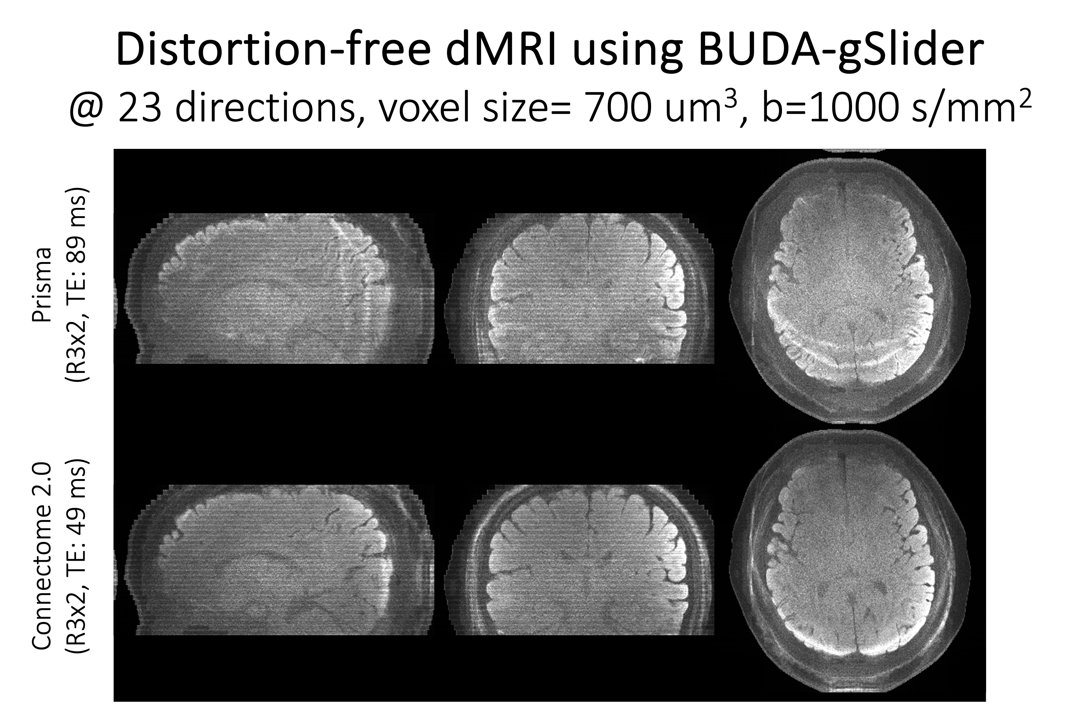
We also acquired the BUDA-gSlider data at 700um isotropic resolution. The imaging parameters are;
Prisma-level gradients: TE=89ms, TR=5200ms, ESP=1140us, Rinplane=3, Rsms=2, BW=1020Hz/px;
Connectome 2.0 gradients: TE=49ms, TR=5200ms, ESP=640us, Rinplane=3, Rsms=2, BW=1724Hz/px.
The b-value is 1000 s/mm2 in 23 directions.
Results
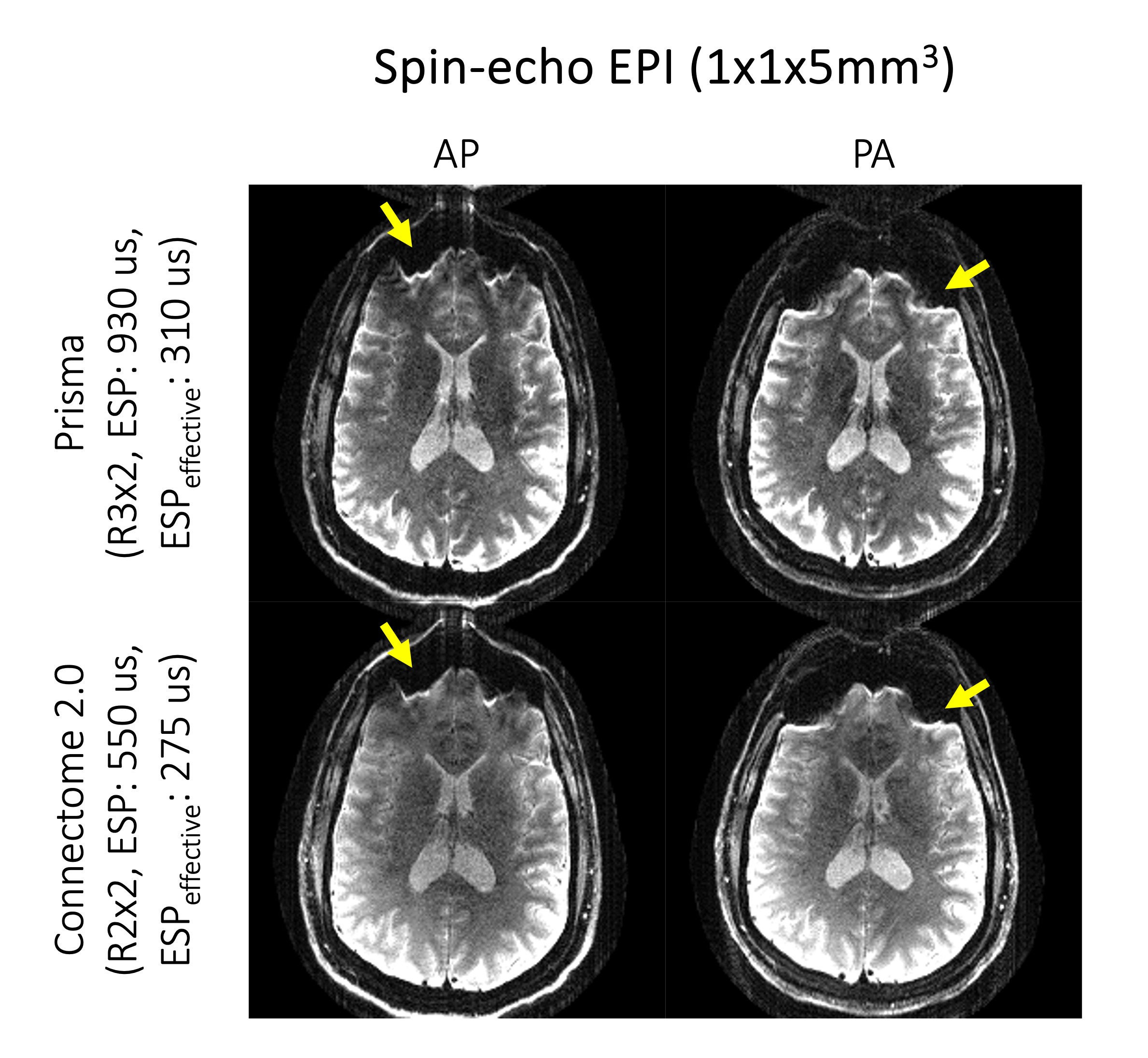
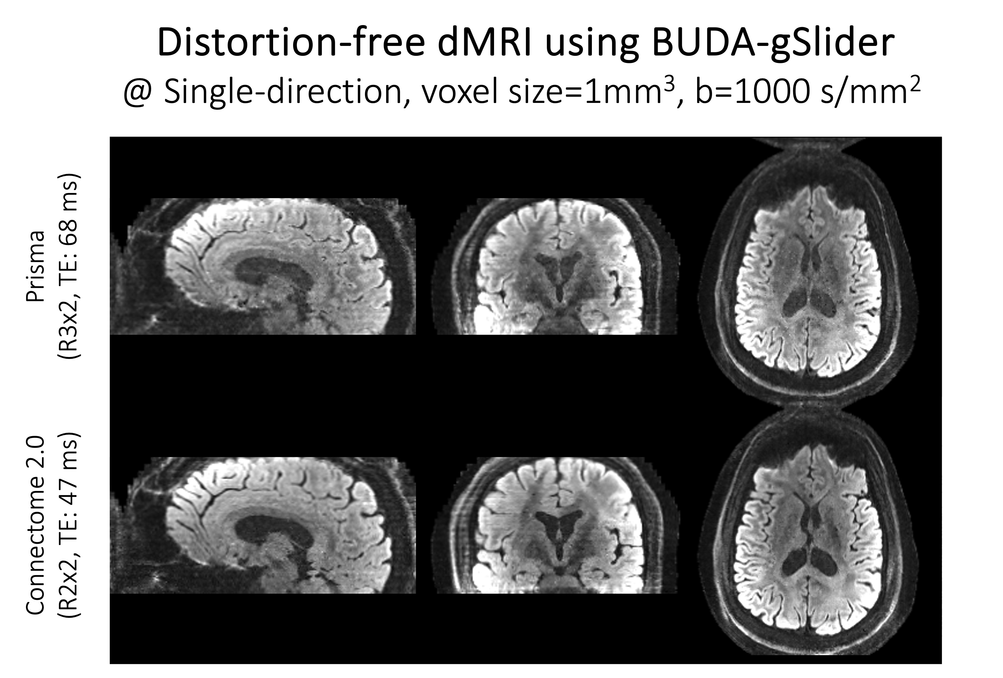
Fig. 2 shows b=0 thick-slab images in blip-up (AP) and blip-down (PA) directions before gSlider super-resolution reconstruction at the resolution of 1x1x5 mm3.Fig. 3 shows BUDA gSlider images in a single diffusion direction at 1mm isotropic resolution, acquired with Prisma-level and Connectome 2.0 gradients.
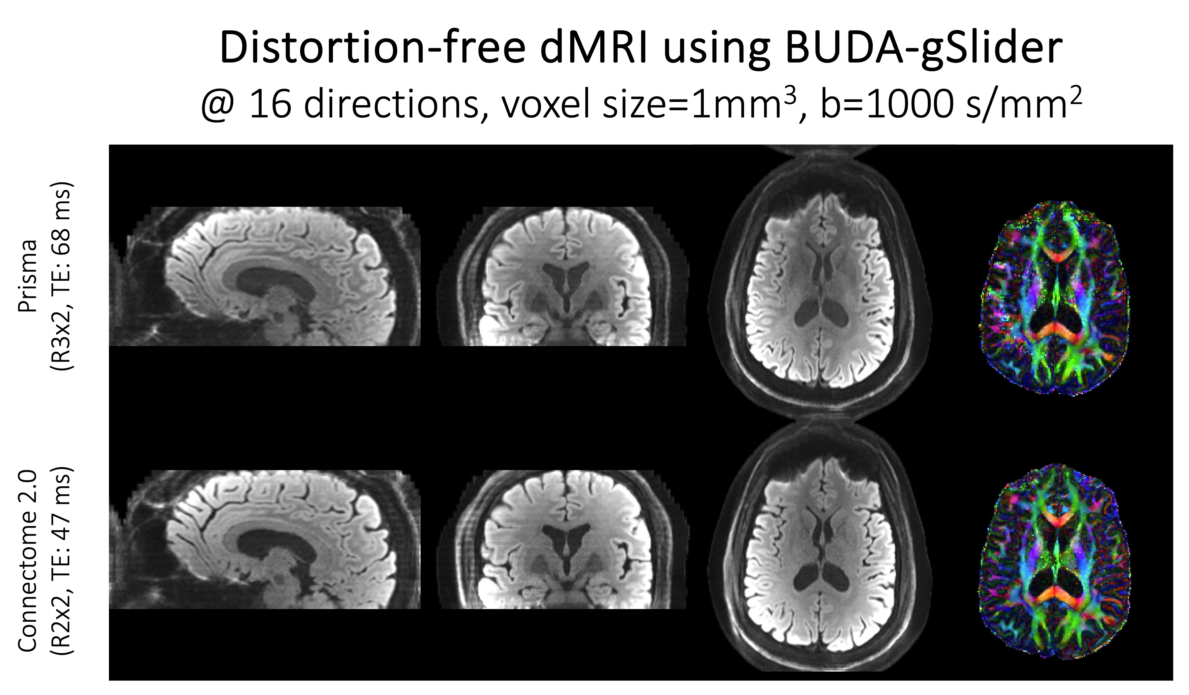
Fig. 4 shows averaged diffusion-weighted images and FA maps in 16 diffusion directions.
Fig. 5 shows diffusion-weighted images averaged in 23 directions at 700 um isotopic resolution with Prisma-level and Connectome 2.0 gradients.
Discussion and Conclusion
In the in vivo experiment at 1 mm resolution, the TE was reduced by 1.45-fold, whereas the EPS was reduced by 1.69-fold. This was achieved by shorter diffusion gradients and a 1.65-fold reduction in the readout bandwidth. For a voxel of T2=70ms, we could expect a 35% SNR gain from the TE difference and a 29% SNR loss from the increased receiver bandwidth, resulting in a 5% SNR gain in addition to the SNR gain from less reduction. This implies we could preserve SNR while reducing the effective EPS, which can mitigate the EPI-related artifacts. In Fig 2, we could observe fewer voxel pile-ups in Connectome 2.0 despite the lower in-plane acceleration factor because effective ESP was even shorter. The actual SNR gains in the images came from using less reduction factor, as well demonstrated in Figs 2, 3, and 4.In the in vivo experiment at 700 um resolution, the TE was reduced by 1.82-fold, whereas the EPS was reduced by 1.78-fold. This was achieved by shorter diffusion gradients and a 1.69-fold reduction in the readout bandwidth. For a voxel of T2=70ms, we expect a 77% SNR gain from the TE difference and a 30% SNR loss from the increased receiver bandwidth, resulting in a 36% SNR gain. The effective ESP was also significantly reduced from 380 us to 213 us, which mitigates the EPI-related artifact. The averaged 23 diffusion direction at 700 um isotropic resolution, of which scan time exceeds 20 min, has lower SNR but could be improved in a longer acquisition with more directions7. Improvements to the fat saturation in the experiment for 700 um resolution will be a part of our future work.
Acknowledgements
This work was supported by research grants NIH R01 EB028797, R01 EB032378, R01 HD100009, R03 EB031175, U01 EB026996, U01 DA055353, P41 EB030006, and the NVidia Corporation for computing support.References
- Huang, S. Y. et al. Connectome 2.0: Developing the next-generation ultra-high gradient strength human MRI scanner for bridging studies of the micro-, meso- and macro-connectome. Neuroimage 243, 118530 (2021).
- Mueller, B. A., Lim, K. O., Hemmy, L. & Camchong, J. Diffusion MRI and its Role in Neuropsychology. Neuropsychol. Rev. 25, 250–271 (2015).
- Afzali, M. et al. The sensitivity of diffusion MRI to microstructural properties and experimental factors. J. Neurosci. Methods 347, 108951 (2021).
- Liao, C. et al. Distortion-free, high-isotropic-resolution diffusion MRI with gSlider BUDA-EPI and multi-coil dynamic B0 shimming. Magn. Reson. Med. 86, 791–803 (2021).
- Kim, T. H., Setsompop, K. & Haldar, J. P. LORAKS makes better SENSE: Phase-constrained partial Fourier SENSE reconstruction without phase calibration. Magn. Reson. Med. 77, 1021–1035 (2017).
- Setsompop, K. et al. Blipped-controlled aliasing in parallel imaging for simultaneous multislice echo planar imaging with reduced g-factor penalty. Magn. Reson. Med. 67, 1210–1224 (2012).
- Haldar, J. P., Liu, Y., Liao, C., Fan, Q. & Setsompop, K. Fast submillimeter diffusion MRI using gSlider-SMS and SNR-enhancing joint reconstruction. Magn. Reson. Med. 84, 762–776 (2020).
Figures