4434
Optimization of Sampling Masks and Reconstruction of Under-sampled Images for SNAP MRI with Model Based Deep Learning Framework1Center for Biomedical Imaging Research, Department of Biomedical Engineering, Tsinghua University, Beijing, China
Synopsis
Keywords: Image Reconstruction, Atherosclerosis, Plaque, Black-blood MRI, Trajectory
Motivation: The two-shot SNAP MRI is effective for carotid plaque diagnosis with extended scan time. To accelerate the scan, under-sampling reconstruction and optimization of sampling locations are considered.
Goal(s): To optimize the sampling masks for IR-TFE and REF-TFE of SNAP MRI respectively and to reconstruct the under-sampled images with higher quality.
Approach: After the parameterization of ky-kz sampling locations for the two shots, a model-based deep learning framework was utilized to achieve the goals.
Results: The framework demonstrated superior performance compared with other under-sampling reconstruction methods. Distinct sampling masks were generated for the two shots after the training process.
Impact: The optimized sampling masks facilitate the acquisition of SNAP MRI with more crucial information. Combined with high-quality under-sampling reconstruction, the utilization of the framework could enhance the clinical applicability, flexibility, and versatility of SNAP MRI.
Introduction
Vulnerable carotid atherosclerotic plaque is a significant cause of ischemic cerebrovascular events1. While the evaluation of luminal stenosis remains widely used for detecting high-risk carotid atherosclerosis2, the compositional features of carotid plaque such as intra-plaque hemorrhage (IPH) could serve as strong predictors for cerebrovascular events3. Black-blood MRI techniques utilize a heavily T1-weighted contrast to capture IPH components4, but they do not usually provide a MR angiography (MRA) for the measurement of luminal stenosis and the anatomical positioning of IPH. Recently, a Simultaneous Non-contrast Angiography and intraPlaque hemorrhage (SNAP) MRI sequence was proposed for carotid artery imaging5. It contains an inversion recovery TFE (IR-TFE) shot and an interleaved reference TFE (REF-TFE) shot. Taking advantage of phase-sensitive reconstruction, it could provide detection of IPH and a co-registered MRA in a single scan. However, the acquisition of REF-TFE shot could extend scan time, which may hinder its clinical application. Several approaches have been used to accelerate SNAP imaging6, 7, but none of them put special focus on the sampling mask optimization for SNAP sequence. In this study, we employed a Model-based Deep Learning (MoDL) method8 to optimize the sampling mask for IR-TFE and REF-TFE of SNAP sequence respectively and to reconstruct the under-sampled images simultaneously.Methods
Network architectureParameterization of ky-kz Sampling Mask
The under-sampling process can be expressed as:$$X=S^{H}F_{kykz}^H F_{kykz}S\widehat{X} $$
where $$$F_{kykz}$$$ is Fourier transform operator built by the sampling locations on ky-kz plane, S is multi-channel sensitivity maps, $$$\widehat{X} $$$ is fully-sampled IR or REF images. The sampling locations in fast Fourier transformation is discrete and non-differentiable. To optimize the sampling mask, we performed 1D continuous Fourier transform along kz direction and then along ky direction. After the operation, for a specific kz, all sampling locations can be represented as a set of continuous numbers between 0 and 1, hence, they are differentiable and trainable.
Network Training
The proposed MoDL framework is shown in Fig 1. The framework alternates between data consistency layer $$$D_{\Theta } $$$ and CNN denoiser $$$C_{\Phi } $$$:$$x_{IR,n+1}=D_{\Theta _{IR}}(z_{IR,n})=(A_{\Theta _{IR}}^H A_{\Theta _{IR}}+I)^{-1}(z_{IR,n}+A_{\Theta _{IR}}^H A_{\Theta _{IR}}\widehat{X_{IR}} )$$$$x_{REF,n+1}=D_{\Theta _{REF}}(z_{REF,n})=(A_{\Theta _{REF}}^H A_{\Theta _{REF}}+I)^{-1}(z_{REF,n}+A_{\Theta _{REF}}^H A_{\Theta _{REF}}\widehat{X_{REF}} )$$$$z_{n+1}=[z_{IR,n+1},z_{REF,n+1}]=C_\Phi ([x_{IR,n+1},x_{REF,n+1}])$$where $$$\widehat{X} $$$ denotes fully-sampled complex images. The data consistency layer was implemented using a conjugate gradient algorithm. Loss function can be expressed as:$$loss=\underset{\Theta_{IR}, \Theta_{REF}, \Phi}{min}\left | \right | C_\Phi [D_{\Theta_{IR}}(A_{\Theta _{IR}}^H A_{\Theta _{IR}}\widehat{X_{IR}}), D_{\Theta_{REF}}(A_{\Theta _{REF}}^H A_{\Theta _{REF}}\widehat{X_{REF}})]-[\widehat{X_{IR}},\widehat{X_{REF}}]\left | \right | _2^2$$CNN weights and sampling locations were updated in the training process, and then kept fixed for inference. Limited by GPU memory, iteration number N was set to 1.
Dataset
A total of 100 3D carotid SNAP MRI data were used in the experiment (80 as training-set and 20 as test-set). MRI scans were performed on a 3T MR scanner (Achieva TX, Philips Healthcare, Best, The Netherlands) with an 8-channel carotid dedicated phased-array surface coil. The detailed scan parameters are displayed on Table 1.
Experiment and Analysis
We firstly trained the proposed model to reconstruct SNAP MRI with different under-sampling factor (4x, 6x and 8x), the performance was measured with PSNR and SSIM. Then we compared its performance with some other under-sampling reconstruction frameworks including MoDL(with fixed sampling mask), U-NET and compressed sensing (total variation regularization) with a 6x under-sampling factor. Notably, for each under-sampling factor, one variable-density Cartesian under-sampling mask on ky-kz plane was randomly generated as the ‘initial mask’ for all the experiments under the same under-sampling factor.
Results
The proposed framework performed well on the reconstruction of test-set with different under-sampling factors, it also provided the best performance compared with other frameworks with a fixed 6x under-sampling ratio, the indexes are presented in Table 2 and the images in Fig 2. As displayed in Fig 3, while the initial masks are the same for IR-TFE and REF-TFE, the proposed model generated different under-sampling masks for the two shots after the training process, the trained masks exhibited smoother edges. The iterative evolution of the masks has converged in the training process.Discussion&Conclusion
Our experiments demonstrated that the proposed model could combine the strengths of CNN and data consistency layer to improve the recovery of under-sampled SNAP images. The evolution of sampling masks in the training process could improve the reconstruction performance, indicating that the trained masks contained more crucial information compared to the initial mask. The distinct trained masks for IR-TFE and REF-TFE also suggested that the two shots have respective focusing areas on the ky-kz plane and may complement each other. These masks can partially reveal the data characteristics of SNAP, and data acquired using these masks may lead to better image quality.Acknowledgements
No acknowledgement found.References
1.Takaya N, Yuan C, Chu BC, Saam T, Underhill H, Cai JM, et al. Association between carotid plaque characteristics and subsequent ischemic cerebrovascular events - A prospective assessment with MRI - Initial results. Stroke. 2006;37(3):818-23.
2.Moore WS, Barnett HJM, Beebe HG, Bernstein EF, Brener BJ, Brott T, et al. Guidelines for Carotid Endarterectomy - a Multidisciplinary Consensus Statement from the Ad-Hoc-Committee, American-Heart-Association. Stroke. 1995;26(1):188-201.
3.Saam T, Hetterich H, Hoffmann V, Yuan C, Dichgans M, Poppert H, et al. Meta-Analysis and Systematic Review of the Predictive Value of Carotid Plaque Hemorrhage on Cerebrovascular Events by Magnetic Resonance Imaging. J Am Coll Cardiol. 2013;62(12):1081-91.
4.Ota H, Yarnykh VL, Ferguson MS, Underhill HR, DeMarco JK, Zhu DC, et al. Carotid Intraplaque Hemorrhage Imaging at 3.0-T MR Imaging: Comparison of the Diagnostic Performance of Three T1-weighted Sequences. Radiology. 2010;254(2):551-63.
5.Wang JN, Börnert P, Zhao HL, Hippe DS, Zhao XH, Balu N, et al. Simultaneous noncontrast angiography and intraPlaque hemorrhage (SNAP) imaging for carotid atherosclerotic disease evaluation. Magnet Reson Med. 2013;69(2):337-45.
6.Wang JN, Chen HJ, Maki JH, Zhao XH, Wilson GJ, Yuan C, et al. Referenceless Acquisition of Phase-Sensitive Inversion-Recovery with Decisive Reconstruction (RAPID) Imaging. Magnet Reson Med. 2014;72(3):806-15.
7.Chen S, Ning J, Zhao XH, Wang JN, Zhou ZC, Yuan C, et al. Fast simultaneous noncontrast angiography and intraplaque hemorrhage (fSNAP) sequence for carotid artery imaging. Magnet Reson Med. 2017;77(2):753-8.
8.Aggarwal HK, Mani MP, Jacob M. MoDL: Model-Based Deep Learning Architecture for Inverse Problems. Ieee T Med Imaging. 2019;38(2):394-405.
Figures
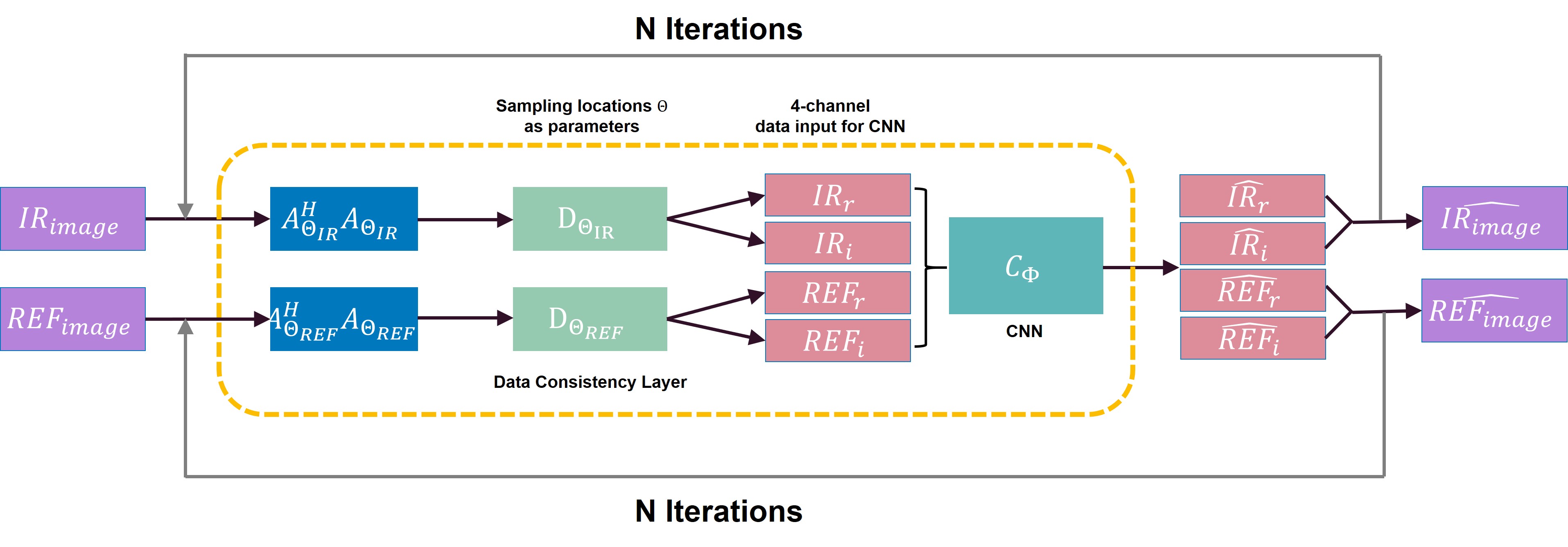
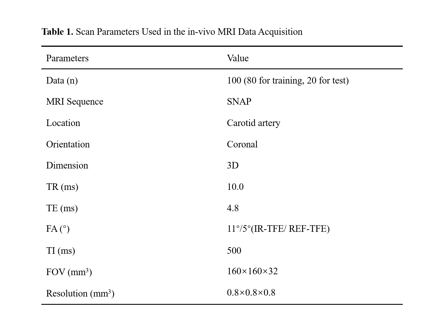
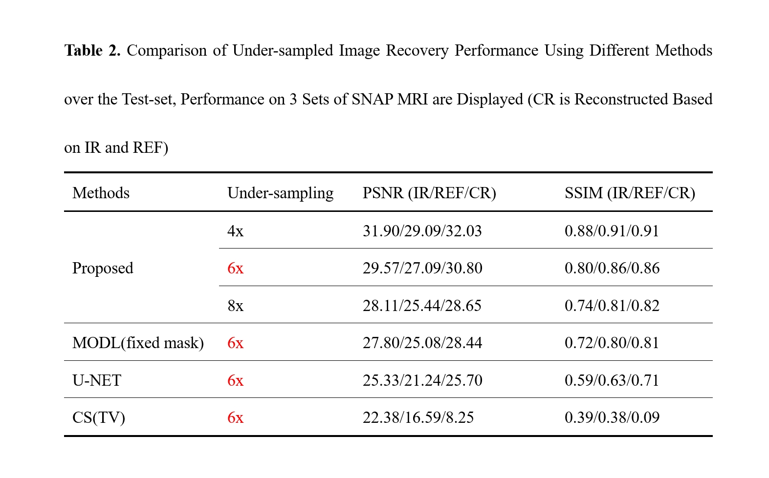
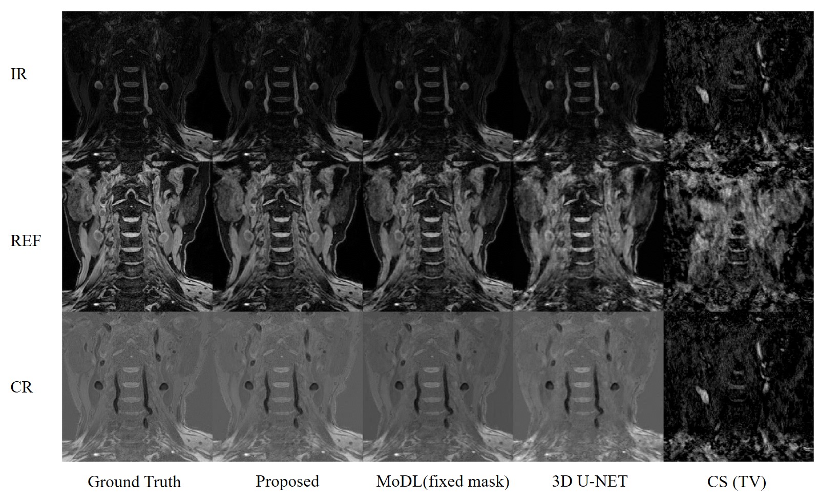
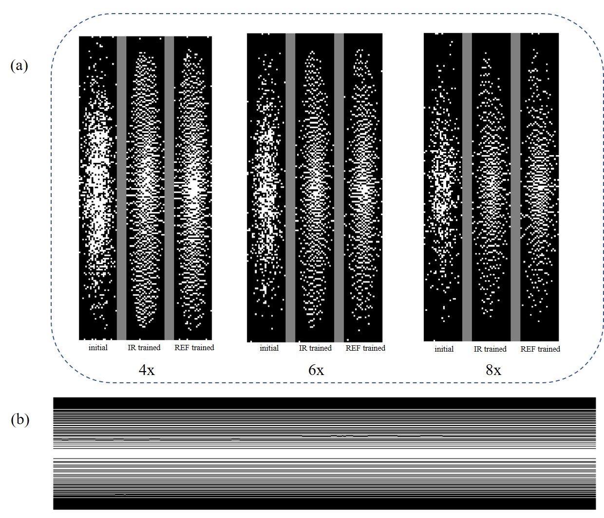
Fig 3. (a) Sampling Mask Used in the Proposed Model with Different Under-sampling Ratio (For each acceleration factor, the left image represents the initial mask, the middle image corresponds to the trained mask for IR shot, and the right image illustrates the trained mask for REF shot.)
(b) Evolution of the Mask of IR shot (6x acceleration, single kz) in the Last 1000 Iterations of the Training Process