4433
Active EMI Elimination at 3T using Routine Receiver Coils and Deep Learning1Division of Medical Physics, Department of Radiology, University Medical Center Freiburg, Faculty of Medicine, University of Freiburg, Freiburg, Germany, 2Laboratory of Biomedical Imaging and Signal Processing, The University of Hong Kong, Hong Kong, China, 3Electrical and Electronic Engineering, The University of Hong Kong, Hong Kong, China
Synopsis
Keywords: Artifacts, RF Arrays & Systems, electromagnetic interference suppression, EMI
Motivation: To reduce siting costs, improve accessibility and relax electromagnetic compatibility (EMC) requirements on in-room equipment in 3T MRI.
Goal(s): To assess a deep-learning based active electromagnetic interference (EMI) elimination algorithm Deep-DSP at 3T.
Approach: Deep-DSP approach previously implemented for 0.055T MRI was applied to 3D imaging at 3 Tesla, performed within the standard shielded room and with deliberately added EMI sources using a 64-channel head coil and four coil arrays located outside the magnet bore for EMI sensing.
Results: Deep-DSP demonstrates excellent performance in phantoms and outperforms a comparison technique in vivo.
Impact: The proposed approach does not require additional dedicated hardware and has potential of substantially reducing siting costs of modern high-performance MR imagers. Furthermore, EMC and RF shielding requirements on the additional equipment in the scanner room may be largely relaxed.
Introduction
Modern clinical MRI machines are installed in dedicated electromagnetically shielded rooms, with all electric signals being either electro-optically decoupled or filtered. Reducing shielding requirements or avoiding Faraday shielding completely may reduce siting costs and improve accessibility of MRI. Furthermore, relaxing EMC requirements for the in-room electronics may simplify scanning of patients requiring life support or allow researchers to introduce a greater variety of experimental equipment.In this project we investigate the performance of a deep-learning based active EMI elimination algorithm previously implemented for 0.055T MRI [1-3] under the conditions of 3T MRI. We deliberately introduce EMI sources in the shielded magnet room and dedicate four additional clinical coil arrays to EMI sensing by positioning them outside the magnet bore.
Methods
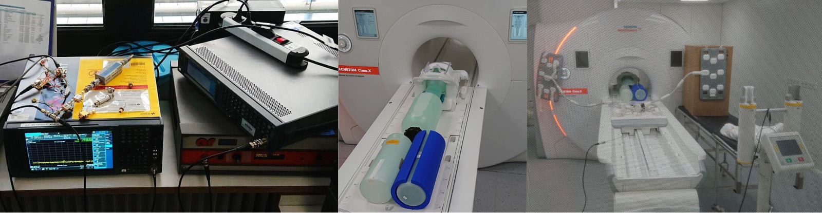
Experiments were performed on a 3T MR system (Magnetom Cima.X, Siemens Healthineers, Erlangen, Germany). A 64-channel head coil was used for imaging. One Body-18 (B18), one SuperFlexLarge (SFL), one SuperFlexSmall (SFS) and the most distal segment of the spine coil were dedicated to EMI sensing; coils ‘B18’ and ‘SFL’ were positioned next to the front side (see Fig. 1), while ‘SFS’ was placed at the bore exit at the service end of the magnet. During imaging data from 100 channels were recorded, of which 40 channels were from the head coil.MPRAGE protocol was used for imaging with a matrix of 256x256x224 without parallel imaging, with the total duration of 9:50 and the isotropic spatial resolution of 0.9mm3; further parameters: TI=900ms, TR=2300ms, excitation flip angle 8°, echo spacing 6.96ms, readout bandwidth 200Hz/pix. Raw data were exported and images reconstructed offline. In vivo imaging was performed in accordance with the IRB-approved protocol and the volunteer gave their written informed consent prior to the experiment.
EMI was induced using a combination of two sources, both located outside the scanner room and connected through BNC connectors in the a penetration panel to two magnetic antennas, positioned separately at the feet end of the patient lift. The first source was an RF signal generator (N5171B, Keysight, Santa Rosa, CA, USA), producing a narrowband sinusoidal waveform close to the Larmor frequency with a peak amplitude of 1.9V. The second broadband signal was produced by a noise source (N9320B, Keysight) connected to two low-noise preamplifiers in series (ZX60-P103LN+, Mini-Circuits, Brooklyn, NY, USA) and a 75-watt RF power amplifier (ZHL-6A+, Mini-Circuits). The gain of the amplifier was adjusted to make the noise level comparable to the image intensity without saturating the receivers. For the reference acquisitions without EMI, the feeding cables were disconnected and the BNC connectors at the penetration panel were terminated.
Our proposed deep learning direct MR signal prediction method, named Deep-DSP, relies on active EMI sensing and deep learning based modeling of the relationships between the EMI sensing coils and MRI receive coils [1-3]. Deep-DSP was implemented and benchmarked against the EDITER algorithm [4]. 50% of the k-space data, located at the periphery, were used for training the Deep-DSP model . For simplicity, only data from 3 head coil channels were processed for demonstration in this study. 6 channels from three other coils (SFL, SFS and spine coils) acted as EMI sensing coils.
Results
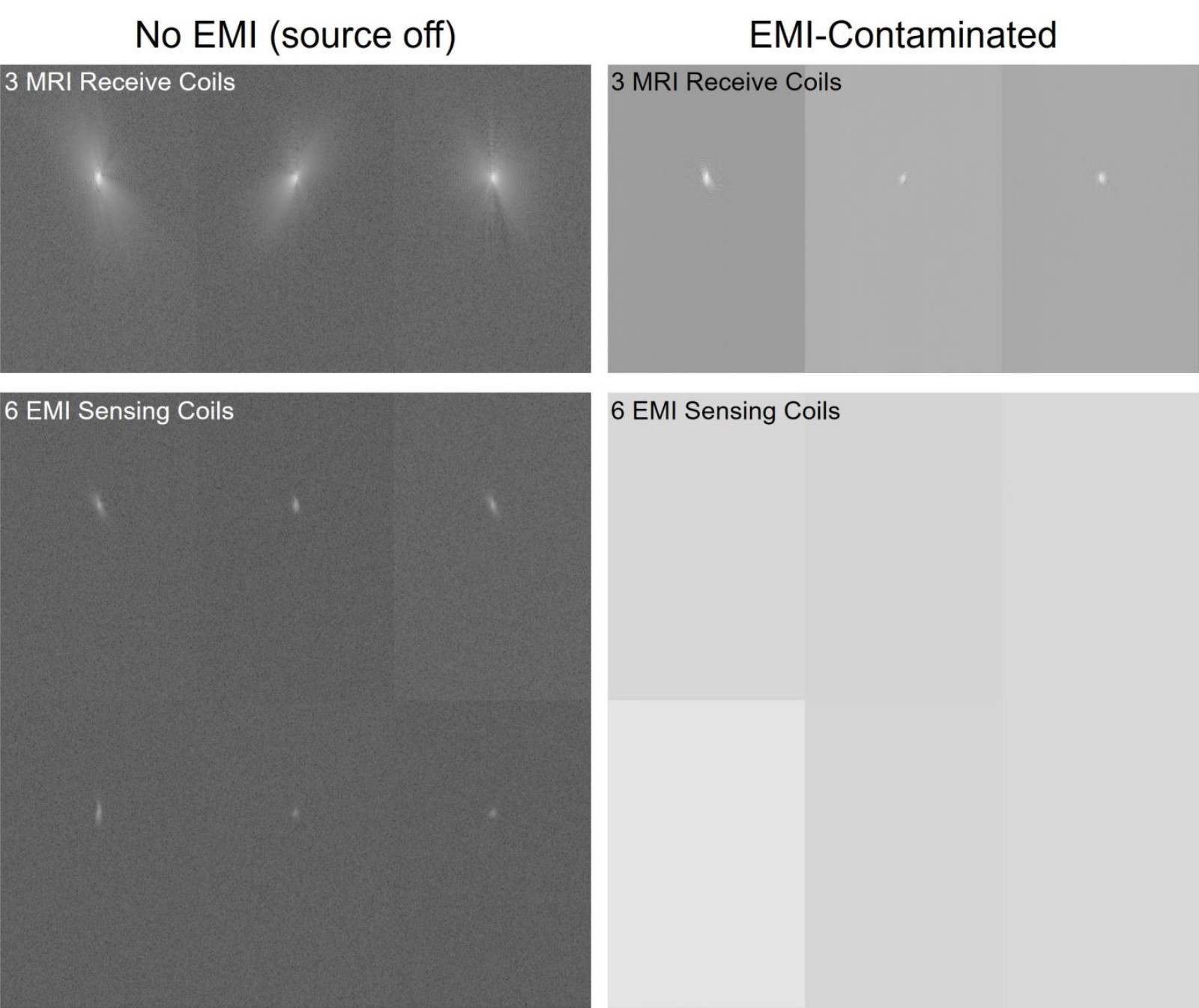
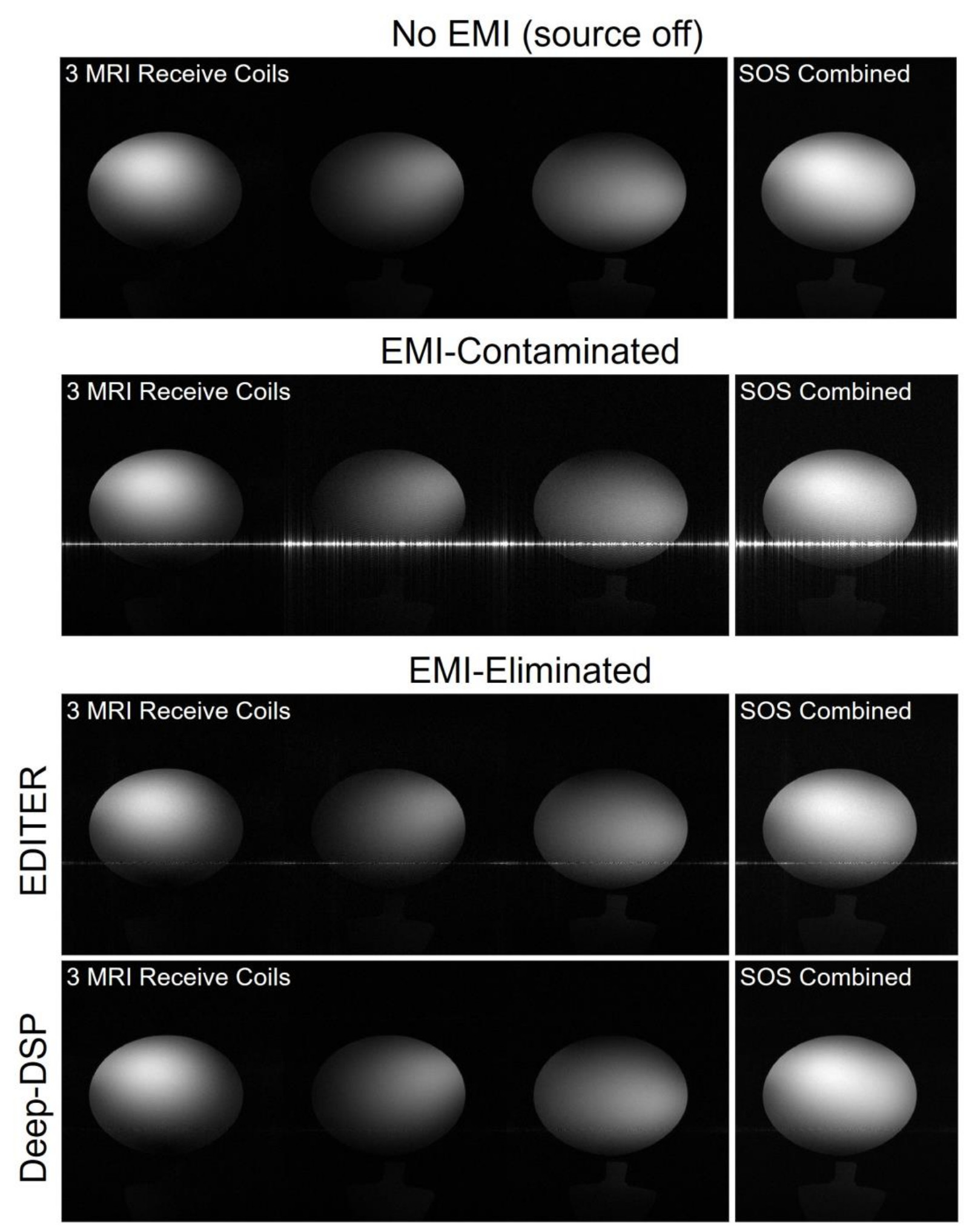
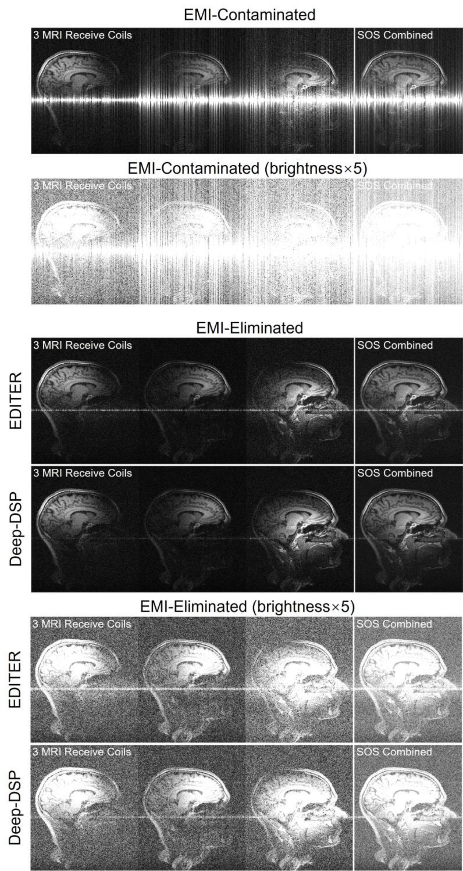
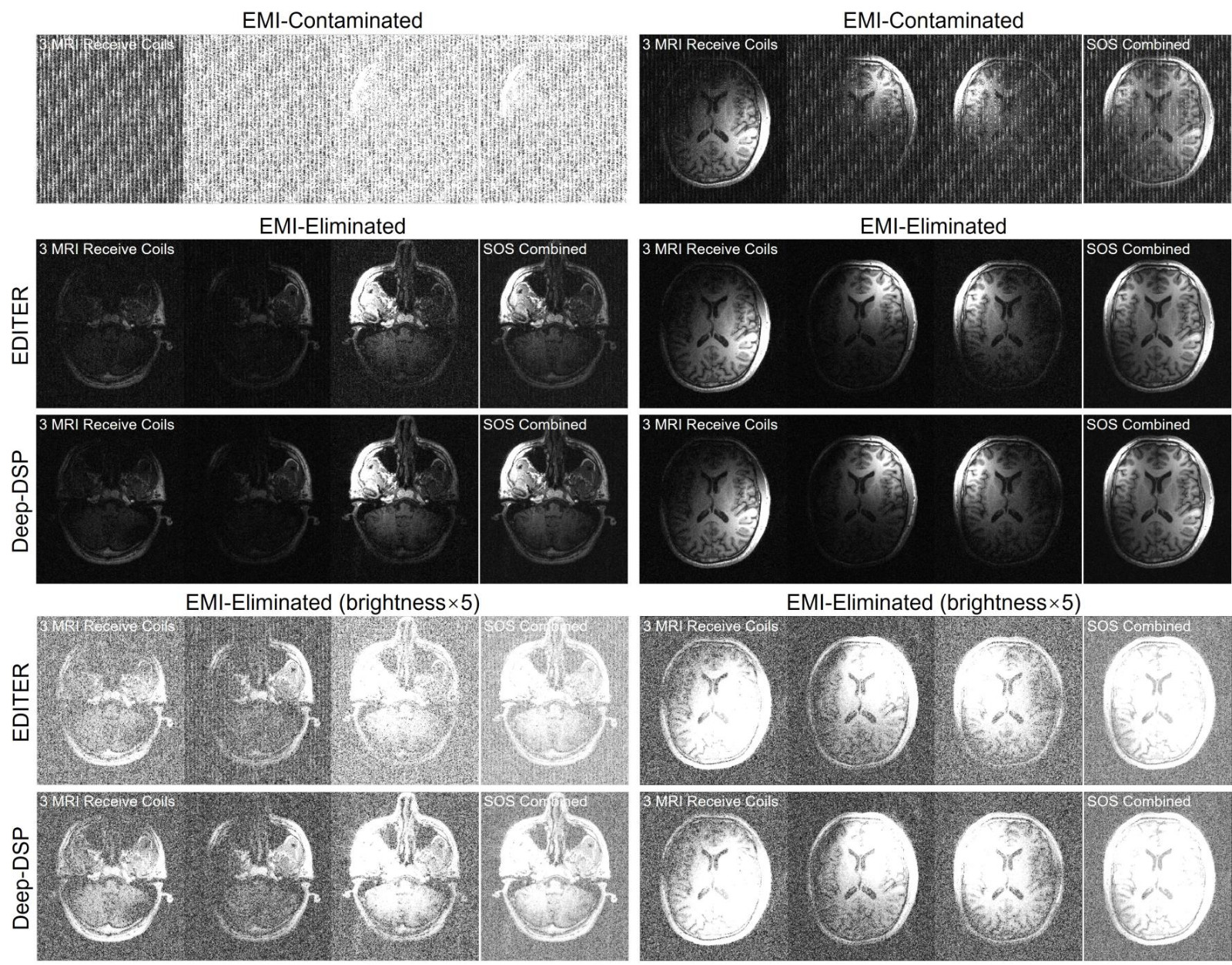
Figure 2 shows k-space data from a phantom acquisition of the selected imaging channels and EMI-sensing channels. Presumably, due to the inter-cable coupling along the analog signal train, also the coils placed externally show minor MR signals in no-EMI case. Figure 3 presents the corresponding images, along with the results of EDITER and Deep-DSP, where only the latter is able to restore the visual appearance of the images, comparable to that without EMI. Figure 4 presents the EMI-elimination results in a sagittal view of the 3D human brain imaging and Figure 5 shows selected axial slices. Deep-DSP clearly outperforms EDITER, but its results still exhibit residual EMI-related artifacts.Discussion
Deep-DSP shows excellent performance in phantoms. Possible reasons for suboptimal results in vivo may be related to the differences, how extremely strong EMI are transported into the bore by the human body. This body-mediated coupling is potentially modulated by physiologic processes (motion, breathing or heartbeat), making EMI elimination more challenging. Positioning some EMI sensing coils on the body might therefore improve the performance of Deep-DSP. Furthermore, previous Deep-DSP implementations relied on dedicated EMI sensing coils, whereas our EMI sensing channels still show some MR signals, potentially affecting Deep-DSP model training and EMI signal characterization.Nonetheless, the proposed approach has a potential of substantially reducing siting costs and increasing accessibility of MR scanner installations and motivates additional research efforts for perfecting the Deep-DSP performance in vivo.
Acknowledgements
This work was supported by research grants NIH R01 EB032378, NIH U24 NS120056, Hong Kong RGC R7003-19F, and Hong Kong RGC HKU17127523.References
1. Liu Y, Leong ATL, Zhao Y, Xiao L, Mak HKF, Tsang A, Lau GKK, Leung GKK, Wu EX. A Low-cost and shielding-free ultra-low-field brain MRI scanner. Nat Commun 2021; 12:1-14. doi:10.1038/s41467-021-27317-1.
2. Zhao Y, Xiao L, Liu Y, Leong AT, Wu EX. Electromagnetic interference elimination via active sensing and deep learning prediction for radiofrequency shielding-free MRI. NMR Biomed. 2023:e4956. doi:10.1002/nbm.4956.
3. Zhao Y, Hu J, Lau V, Xiao L, Leong ATL, Wu EX. Robust Electromagnetic Interference (EMI) Elimination for RF Shielding-Free MRI via Active EMI Sensing and Deep Learning MRI Signal Prediction. In: Proceedings of the 31st Annual Meeting of ISMRM, 2023, p 6357.
4. Srinivas SA, Cauley SF, Stockmann JP, Sappo CR, Vaughn CE, Wald LL, Grissom WA, Cooley CZ. External Dynamic InTerference Estimation and Removal (EDITER) for low field MRI. Magn Reson Med 2022; 87:614-628. doi:10.1002/mrm.28992.
Figures