4428
Deep learning based chemical shift artifact reduction in Zero Echo Time (ZTE) MRI1GE HealthCare MR Clinical Solutions, Atlanta, GA, United States, 2GE HealthCare MR Clinical Solutions, San Diego, CA, United States, 3GE HealthCare MR Clinical Solutions, Munich, Germany, 4GE HealthCare MR Clinical Solutions, New York, NY, United States, 5GE HealthCare MR Clinical Solutions, Calgary, AB, Canada
Synopsis
Keywords: Artifacts, Artifacts, Chemical-Shift, Off-resonance, Deep-Learning, radial, ZTE
Motivation: Chemical Shift artifacts in non-Cartesian MRI scans can lead to blurring and other artifacts at tissue interfaces. ZTE scans are particularly sensitive to this issue.
Goal(s): Addressing this artifact can enable the generation of more accurate ZTE images. Additionally, subsequent post-processing tasks like bone volume rendering, pseudo CT etc., can benefit from mitigating this artifact.
Approach: We introduce a deep learning based method to address this artifact and demonstrate its performance on phantom and in-vivo cases.
Results: The results demonstrate that gross chemical shift artifact can be corrected using the proposed method.
Impact: ZTE suffers from poor intrinsic SNR and chemical shift related blurring. Scanning at high field helps SNR but makes blurring more serious. Our proposed method helps mitigate chemical shift artifacts and opens up new possibilities for ZTE imaging.
Introduction
Zero Echo Time (ZTE) MRI enables the imaging of short T2 species and can support virtually silent scanning. These features make it attractive when imaging rapidly decaying tissue (like bone or lung parenchyma) or where low acoustic noise is desirable. One source of image quality degradation in ZTE stems from the fat-water chemical shift (CS). ZTE sequences typically acquire data in the form of center-out radial spokes and fat in the image accrues phase during the readout. This phase accrual manifests as a high frequency artifact and is particularly prominent at tissue interfaces.Recent work in mitigating these artifacts have focused on separating fat and water signal from the acquired data1 or split the data into in-phase and out-phase components2. These approaches either suffer from a poor conditioning of the inversion problem or require the acquisition of additional data. Another approach to mitigate CS artifact is to scan at a frequency between water and fat, however this approach leads to blurring in both water and fat components and may be undesirable3. In this work, we propose a deep learning (DL) based approach to mitigate CS artifacts in ZTE MRI.
Methods & Results
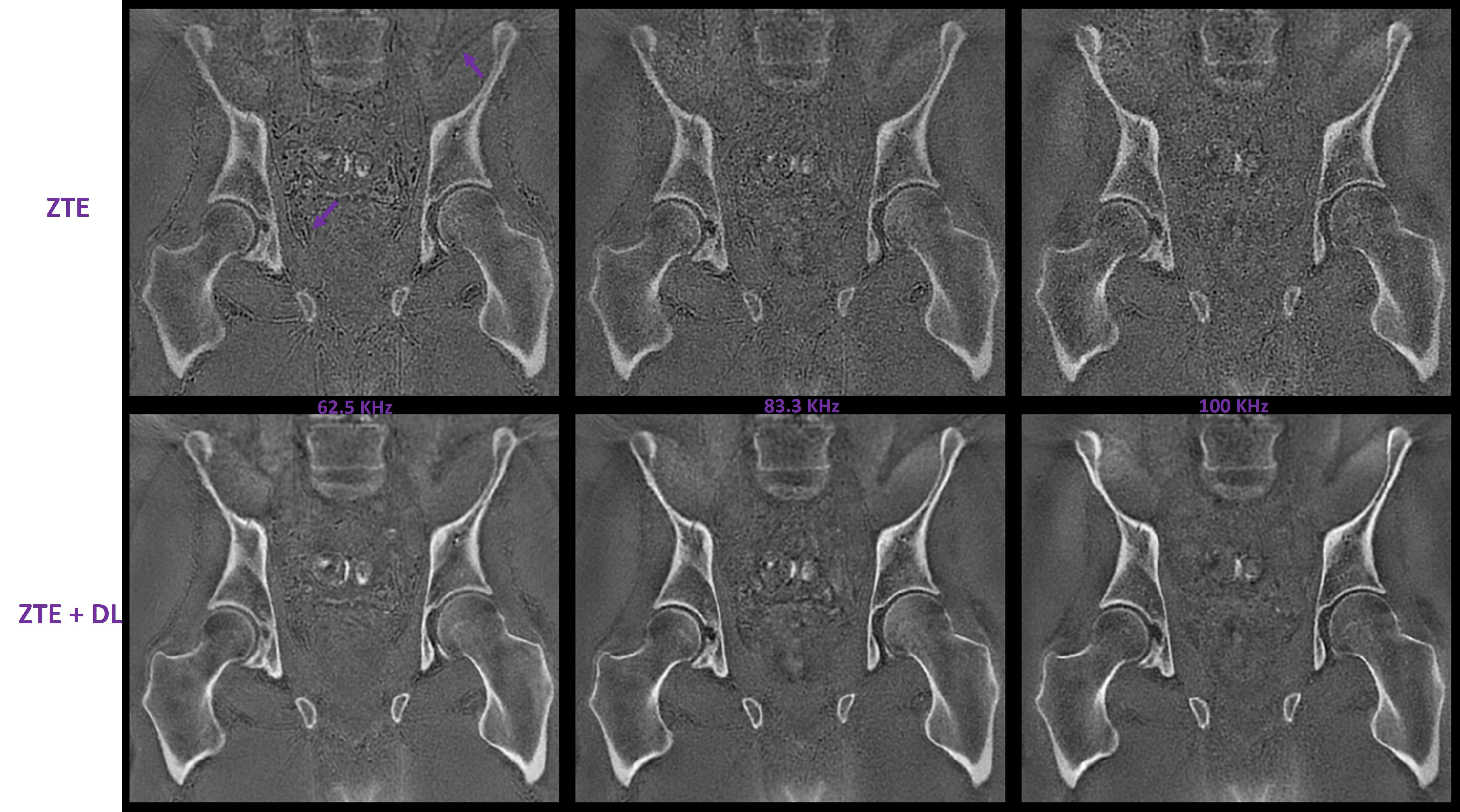
DL based CS correction: At typical field strengths used on clinical scanners, the 3.5 ppm CS between fat and Water is on the order of 200 – 1000 Hz and is in the range of commonly used bandwidth / pixel (BWP). Depending on the BWP, the artifact can appear as either blurring or destructive interference at tissue interfaces3. Figure 1 illustrates the appearance of these artifacts at three typical BWPs in the top row. Note that at 3T, the 3.5 ppm shift corresponds to about 420 Hz of off-resonance.A database of 10,000 labeled image pairs was used in training a DL model in a supervised manner4. The network is designed to remove CS, noise and recover fine details. The trained model is robust to field strength, under-sampling rates and noise levels. Conventionally reconstructed images are processed using the trained DL model and then pass through the subsequent post-processing pipelines. Bottom row of Figure 1 illustrates the impact of the DL method at different BWP.
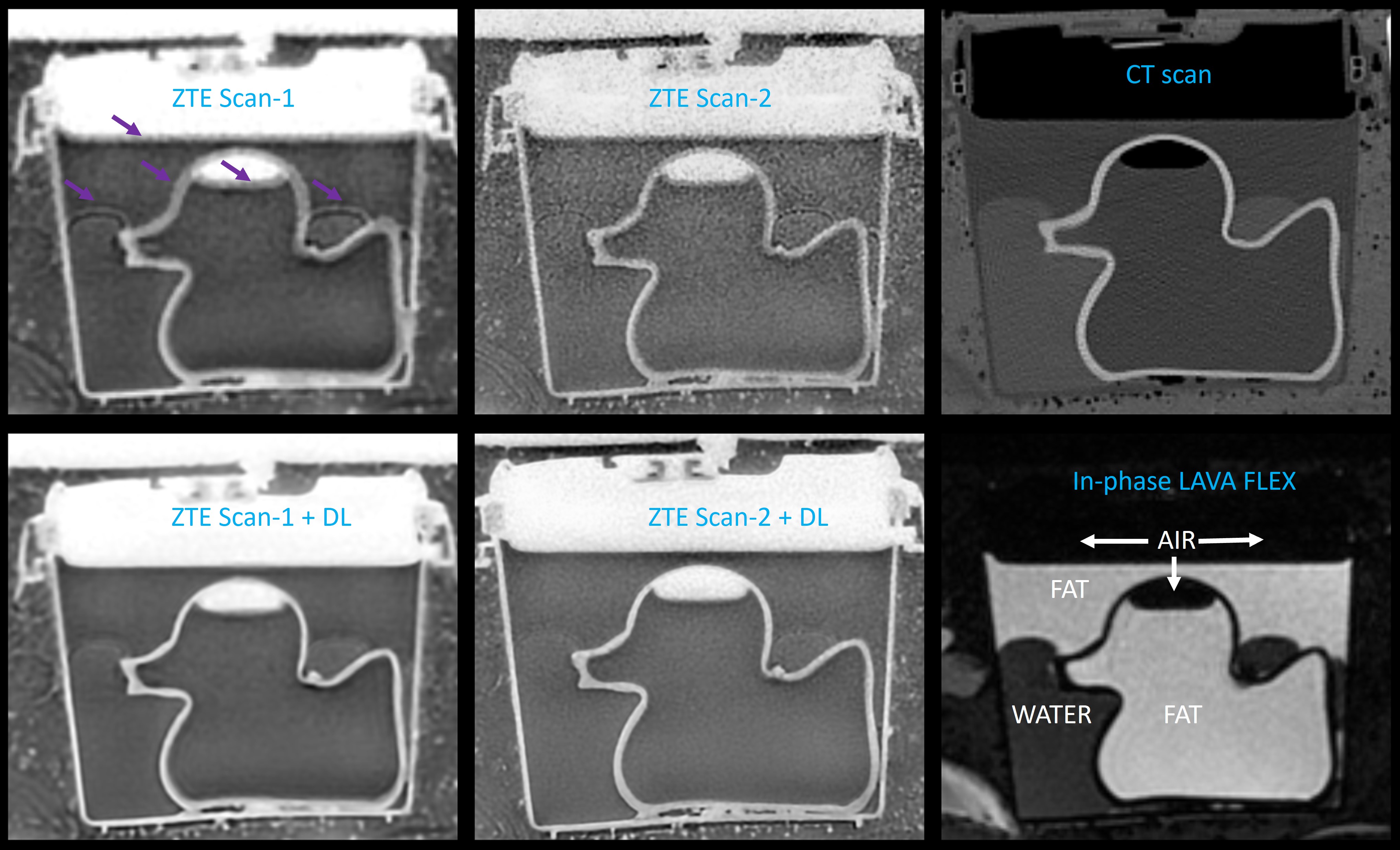
Phantom Experiments: A phantom was developed to demonstrate CS artifacts. To build the phantom, a rubber duck filled with peanut oil is submerged in a plastic cup. The cup is partially filled with water and peanut oil is filled on top of the water. Two ZTE scans were acquired at 3T on water resonance: Scan-1 (256 x 256 matrix) is acquired at 1mm iso and uses a BWP of 390.625 while Scan-2 (512 x 512 matrix) is acquired at 0.625mm iso and uses a BWP of 325.527 Hz/pixel. CT scan and LAVA FLEXTM scan were also collected for this phantom. Figure 2 shows the results of the DL method in correcting the CS artifacts on the phantom. Arrows highlight prominent regions of CS artifact, and the DL method successfully fixes these artifacts, and the resultant images are in good agreement with the CT scan.
In-vivo Experiments: In-vivo scans of hips, Head and Shoulder were acquired on a 3T scanner. The relevant acquisition parameters in these cases are provided in the Figure captions. The most relevant parameter, BWP, is typical for what is used in clinical settings.
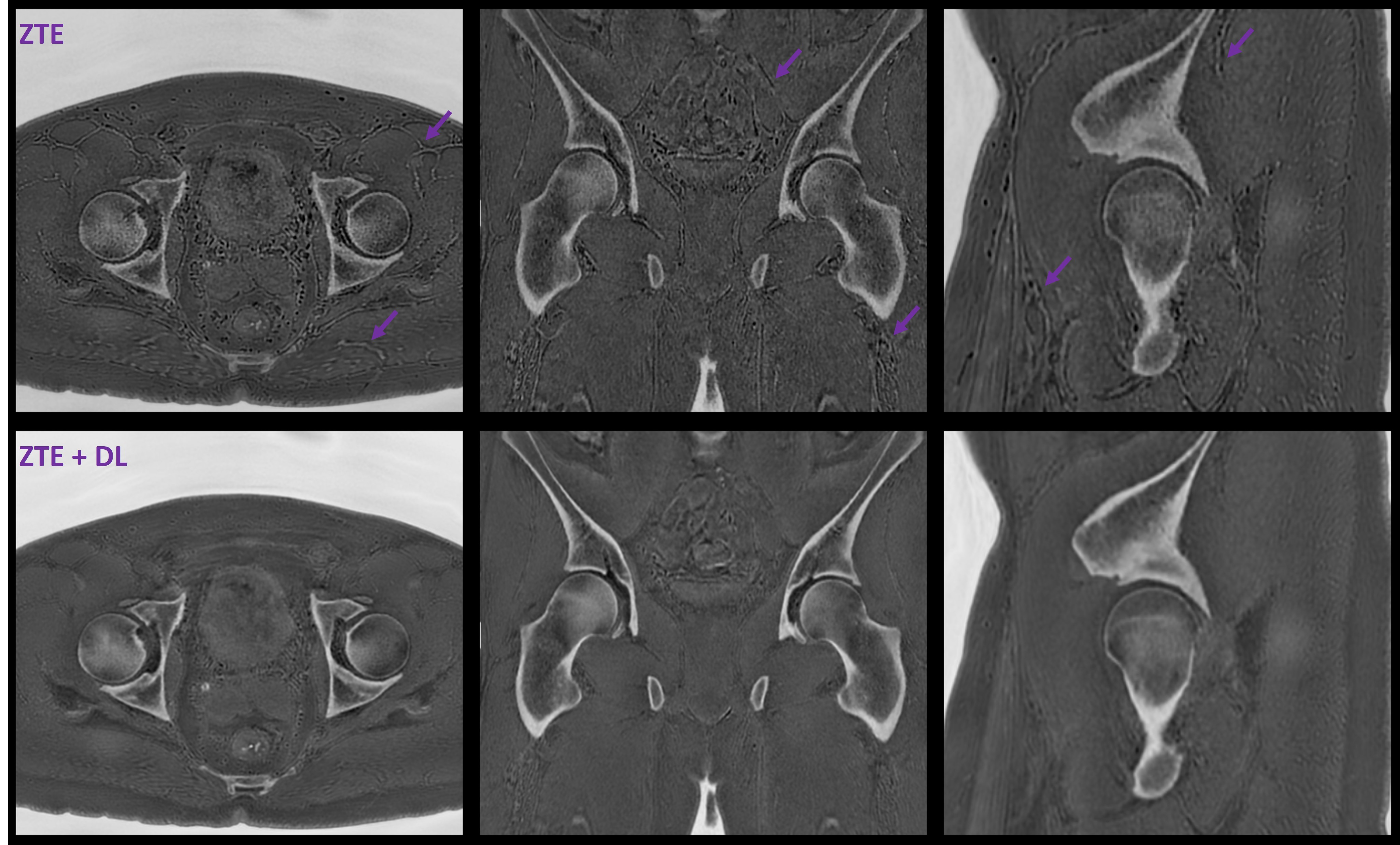
Figure 3 shows the results of a high-resolution hip oZTEoTM with arrows highlighting areas with strong CS artifacts. In this case, the artifact manifests quite strongly as destructive interference at tissue interfaces and the DL reconstruction effectively mitigates it.
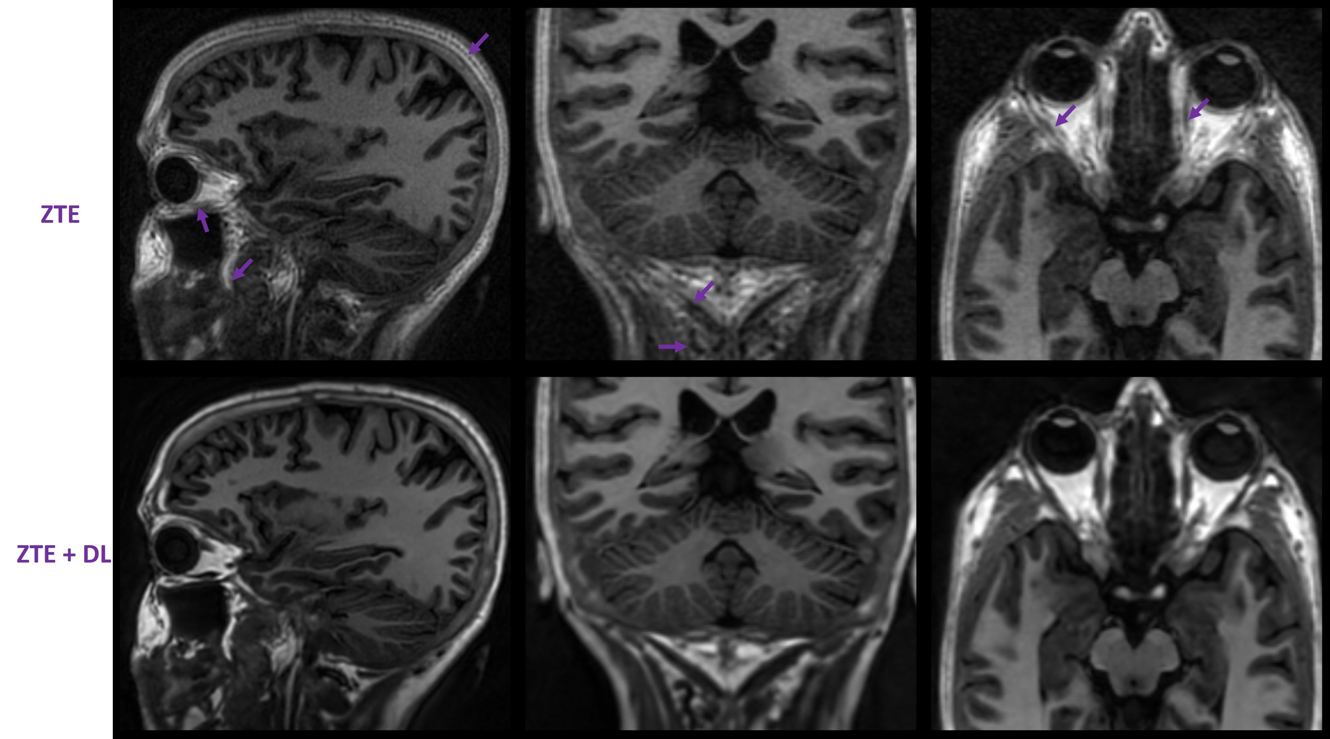
Figure 4 shows the results on T1 weighted brain where the effects of CS off-resonance on the orbits and neck are readily evident. Arrows highlight areas of note and their improvement with the use of the DL method.
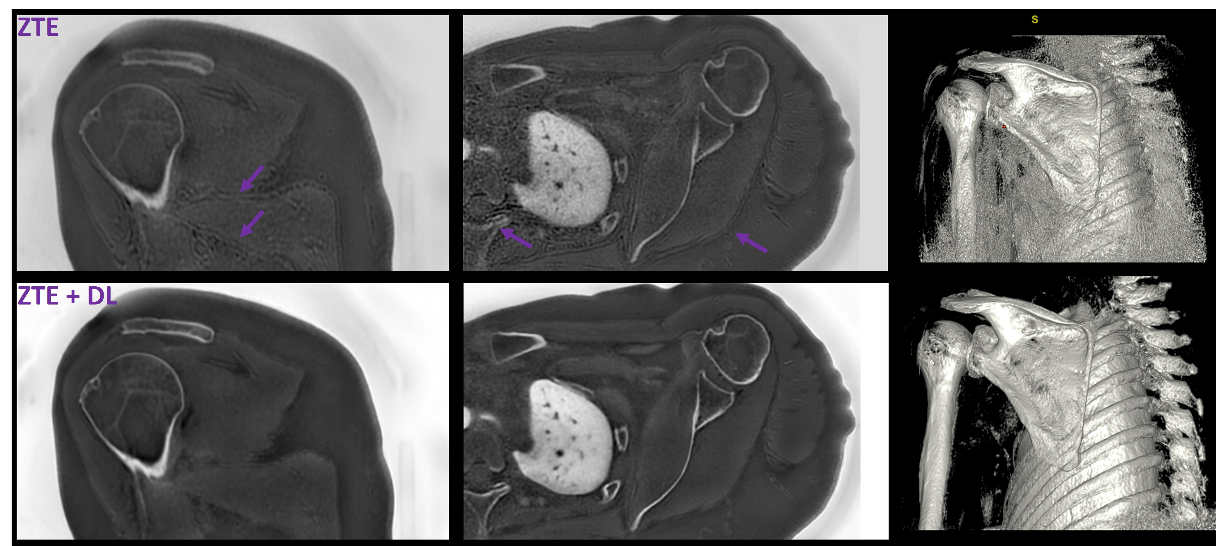
Figure 5 shows the improvements in shoulder oZTEoTM and highlights the application for better volume rendering.
Conclusion
ZTE forms the basis of several novel MR imaging applications and is becoming increasingly popular. A major artifact associated with ZTE, because of the radial sampling, is its sensitivity to off-resonance. Chemical-shift is a significant source of off-resonance in-vivo and can lead to either blurring or destructive interference artifacts at tissue interfaces. While high BW scanning can mitigate these artifacts, it can lead to poor SNR efficiency. We demonstrate in this work that it is possible to mitigate the effects of chemical shift off-resonance using a deep-learning based method. The reduction in these artifacts leads to images that are more accurate (reduces spurious edge enhancement) and is expected to be useful for subsequent post-processing tasks.Acknowledgements
The authors gratefully appreciate support from Emory University that made the phantom comparisons possible.References
- Froidevaux et al., Signal-model-based water-fat separation in Zero Echo Time (ZTE) MRI. ISMRM 2017 Abstract 0722
- Engstrom et al., In‐phase zero TE musculoskeletal imaging. MRM 2020; 83: 195-202
- Weiger et al., Short-T2 MRI: Principles and recent advances. Progress in Nuclear Magnetic Resonance Spectroscopy 114–115 (2019) 237–270
- Lebel et al., Performance characterization of a novel deep-learning based MR image reconstruction pipeline. https://arxiv.org/abs/2008.06559v1
Figures