4427
3D high resolution, distortion-free, reduced field of view diffusion-prepared MRI at 3T1King's College London, London, United Kingdom, 2Siemens Healthcare Limited, Frimley, United Kingdom, 3Siemens Healthcare, Courbevoie, France
Synopsis
Keywords: Pulse Sequence Design, Diffusion/other diffusion imaging techniques
Motivation: High resolution distortion-free diffusion-prepared imaging (DiffPrep) is desirable for high-precision radiotherapy or surgical planning.
Goal(s): To demonstrate 3D high resolution distortion-free DiffPrep using a reduced field of view (RFOV) approach and gradient echo (GRE) readout.
Approach: A DiffPrep sequence was developed with magnitude stabilisers, RFOV excitation, fat suppression, 3D-GRE readout and 2D phase-correction navigator. The proposed approach using selective excitation of the tip-down pulse was compared against non-selective excitation at 3T in a phantom and in the spinal cord of a healthy volunteer.
Results: Preliminary results presented in a phantom and in-vivo demonstrate successful outer volume signal suppression using the reduced FOV approach.
Impact: The sequence introduced in this work enables 3D RFOV distortion-free DiffPrep with a GRE readout. This sequence could be advantageous for applications requiring accurate target delineation, such as radiotherapy planning or surgical planning.
Several applications of diffusion imaging, such as for surgical planning, radiotherapy planning and monitoring of treatment response, require accurate geometric fidelity1,2. Diffusion imaging is conventionally performed using diffusion-sensitizing gradients combined with 2D single-shot spin-echo EPI (SS-EPI) readout, which minimises artefacts due to bulk motion but suffers from distortions, especially at higher field strengths. Diffusion-prepared imaging (DiffPrep) has been proposed in combination with turbo spin echo (TSE) sequences3 for distortion-free diffusion imaging however TSE acquisitions suffer from SAR limitations, especially at 3T. We propose a novel 3D reduced FOV (RFOV) DiffPrep sequence which uses a gradient echo readout. By minimizing the shot length, RFOV diffusion imaging reduces T1 contamination effects, maximises diffusion weighting and facilitates the acquisition of a low-resolution 2D phase navigator at the end of the readout.
Methods
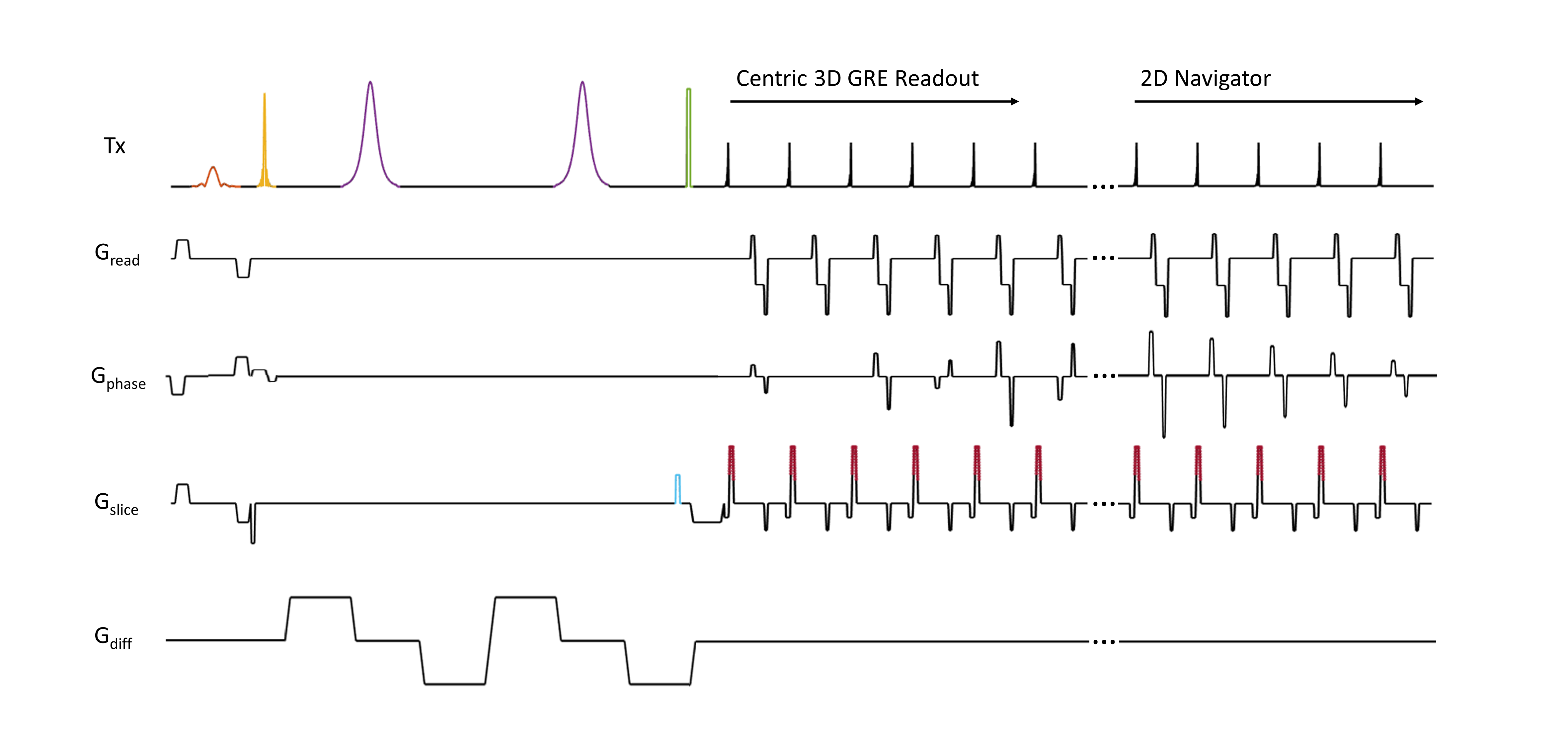
Pulse Sequence and phase correction: The sequence (Figure 1) consists of a twice-refocused diffusion preparation scheme4, paired with a centric-encoded 3D gradient-echo readout, followed by a linearly-encoded 2D phase correction navigator. The diffusion preparation includes a fat saturation pulse immediately before the tip-down pulse, which is applied simultaneously with a slab-selective gradient in the phase-encoding direction, thereby restricting excitation to water protons within a reduced FOV. The excitation is followed by a pair of bipolar diffusion gradients, each surrounding a 180° adiabatic refocusing pulse. A magnitude stabiliser5 converts phase-induced magnitude errors that would normally occur after the preparation module back into intershot phase inconsistencies, correctable with a phase navigator6. A magnitude stabiliser gradient is applied in the slice direction directly before the non-selective tip-up pulse to fully dephase spins across the slice. Half of the magnetisation from excited water protons is thereby restored to the longitudinal axis. Finally, a spoiler gradient is executed to dephase any remaining transverse magnetisation including outer-volume and/or fat protons. The original phase of the spins is restored by a magnitude stabiliser rewinder gradient, which is played out after each RF pulse. The magnitude stabiliser rewinder also acts as a spoiler gradient for the outer volume magnetisation. Each imaging shot acquires all phase encoding lines for one partition.
For the reconstruction, a Fourier transform is applied to each phase navigator, followed by a Hamming filter. A Fourier transform is then applied to each imaging shot, followed by a phase correction using the phase navigator for each receiver coil6. An inverse Fourier transform is then applied to the imaging shot.
Experiments: All images were acquired on a 3T PET-MR scanner (Biograph mMR, Siemens Healthineers, Erlangen, Germany). The proposed sequence was acquired twice in a phantom (CaliberMRI diffusion phantom) to assess the effectiveness of the RFOV approach: once with a selective and once with a non-selective tip-down pulse. The following parameters were applied: b-value: 500 s/mm2, Number of diffusion directions: 3, T2 prep duration: 65ms, TE/TR: 2.0ms/4.4ms, shot TR: 2000ms, FOV: 200mm x 100mm, acquired matrix size: 96 x 48, acquired slice thickness 4mm, number of partitions: 8, number of averages: 5. An in-vivo scan was acquired in the spinal cord of a healthy volunteer following IRB approval and informed consent. The proposed sequence was acquired with the selective and non-selective tip-down excitation pulse with the same parameters as in the phantom measurement but with the exception of FOV: 200mm x 50mm, acquired matrix size: 96x24 and number of partitions: 12.
Results
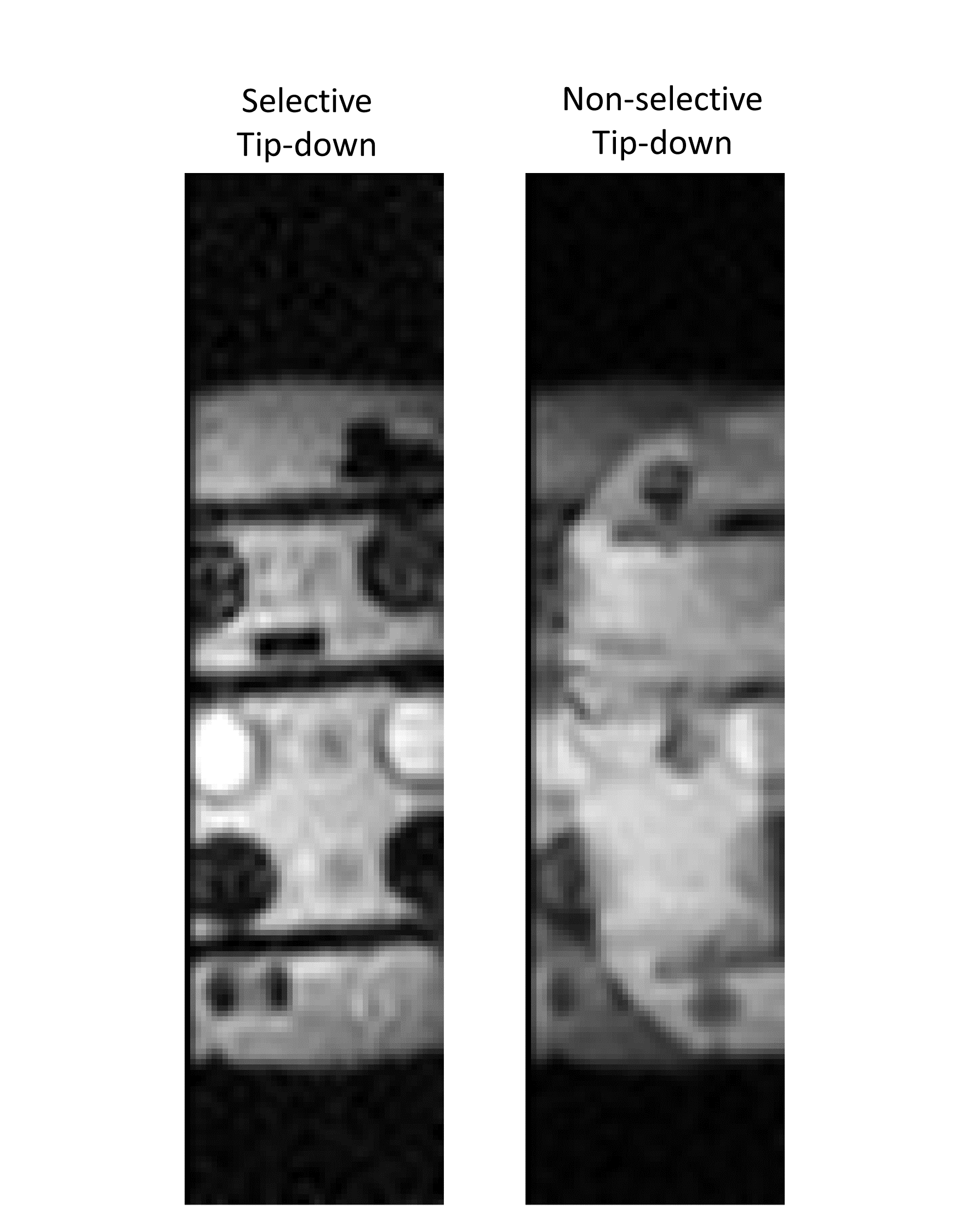
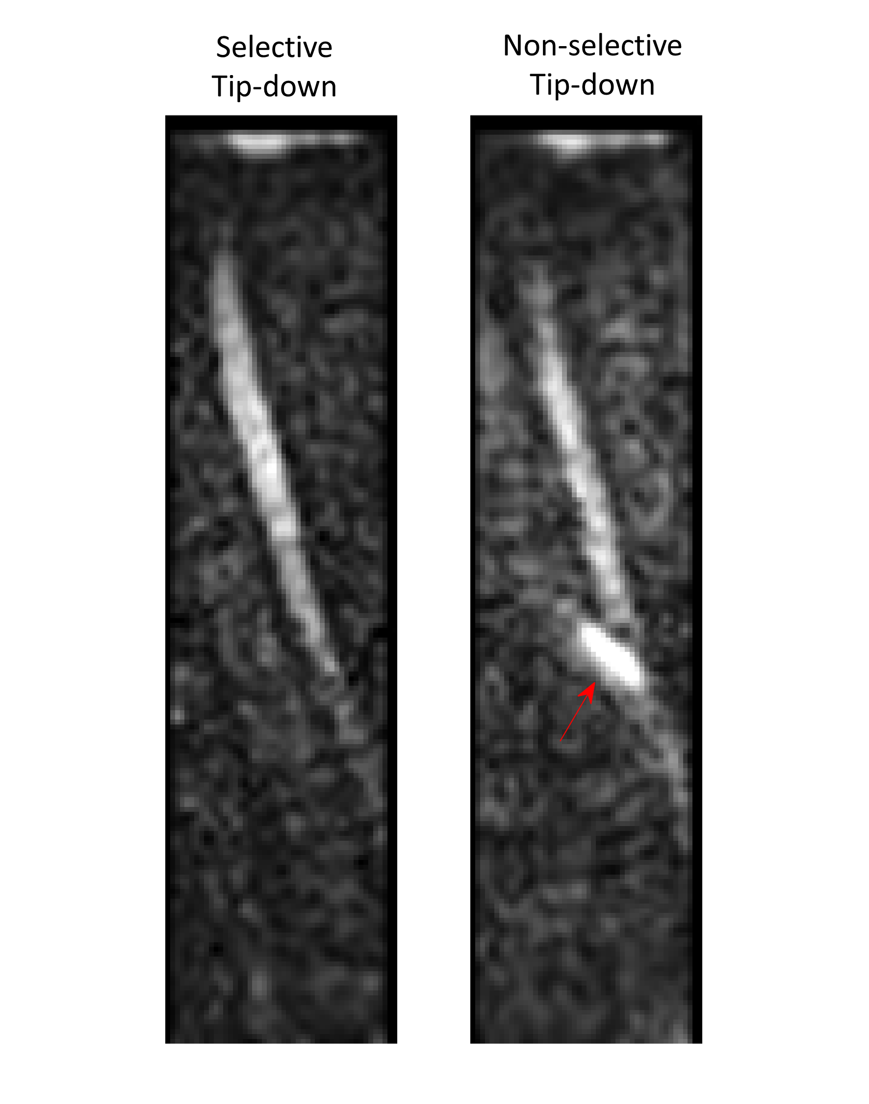
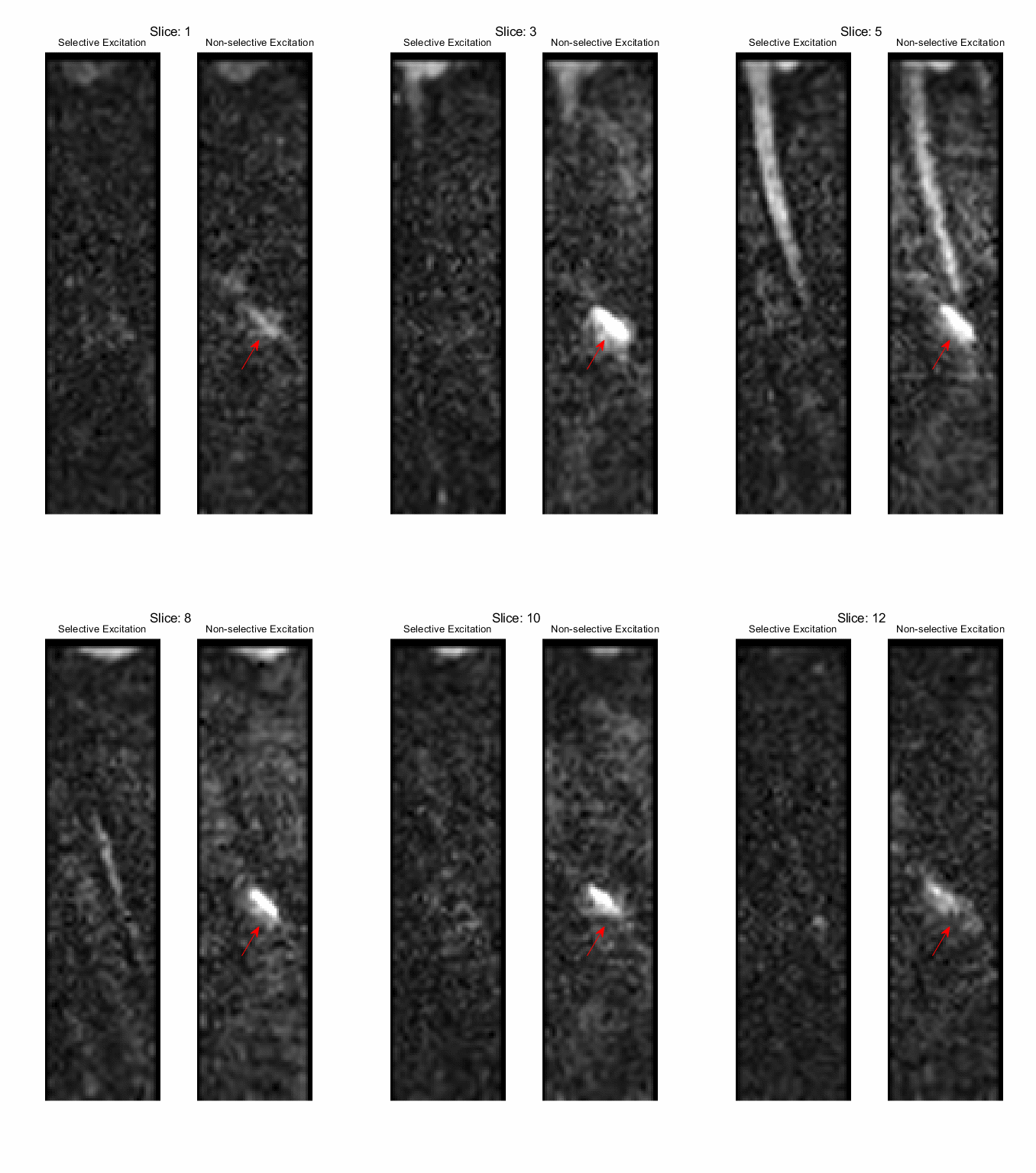
Images from the phantom study are shown in Figure 2, demonstrating robust suppression of outer volume signal using the RFOV approach described. When the selective tip-down pulse is replaced by a non-selective pulse, the outer volume of the phantom is excited, producing severe wrap in the resultant image. The RFOV approach was also effective for DiffPrep imaging of the spinal cord as shown in Figure 3. Outer volume signal is suppressed demonstrating that fat signal is also suppressed using this approach.
Discussion and Conclusion
We have shown initial results for a 3D RFOV diffusion-prepared GRE sequence using the combination of magnitude stabilisers, slab-selective tip-down excitation pulse and a 2D phase navigator to tackle the challenges associated with high resolution diffusion imaging. Future work will focus on correction of T1 effects for accurate ADC mapping with this technique.
Acknowledgements
No acknowledgement found.References
1. Walker A, Liney G, Metcalfe P, Holloway L. MRI distortion: considerations for MRI based radiotherapy treatment planning. Australasian physical & engineering sciences in medicine. 2014;37:103-113.
2. Pappas EP, Alshanqity M, Moutsatsos A, et al. MRI-related geometric distortions in stereotactic radiotherapy treatment planning: evaluation and dosimetric impact. Technology in cancer research & treatment. 2017;16(6):1120-1129.
3. Zhang Q, Coolen BF, Versluis MJ, Strijkers GJ, Nederveen AJ. Diffusion‐prepared stimulated‐echo turbo spin echo (DPsti‐TSE): An eddy current‐insensitive sequence for three‐dimensional high‐resolution and undistorted diffusion‐weighted imaging. NMR in Biomedicine. 2017;30(7):e3719. doi:10.1002/nbm.3719
4. Reese TG, Heid O, Weisskoff RM, Wedeen VJ. Reduction of eddy‐current‐induced distortion in diffusion MRI using a twice‐refocused spin echo. Magnetic Resonance in Med. 2003;49(1):177-182. doi:10.1002/mrm.10308
5. Alsop DC. Phase insensitive preparation of single‐shot RARE: Application to diffusion imaging in humans. Magnetic Resonance in Med. 1997;38(4):527-533. doi:10.1002/mrm.1910380404
6. Miller KL, Pauly JM. Nonlinear phase correction for navigated diffusion imaging. Magnetic Resonance in Med. 2003;50(2):343-353.
Figures