4426
Promotion of Reconstruction Performance for Equally Spaced Under-sampled Signals Using Amplitude Modulation Pulses1Utsunomiya University, Utsunomiya, Japan
Synopsis
Keywords: Image Reconstruction, Image Reconstruction
Motivation: In compressed sensing, it is impossible to separate folded-over images using equally spaced under-sampled signal when the subject is a real-valued image.
Goal(s): Our goal was to enable image reconstruction using equally spaced under-sampled signal, and to generalize it to phase-varied images.
Approach: Amplitude modulation in the phase-encoding direction makes the folded image complex-valued and thus facilitates separation of overlapping images in the image space.
Results: The image reconstruction was successful up to a speedup factor of 4 with U-Net. Phase images were successfully reconstructed by partial continuous signal acquisition and phase correction.
Impact: Reconstruction performance is improved by applying specific amplitude-modulated pulses prior to signal acquisition in equally spaced under-sampling CS, which is advantageous for sharp image reconstruction.
Introduction
In compressed sensing MRI, equally spaced signal under-sampling has the advantage of less image blurring, but strong aliasing artifacts make image reconstruction difficult. Although deep learning reconstruction (DRL) has made it possible to reconstruct images from equally spaced under-sampled signals [1], it is still common to collect the signals in central portion of the k-space without under-sampling. We propose a new imaging scheme that promotes image reconstruction performances for equally spaced under-sampled signals in DRL using amplitude modulation pulses (AmpDRL). The purpose of this study is to clarify the degree of image quality improvement by this method and the application of this method to phase varied images.Methods
A gradient field $$$G_{y1}$$$ is applied for a time $$$t_1$$$ in the phase-encoding direction (y-direction) after an RF pulse is applied. Let the spin density be $$$\rho(x,y)$$$, MR signal can be written as Eq.(1) by variable transformation $$$k_{m1}=\gamma G_{y1} t_1$$$,$$v(k_x,k_y)= \int \hspace{-2.0mm} \int^{\infty}_{-\infty} \rho(x,y) e^{-j k_{m1} y} e^{-j \left(k_x x+k_y y \right)} dxdy ... (1) $$
The reconstructed image is obtained by compensating the phase modulation term $$$\exp(j k_{m1} y)$$$ by the inverse Fourier transformed image.
$$\rho(x,y) = e^{j k_{m1} y} {\rm IFT} [v(k_x,k_y)] ... (2) $$
Equation (2) can be written as Eq. (3) by expressing a discrete signal equation using a continuous equation.
$$\rho_{rec}(m,n)=\rho(m,n) + \rho\left(m,\left(n \mp 1 \frac{N}{s}\right) \right) e^{\pm j \left( 1 \frac{a}{s} \right) \pi } + \rho\left(m,\left(n \mp 2 \frac{N}{s}\right) \right) e^{\pm j \left( 2 \frac{a}{s} \right) \pi} + \cdot \cdot \cdot \rho\left(m,\left(n \mp p \frac{N}{s}\right) \right) e^{\pm j \left( p \frac{a}{s} \right) \pi} +\cdot \cdot \cdot (3) $$
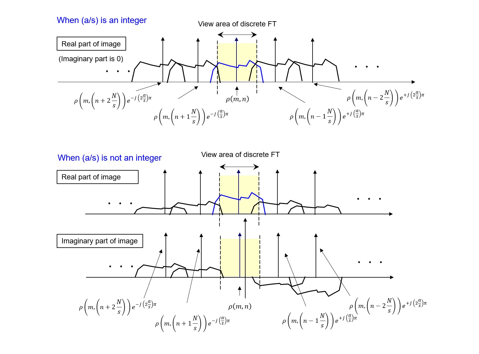
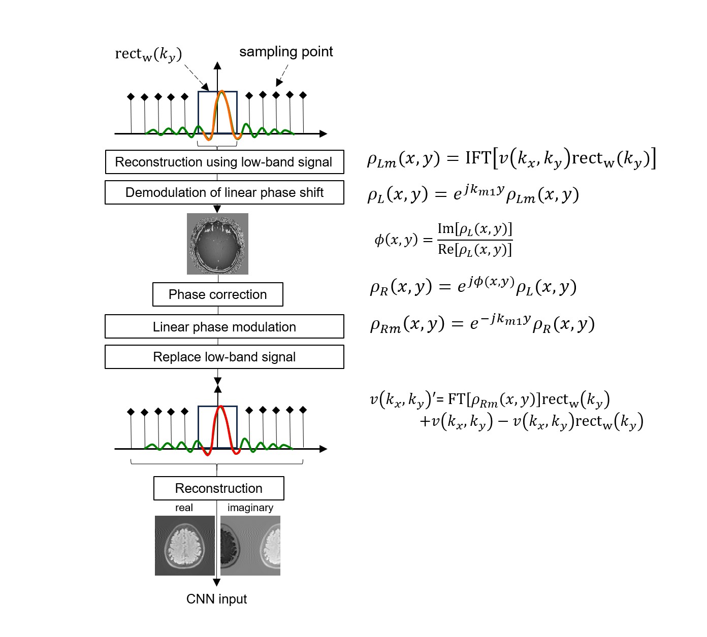
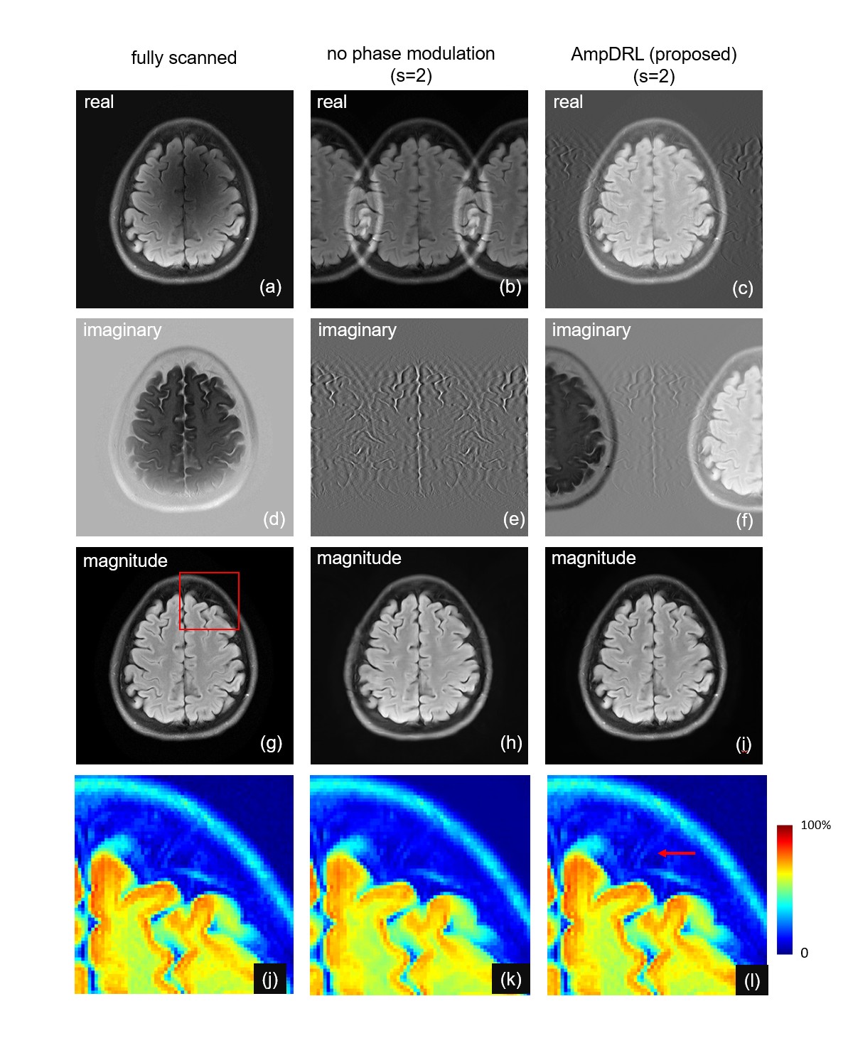
In equation (3), $$$s$$$ and $$$p$$$ are reduction factor and natural number, respectively, and the reconstructed image is assumed to consist of (m,n) points in the discrete signal representation. The phase modulation factor corresponding to $$$k_{m1}$$$ is $$$a$$$. When $$$p a/s$$$ is an integer, the phase shift of the reconstructed image $$$(p a/s) \pi$$$ is a multiple of $$$\pi$$$, as shown in the Fig.1 (a), so the reconstructed image has only the real part. In this case, folded-over images cannot be separated. On the other hand, if $$$p a/s$$$ is not an integer, the reconstructed image has an imaginary part as shown in Fig.1(b). Since the images overlap differently in the real and imaginary parts, there is a possibility that the folded images can be separated. The algorithm for complex-valued images is shown in the Fig.2. The center of the k-space is collected continuously without under-sampling, and the image $$$\rho_{Lm} (x,y)$$$ is reconstructed using only the continuously collected signal. Multiplying $$$\rho_{Lm} (x,y)$$$ by the phase $$$\exp(j k_{m1} y)$$$ yields $$$\rho_{L} (x,y)$$$. Here, after phase correction, the phase modulation $$$\exp(j k_{m1} y)$$$ is again given to obtain $$$\rho_{Rm}(x,y)$$$. Fourier transform of $$$\rho_{Rm}(x,y)$$$ is replaced by the continuously collected part of the k-space signal.
Results
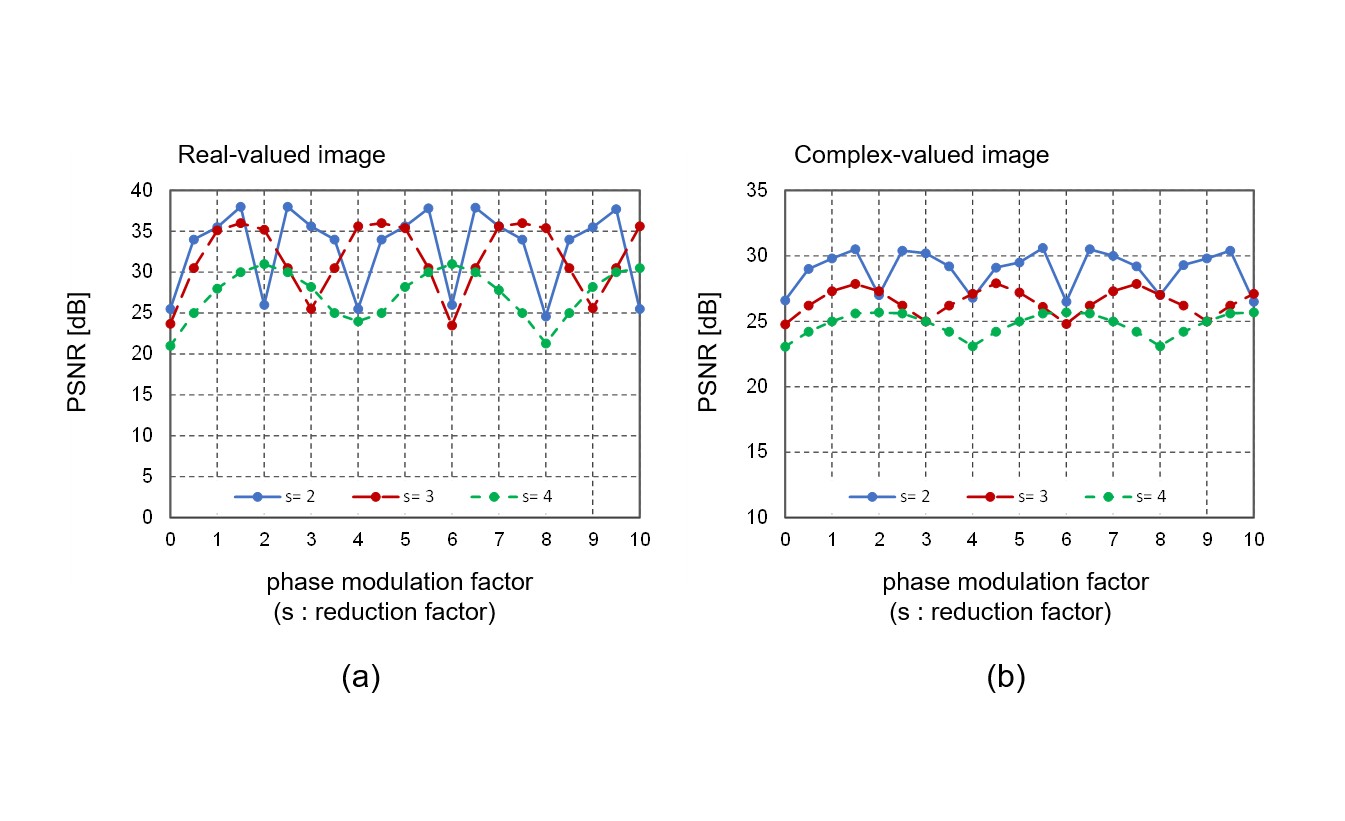
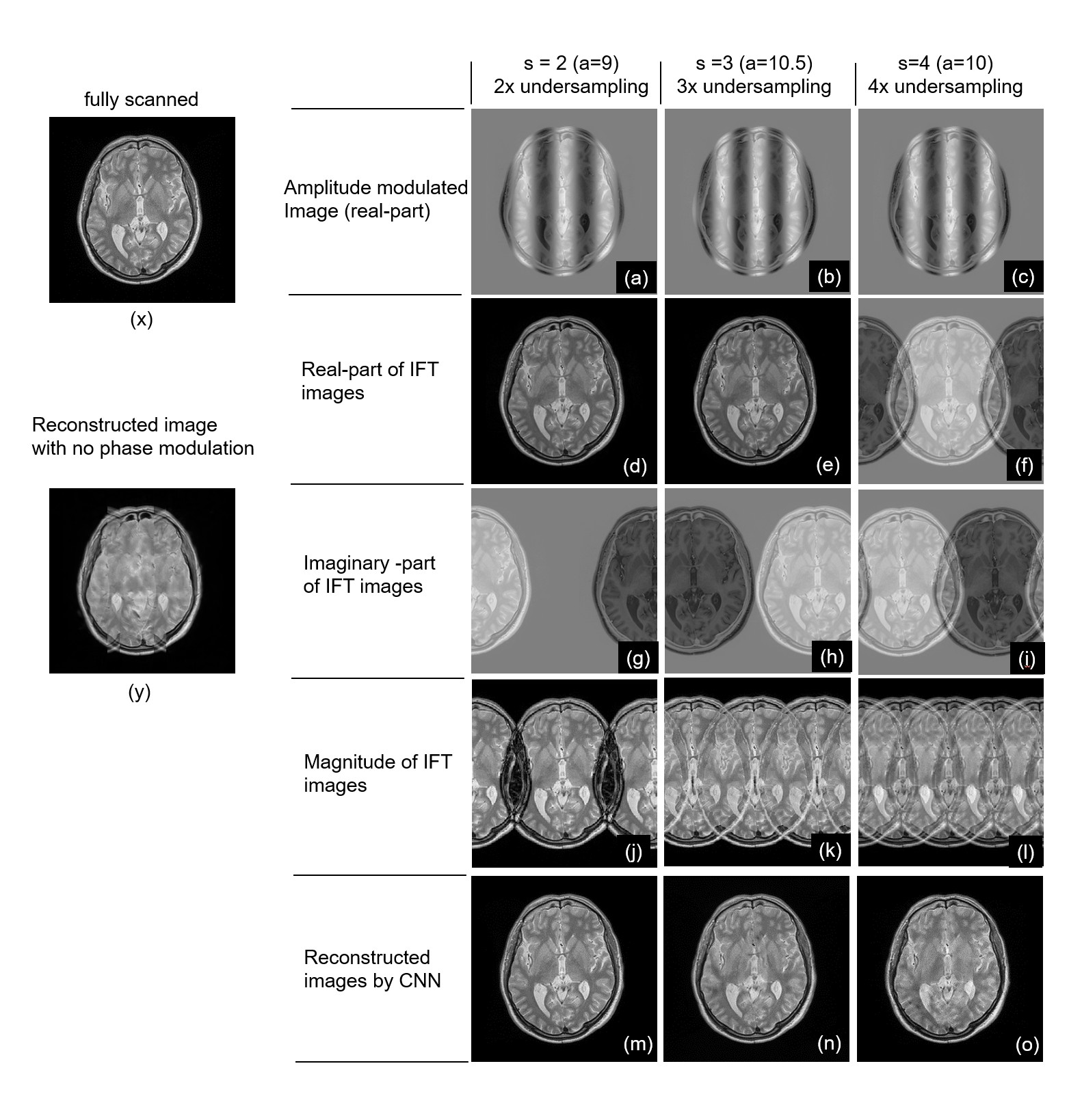
In the simulation experiments, 500 real-valued images from the IXI dataset (PDW, 256x256 pixels) [2] were used to train U-Net. 100 images were used for validation and 100 for testing. When the phase modulation factor is normalized to $$$(a/N) \pi$$$, Fig. 3(a) shows the relationship between $$$a$$$ and PSNR for different reduction factor s=2, 3, and 4. In all cases, PSNR become low when a is a multiple of reduction factor s and take maximum value at intermediate values. The maximum PSNR tends to decrease as s increases. Figure 4 shows the reconstructed images for s=2, 3, and 4. The reconstruction fails in the area where the folding of image occurred as shown in Fig.4(y). The fold-over artifacts are well separated in (m) to (o) images. Figure 5 shows the simulation results of phase varied images. The fastMRI dataset [3] converted to 256x256 pixels were used. Because the phase correction contains some errors, the results differ from those of the real-valued images. Artifacts were observed in (c), (f), but the images are close to Fig.4 (d), (g) in the real-valued case. The relationship between $$$a$$$ and PSNR for the phase-varied images is shown in Fig. 3(b). The reconstructed image (i) shows better structural preservation than (h). The possibility of image quality improvement was also demonstrated with phase-varied images. Since this method requires phase modulation on the subject, it cannot be applied to diffusion-weighted images or fluid measurements in which information is woven into the phase.Conclusion
Amplitude modulation prior to data acquisition sequence shows the possibility of promoting the reconstruction performance using an equally spaced under-sampled signal.Acknowledgements
We acknowledge Imperial College London and NYU for providing the IXI data set and fastMRI Dataset.References
[1] Hammernik K, Knoll F, Sodickson DK, Pock T. On the influence of sampling pattern design on deep learning- based MRI reconstruction. 25th ISMRM, Hawaii, 2017; 644
[2] Imperial College London. IXI dataset. https://brain-development.org/ixi-dataset/
[3] NYU Langone Health fastMRI Dataset. https://fastmri.med.nyu.edu/
Figures