4424
Dual-polarity SENSE with calibration refinement enables robust Nyquist ghost correction on a high-performance gradient system1Center for Biomedical Imaging Research, Department of Biomedical Engineering, School of Medicine, Tsinghua University, Beijing, China, 2Athinoula A. Martinos Center for Biomedical Imaging, Massachusetts General Hospital, Charlestown, MA, United States, 3Department of Radiology, Harvard Medical School, Boston, MA, United States
Synopsis
Keywords: Artifacts, Artifacts
Motivation: Echo-planar imaging (EPI) is prone to Nyquist ghosts, which are exacerbated on scanners with high-performance gradients. While dual-polarity GRAPPA (DPG) helps alleviate these artifacts, it does not permit regularized multi-shot reconstruction.
Goal(s): To introduce a SENSE-based method to address Nyquist ghosts on high-performance gradient systems and to lend itself to regularized multi-shot reconstruction.
Approach: We propose dual-polarity SENSE (DPS) for ghost correction where phase differences between even and odd lines are captured in ESPIRiT coil sensitivity estimates based on a tailored calibration scan.
Results: DPS effectively reduces Nyquist ghost artifacts in phantom and in vivo data on high-performance gradient systems.
Impact: We provide a robust SENSE-based Nyquist correction method that can be integrated with advanced multi-shot EPI reconstruction techniques and can address challenging Nyquist ghosts on high-performance gradient systems.
Introduction
EPI is inherently prone to Nyquist ghosts due to its fast gradient switching. With the advent of scanners featuring ultra-high field or high-performance gradients, Nyquist ghosts are exacerbated since they can no longer be adequately corrected with linear phase correction (LPC). To address this issue, approaches including DPG1 or RAC-LORAKS2 have emerged to capture the 2-dimensional phase differences between readouts with opposite polarities (RO+ and RO-), yielding improved ghost correction performance. However, these methods usually rely on k-space-based reconstruction, while emerging multi-shot EPI techniques (e.g. BUDA3, EPTI4) employ SENSE-based reconstruction.To render improved ghost correction applicable in SENSE-based reconstructions that admit advanced regularizers, we introduce dual-polarity SENSE (DPS). DPS involves a tailored data calibration acquisition from which high-fidelity reference data with RO+ and RO- images are derived. These are input to ESPIRiT5 sensitivity estimation whereby 2-dimensional phase differences between the readout polarities are captured. Phantom and in vivo data acquired on the Connectome 2.0 with high-performance gradients demonstrate robust ghost correction using DPS.
Methods
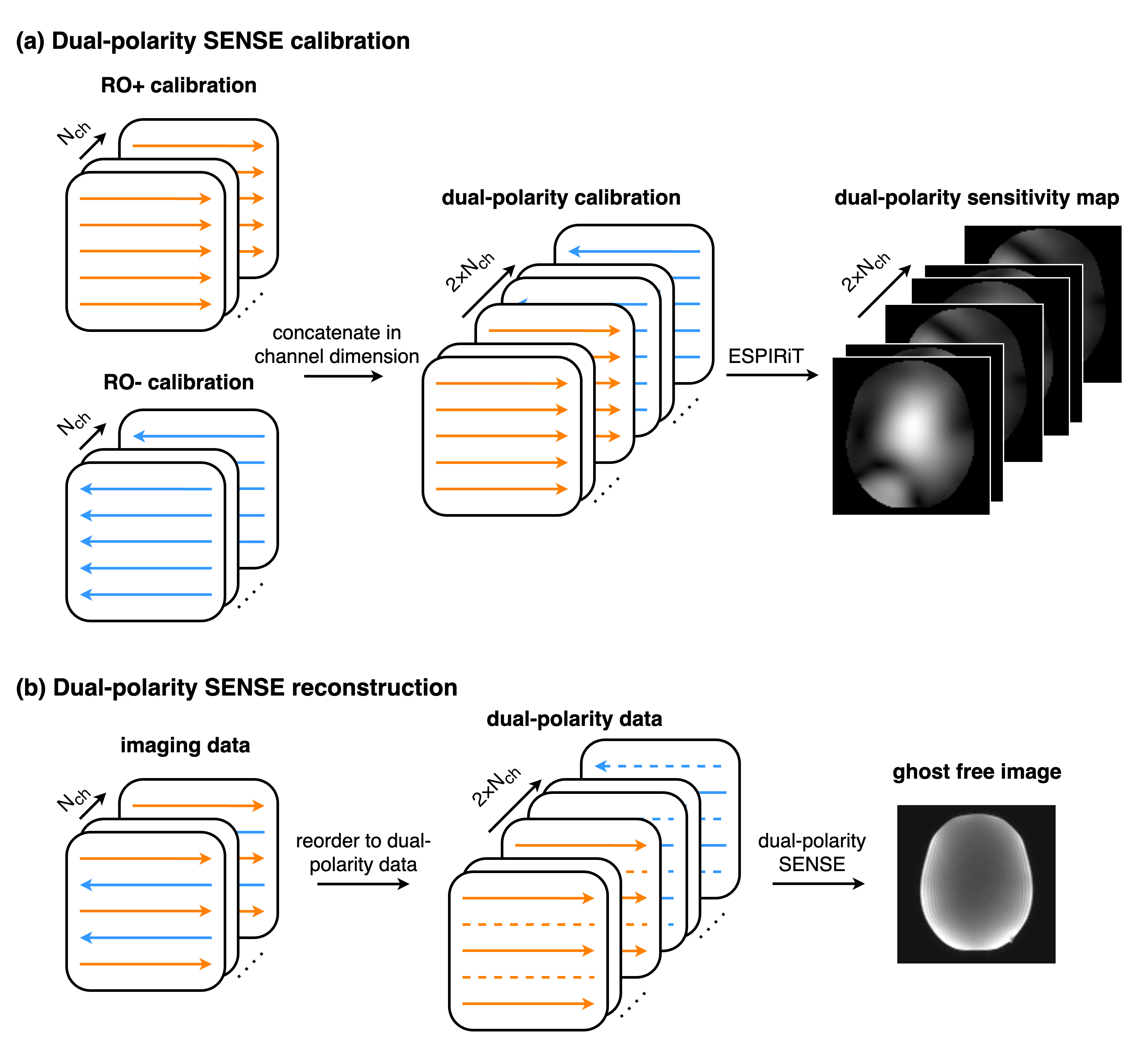
Dual-polarity SENSEFig 1 presents the DPS workflow. DPS employs GESTE6 calibration, which comprises RO+ and RO- data, each with Nch channels. The RO+ and RO- data are first concatenated in the channel dimension, forming dual-polarity calibration with 2×Nch channels. ESPIRiT is applied to calculate the dual-polarity sensitivity map.
The reconstruction process for DPS involves splitting and reordering the RO+ and RO- data from each channel of the imaging data, generating a dual-polarity dataset. The undersampled dual-polarity data is then SENSE reconstructed by the previously obtained dual-polarity sensitivity map to form a ghost-free volume.
Calibration refinement
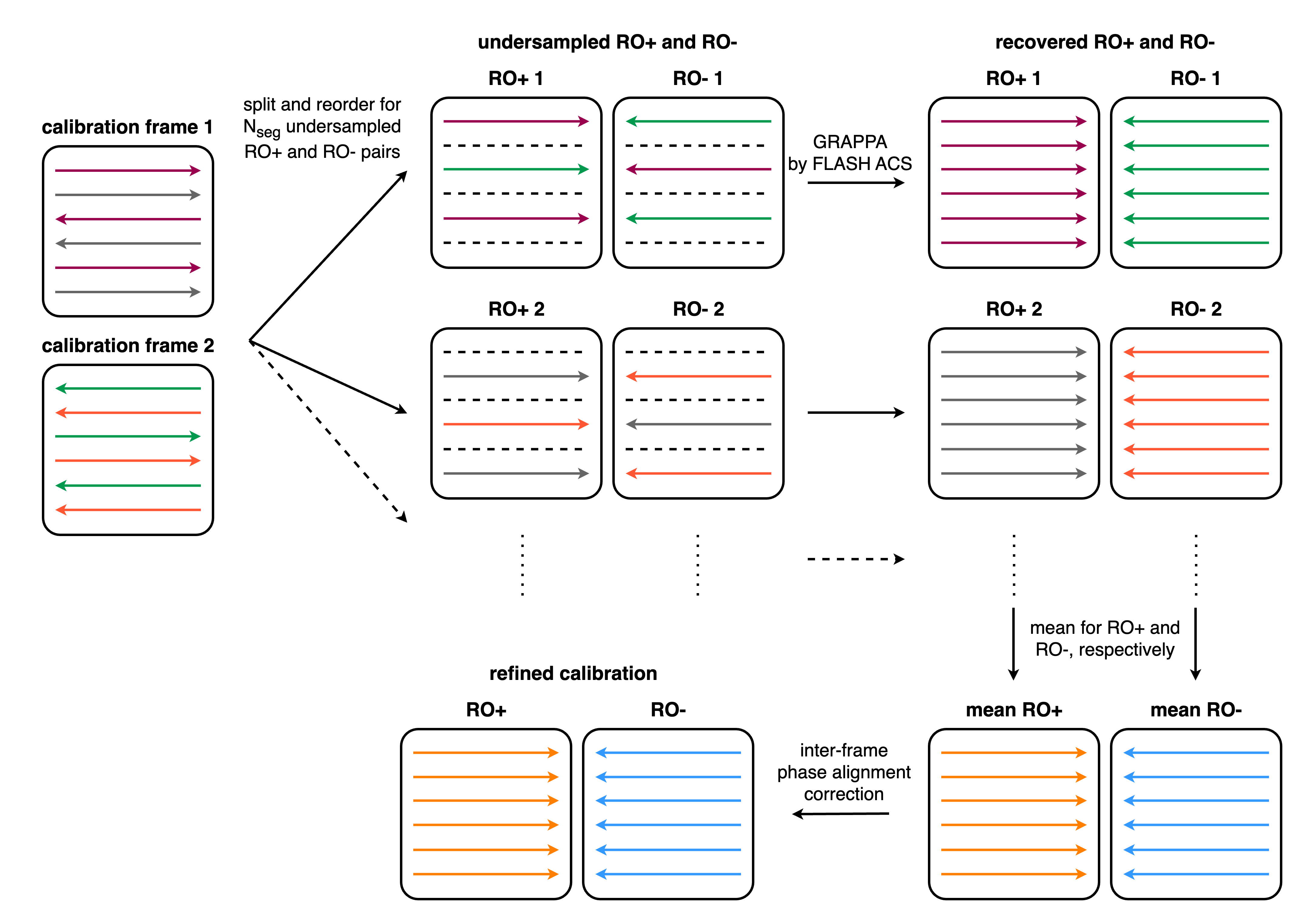
The performance of DPS depends on the quality of GESTE calibration from which the sensitivity maps are derived. Although GESTE data includes RO+ and RO- volumes that only include readouts with the same polarity, they may still contain residual ghosts. We therefore propose a calibration refinement to improve the calibration quality, as depicted in Fig 2.
GESTE calibration with Nseg segments is initially divided into Nseg pairs of undersampled RO+ and RO- data. GRAPPA reconstruction is then applied to recover these data, with the GRAPPA kernels trained on separately acquired FLASH ACS. A pair of mean RO+ and RO- data is obtained by averaging all the Nseg pairs. The inter-frame phase alignment correction7 is used for final refinement.
Data acquisition and evaluation
Data from a head phantom and a volunteer were acquired on the Connectome 2.0 scanner8 with a 72-channel head coil. Data sampling was performed using a spin-echo EPI DWI sequence with GESTE and FLASH calibration.
Phantom parameters: 2 mm isotropic resolution, 110×110 matrix, TE = 83/61/54 ms for R = 1/2/3, TR = 2900 ms, ESP = 0.40 ms, and b = 0 and 1000 s/mm2.
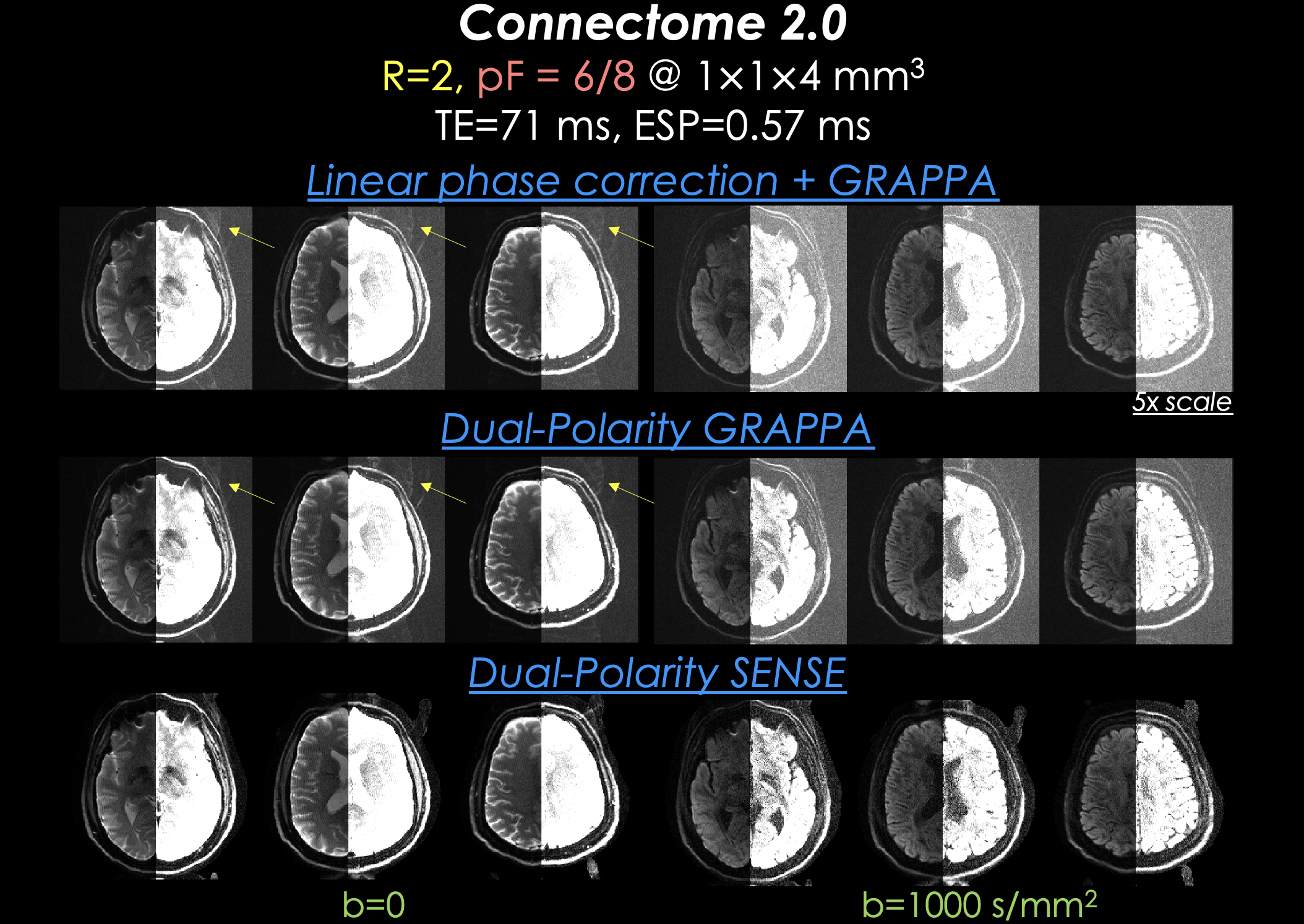
In vivo parameters: 1×1×4 mm3 resolution, 220×220 matrix, R = 2, pF = 6/8, TE/TR = 71/3000 ms, ESP = 0.57 ms, and b = 0 and 1000 s/mm2.
We applied DPS for data reconstruction and compared it with DPG and LPC+GRAPPA. Both DPG and DPS used our proposed calibration refinement.
Results
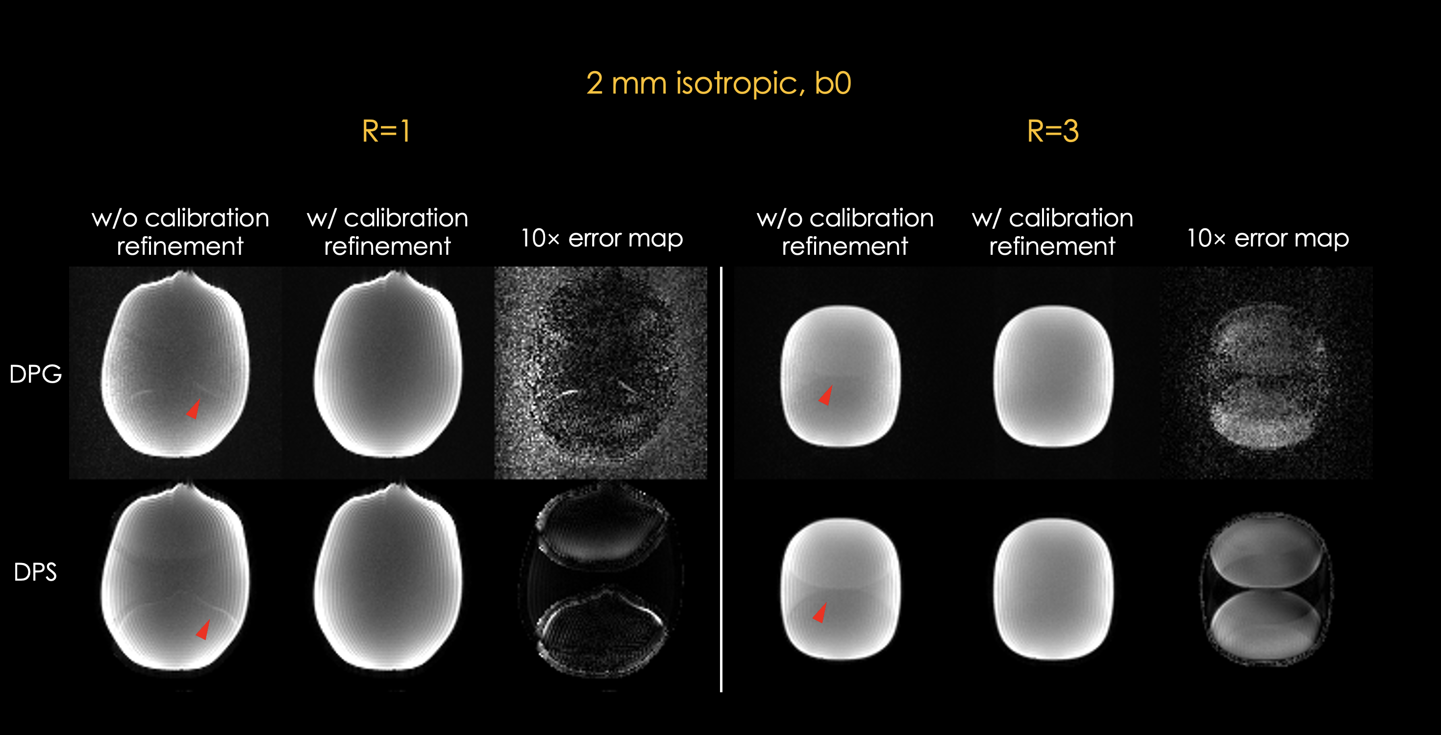
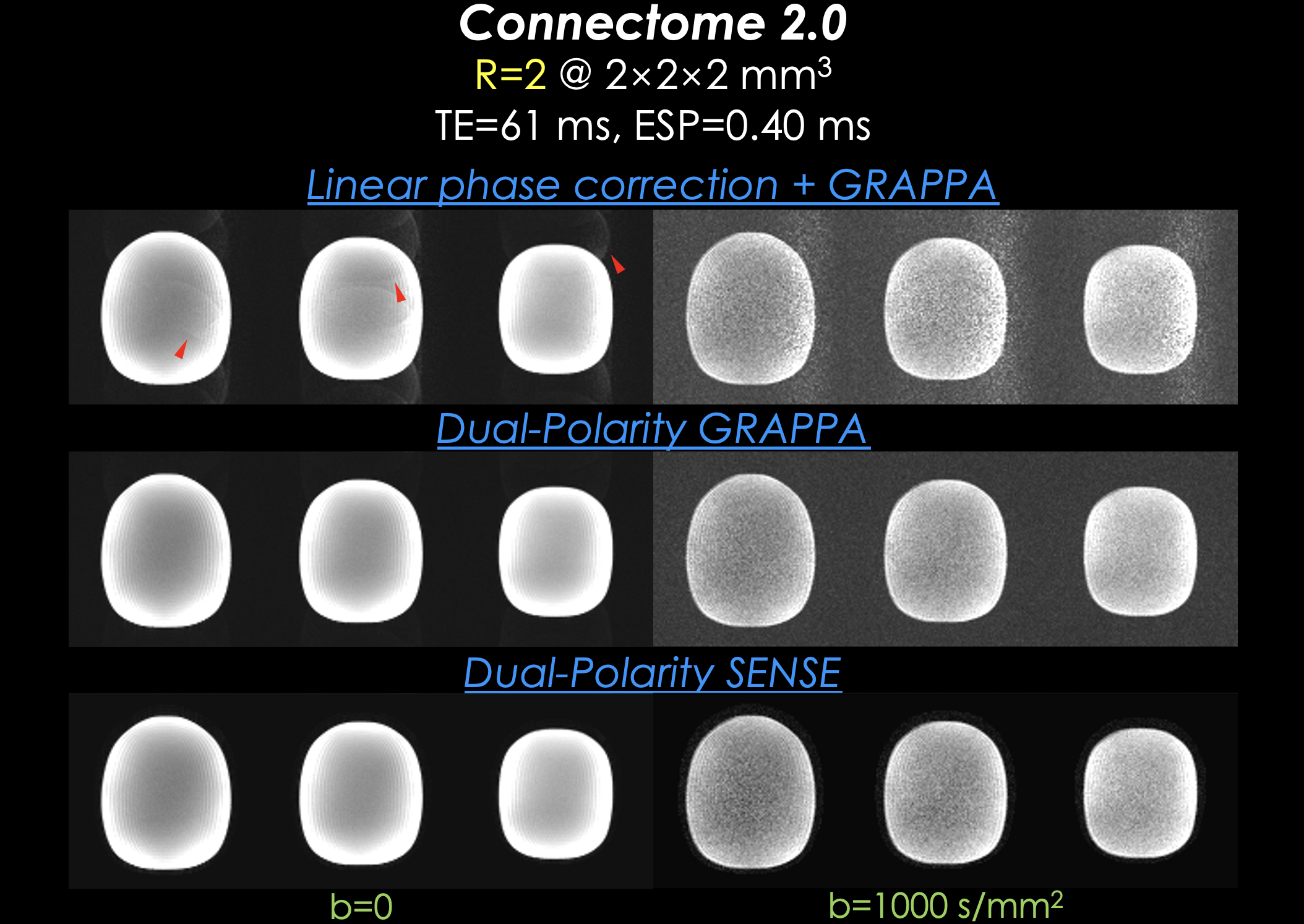
Fig 3 displays the phantom results for DPG and DPS with and without calibration refinement. Results using the original calibration show residual Nyquist ghosts (arrowheads), while those with refined calibration are ghost-free.Fig 4 presents the R = 2 phantom results. For the b0 images, LPC+GRAPPA exhibits noticeable ghosts (arrowheads), whereas DPG and DPS show no visible ghosts. For the b1000 images, due to its lower signal levels, none of the methods have any visible ghosts, but LPC+GRAPPA displays higher noise levels compared to the other two methods.
Fig 5 showcases the in vivo results. No apparent ghosts are visible in the brain parenchymas for all three methods. Residual ghosts are still visible in the background of LPC+GRAPPA and DPG (arrows). DPS results exhibit almost no ghosts in the background, likely due to its use of sensitivity maps with masks. For the b1000 images, DPG and DPS demonstrate lower noise levels compared to LPC+GRAPPA, consistent with the findings in the phantom results.
Discussion and Conclusion
The experiments demonstrate that DPS can correct Nyquist ghosts in data acquired on high-performance gradient systems such as the unique Connectome 2.0 with Gmax = 500 mT/m and Slew = 600 T/m/s specifications. It outperformed LPC+GRAPPA and offered comparable results to DPG. Additionally, the proposed calibration refinement effectively boosts the reconstruction quality of both DPG and DPS. Future steps include collecting data at ultra-high fields and exploring the combination of DPS with methods including LORAKS to extend it to multi-shot imaging.Acknowledgements
This work was supported by research grants NIH R01 EB028797, U01 EB025162, P41 EB030006, U01 EB026996, R03 EB031175, R01 EB032378, UG3 EB034875, and NVidia Corporation for computing support.References
1. Hoge WS, Polimeni JR. Dual-polarity GRAPPA for simultaneous reconstruction and ghost correction of echo planar imaging data. Magnetic Resonance in Medicine 2016;76(1):32-44.2. Lobos RA, Hoge WS, Javed A, et al. Robust autocalibrated structured low‐rank EPI ghost correction. Magnetic Resonance in Medicine 2021;85(6):3403-3419.
3. Liao C, Bilgic B, Tian Q, et al. Distortion-free, high-isotropic-resolution diffusion MRI with gSlider BUDA-EPI and multicoil dynamic B0 shimming. Magnetic Resonance in Medicine 2021;86(2):791-803.
4. Dong Z, Wang F, Reese TG, Bilgic B, Setsompop K. Echo planar time‐resolved imaging with subspace reconstruction and optimized spatiotemporal encoding. Magnetic Resonance in Medicine 2020;84(5):2442-2455.
5. Uecker M, Lai P, Murphy MJ, et al. ESPIRiT-an eigenvalue approach to autocalibrating parallel MRI: Where SENSE meets GRAPPA. Magnetic Resonance in Medicine 2014;71(3):990-1001.
6. Hoge WS, Tan H, Kraft RA. Robust EPI Nyquist ghost elimination via spatial and temporal encoding. Magnetic Resonance in Medicine 2010;64(6):1781-1791.
7. Chang YV, Zhou K, Hoge WS, et al. Inter-frame phase alignment for Echo Planar Imaging calibration data acquired with opposite read-out polarities. Proc ISMRM. 2019, 27: 928.
8. Huang SY, Witzel T, Keil B, et al. Connectome 2.0: Developing the next-generation ultra-high gradient strength human MRI scanner for bridging studies of the micro-, meso- and macro-connectome. NeuroImage 2021;243:118530.
Figures