4423
Improve conspicuity of distal small vessels in velocity-selective ASL MRA using variable flip angle readout1Institute of Science and Technology for Brain-inspired Intelligence, Fudan University, Shanghai, China, 2MR Research Collaboration Team, Siemens Healthineers Ltd., Shanghai, China, 3Yangzhou Institute of Precision Medicine for Kidney Diseases, Yangzhou, China, 4Key Laboratory of Computational Neuroscience and Brain-Inspired Intelligence (Fudan University), Ministry of Education, Shanghai, China, 5Human Phenome Institute, Fudan University, Shanghai, China
Synopsis
Keywords: Data Acquisition, Vessels
Motivation: The conventional velocity-selective ASL-based (VSASL) MRA's acquisition efficiency is low since a long waiting time is used for blood refreshing. A large turbo factor for acquisition window can be used, but will lead to image blurring and degraded depiction of small vessels.
Goal(s): To explore the potential of variable flip angle (VFA) strategy for improving conspicuity of distal small vessels in VSASL MRA.
Approach: Four VFA strategies were designed and compared with constant FA in vessel visualization. Vessel sharpness of ACA, MCA and PCA was calculated.
Results: Compared to constant FA, VFA has higher sharpness and can reduce the blurring of small vessels.
Impact: The sharpness was higher for VFA than constant FA, suggesting the effectiveness of the VFA strategy in reducing blurring and improving the conspicuity of distal small vessels for VSASL MRA.
Introduction
MR angiography (MRA) has been widely used for diagnosis of cerebrovascular diseases in clinical practice. Compared to time-of-flight (TOF) MRA, velocity selective arterial spin labeling-based (VSASL) MRA, which usually employs a multi-shot turbo gradient echo readout, can visualize more distal branches of cerebral vessels1. To increase acquisition efficiency, a long acquisition window following the VS preparation, or a large turbo factor, is desired. However, this will lead to image blurring and degraded depiction of the small vessels, since the high-frequency k-space data are acquired under a low ASL signal (assuming a centric-out sampling trajectory is used). Variable flip angle (VFA) strategy has been demonstrated to reduce image blurring in fast spin echo readout2. In this preliminary study, we aim to explore the potential of the VFA strategy for improving the conspicuity of distal small vessels in VSASL MRA.Methods
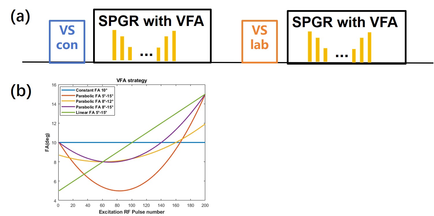
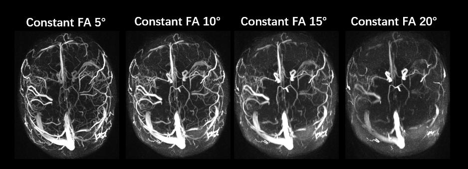
Flip angle design: As shown in Figure 1(a), the VSASL MRA consists of a VS saturation preparation followed by a 3D spoiled gradient echo (SPGR) readout. We first compared the images acquired with different constant FAs. According to the results shown in Figure 2, better visualization of the vessels was obtained when the flip angle was 10°. Therefore, a constant flip angle of 10° was used as the baseline. Then four VFA strategies were designed, three of which have a parabolic relationship between FA and the number of RF pulses (the FA range was 5-15°,8-12° and 8-15°, respectively) and the fourth has a linear relationship (FA range: 5-15°). The specific VFA profiles are shown in Figure 1(b).In vivo experiment: Ethical approval was obtained for the study. Two healthy volunteers were recruited. All experiments were performed on a Siemens Prisma 3T MRI scanner with a 32-channel head coil. Imaging parameters of the VSASL MRA sequence were: BIR-8 VS preparation was used, FOV = 200×200×102 mm3; TR/TE = 5.20/1.98 ms; voxel size = 0.76×0.76×0.8 mm3; lines per shot = 200; VS cutoff velocity = 3.11 cm/s; shot length = 1500 ms; total scan time = 4 min 18 s.
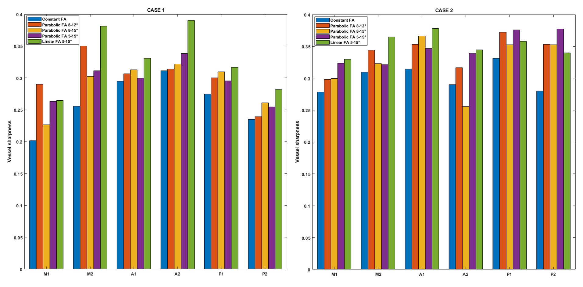
Image analysis: The visual comparison of VSASL MRA images with different FA strategies was performed. In addition, we measured the sharpness of vessel edges on the MIP images. Specifically, a line was drawn across the vessel to obtain an edge spread function of intensity, and vessel sharpness was defined as the maximum gradient value of the edge spread function3. The sharpness of the middle cerebral artery (MCA, M1 and M2), anterior cerebral artery (ACA, A1 and A2), and posterior cerebral artery (PCA, P1 and P2) was measured.
Results and Discussion
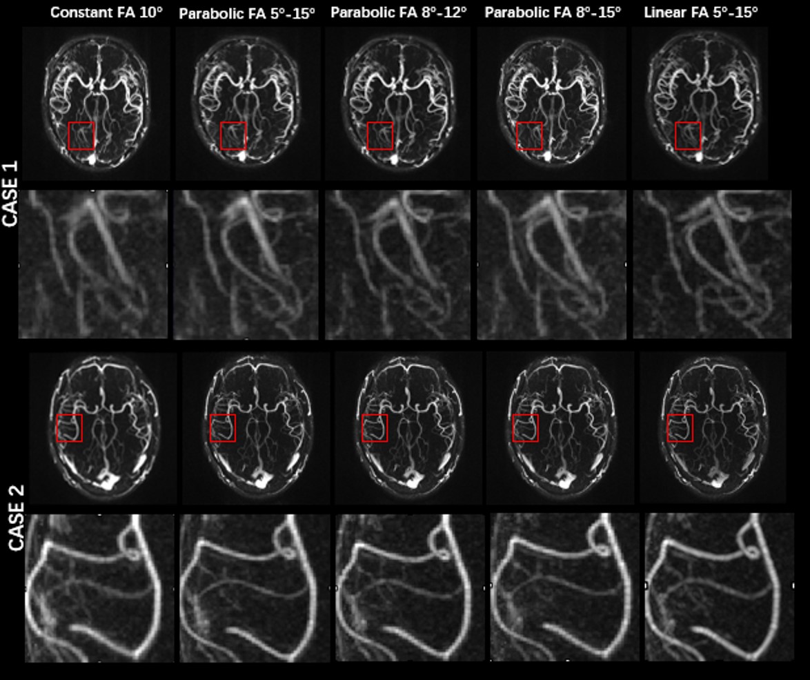
As shown in Figure 3, compared to the constant FA, VFA can reduce the blurring of the small vessels. The sharpness was higher for VFA than the constant FA (Figure 4), suggesting the effectiveness of the VFA strategy in reducing the blurring and improving the conspicuity of distal small vessels for VSASL MRA. However, it should be noted that the current VFA design is heuristic, and the optimal VFA strategy is yet to be developed, which probably needs to take the imaging parameters and tissue properties (e.g. T1, blood velocity) into account. Besides, further validation on more subjects, including patients, is needed.Conclusion
We demonstrated that the VFA strategy could reduce vessel blurring and improve the conspicuity of distal small vessels for VSASL MRA. Further development of the method to find an optimal VFA strategy is needed.Acknowledgements
This work was supported by Natural Science Foundation of Shanghai (22ZR1403900).References
1. Qin Q, et al. Velocity-selective magnetization-prepared non-contrast-enhanced cerebral MR angiography at 3 Tesla: Improved immunity to B0/B1 inhomogeneity. Magn Reson Med. 2016 Mar;75(3):1232-41.
2. Zhao L, et al. Controlling T2 blurring in 3D RARE arterial spin labeling acquisition through optimal combination of variable flip angles and k-space filtering. Magn Reson Med. 2018 Oct;80(4):1391-1401.
3. Fok WYR, et al. Accelerated free-breathing 3D whole-heart magnetic resonance angiography with a radial phyllotaxis trajectory, compressed sensing, and curvelet transform. Magn Reson Imaging. 2021 Nov;83:57-67.
Figures