4421
Correlations between cognitive reserve, brain and cerebrospinal fluid volume in mild cognitive impairment patients1lanzhou university second hospital, lanzhou, China
Synopsis
Keywords: fMRI Analysis, Alzheimer's Disease, cognitive reserve, brain volume, cognitive function, cerebrospinal fluid, mild cognitive impairment
Motivation: Cognitive reserve (CR), which can preserve cognitive function despite underlying neuropathology, explains at least some variability in cognitive performance.
Goal(s): To explore the effect of CR on brain and CSF volume in patients with MCI and healthy elders (HC).
Approach: The study population included 31 HC and 50 MCI patients were collected in this study to obtain high-resolution 3D-T1 structure images, cognitive function and composite CR scores.
Results: The results showed that cognitive reserve was significantly associated with cognitive performance. Some brain and CSF volumes correlate with cognitive reserve. Cognitive reserve affects cognitive performance by reducing the volume of the right parahippocampal gyrus.
Impact: Our study provides evidence that CR protects neurocognitive function in the preclinical stages of AD, particularly in the right parahippocampal gyrus, through mechanisms that modulate brain and CSF volume.
Introduction
It is widely believed that improving the flexibility and adaptability of brain networks through education and regular cognitive engagement, termed cognitive reserve (CR), can help slow age- and disease-related brain changes [1,2]. CR can preserve cognitive function despite underlying neuropathology, explains at least some variability in cognitive performance, has been claimed as a factor mitigating the clinical manifestations of Alzheimer disease (AD)[3].CR partly explains cognitive variability in the presence of pathological brain aging. Compared with low cognitive reserve(LCR), high cognitive reserve(HCR)have a later onset of mild cognitive impairment (MCI) and Alzheimer disease symptoms and greater neuropathology at similar cognitive and clinical levels. The present study follows a previous report showing the faster conversion from MCI to Alzheimer disease for LCR patients than comparable HCR, as predicted by a CR.Our study to explore the effect of CR on brain and cerebrospinal fluid (CSF)volume in patients with MCI and healthy controls(HC)[4].Methods
Our study population included 31 HC and 50 MCI patients,which were collected in this study to obtain high-resolution 3D-T1 structure images, cognitive function and composite CR scores. Educational attainment, leisure time and working activity ratings from two groups were used to generate cognitive reserve index questionnaire (CRIq)scores. The different volumes of brain regions and CSF were obtained using uAI research portal in both groups, which were then taken as the regions of interest (ROI), the correlation analyses between ROIs and CRIq scores were conducted. Lastly,The mediating analysis between brain and cerebrospinal fluid volume and cognitive performance and cognitive reserve was carried out to observe whether the brain and cerebrospinal fluid volume mediated the relationship between cognitive reserve and cognitive performance.Results
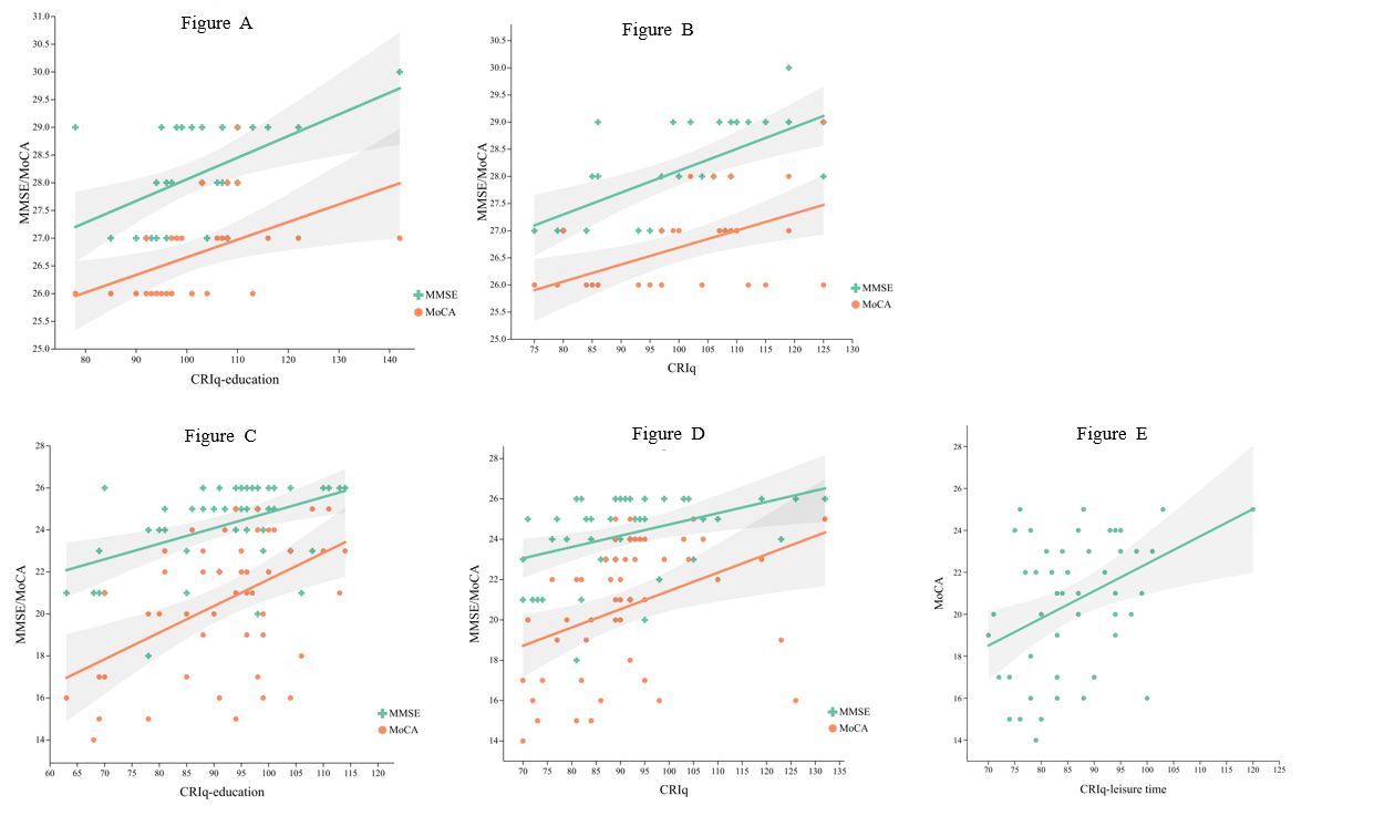
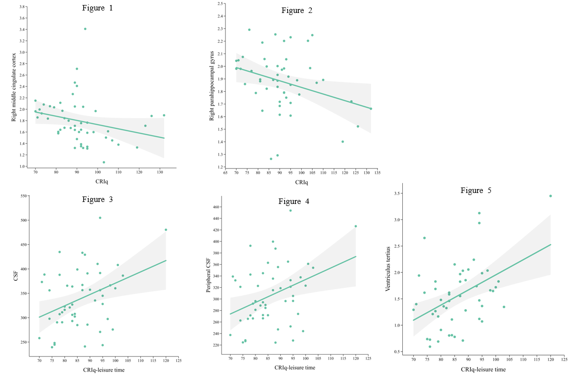
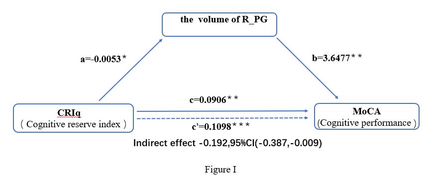
The scores of CRIq, CRIq-leisure time, and CRIq-education in NC group were significantly higher than patients in MCI group, and the MoCA and MMSE scores were positively correlated with the CRIq, CRIq-education in both groups, and were positively correlated with CRIq-leisure time in MCI group and CRIq-working activity in NC group respectively. In MCI group, the volume of the right middle cingulate cortex and the right parahippocampal gyrus were negatively correlated with the CRIq and CRIq-working activity scores, and the volume of the CSF, peripheral CSF and ventriculus tertius were positively correlated with the CRIq- leisure time score. Using MoCA score as the dependent variable, CRIq as the independent variable, and the volume of R_PG as the moderating variable, we found that the relationship between CRIq and cognitive performance was partially mediated by changes in the volume of the right parahippocampal gyrus in MCI.Discussion
CR protects against cognitive decline in both healthy aging and dementia. Previous research[5] has shown that HCR may contribute to CR by demonstrating later onset of AD symptoms and greater neuropathology for HCR patients despite having a similar cognitive function as their LCR peers. This pattern has also been reported during the first stages of neurodegeneration for MCI. This prodromal phase is crucial to investigate CR factors because it is at this stage when future dementia can be predicted. According to the Stern CR model[6], patients with increased CR should convert faster to dementia, a point recently demonstrated for some CR studies. However, no brain data were reported in that paper so the potential reasons for earlier conversion from MCI to AD for the HCR patients remained speculative.Using uAI to calculate brain and cerebrospinal fluid volume, We correlated the results with cognitive reserve.Then, the volume rresults with a difference was used as a mediator variable ,MoCA score as the dependent variable, CRIq as the independent variable, that conducted mediation analysis. It was concluded that the relationship between CRIq and cognitive performance was partially mediated by changes in the volume of the right parahippocampal gyrus in MCI. For patients with mild cognitive impairment, cognitive reserve affects cognitive performance by reducing the volume of the right right parahippocampal gyrus, suggesting that part of the brain gray matter volume or cerebrospinal fluid volume may be a potential neural mechanism in the pathophysiology of cognitive reserve. Therefore, It is promising to explain reasons for earlier conversion from MCI to AD for the HCR patients.
Conclusion
People with higher CR show better levels of cognitive function, and MCI patients with higher CR showed more severe volume atrophy of the right middle cingulate cortex and the right parahippocampal gyrus, but more CSF at a given level of global cognition. For patients with mild cognitive impairment, cognitive reserve affects cognitive performance by reducing the volume of the right right parahippocampal gyrus.Acknowledgements
We would like to thank our patients for their role in the research design.References
[1] STERN Y. Cognitive reserve in ageing and Alzheimer's disease [J]. Lancet Neurol, 2012, 11(11): 1006-12.
[2] STERN Y, BARNES C A, GRADY C, et al. Brain reserve, cognitive reserve, compensation, and maintenance: operationalization, validity, and mechanisms of cognitive resilience [J]. Neurobiol Aging, 2019, 83: 124-9.
[3] BRENNER E K, THOMAS K R, WEIGAND A J, et al. Cognitive reserve moderates the association between cerebral blood flow and language performance in older adults with mild cognitive impairment [J]. Neurobiology of Aging, 2023, 125: 83-9.
[4] ZHU W, GAO Z, LI H, et al. Education reduces cognitive dysfunction in Alzheimer’s disease by changing regional cerebral perfusion: An in-vivo arterial spin labeling study [J]. Neurological Sciences, 2023, 44(7): 2349-61.
[5] CALVO N, ANDERSON J A E, BERKES M, et al. Gray Matter Volume as Evidence for Cognitive Reserve in Bilinguals With Mild Cognitive Impairment [J]. Alzheimer Dis Assoc Disord, 2023, 37(1): 7-12.
[6] STERN Y. Cognitive reserve and Alzheimer disease [J]. Alzheimer Dis Assoc Disord, 2006, 20(3 Suppl 2): S69-74.
Figures