4418
Evolution of resting-state network efficiency after stroke: an individual confrontation with the norm.1I2FH, CHU Montpellier, Montpellier, France, 2Neuroradiology, CHU Montpellier, Montpellier, France, 3Charles Coulomb Laboratory, UM Montpellier, Montpellier, France, 4Physical Rehabilitation Medicine, CHU Montpellier, Montpellier, France, 5Euromov DHM, UM Montpellier, Montpellier, France
Synopsis
Keywords: Functional Connectivity, fMRI (resting state), rehabilitation
Motivation: To provide personalized rehabilitation after stroke, we need to identify brain biomarkers that inform us about what differs from a normal organization to target rehabilitation accordingly.
Goal(s): Evaluate the potential of a norm-based functional brain network organization analysis in the individual follow-up post-stroke.
Approach: Compare fMRI resting-state network functioning of 21 people post-stroke before and after rehabilitation with the norm, based on 569 controls, while taking into account the motor deficit.
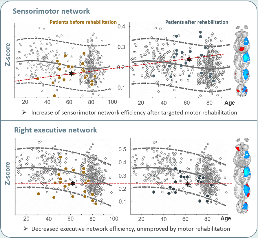
Results: Using a norm, we showed that targeted motor rehabilitation improves the motor network efficiency for recovering patients, whereas executive network efficiency remained suboptimal, potentially negatively interfering with motor recovery.
Impact: Comparing people with a norm, not only post-stroke but also with other central neurological deficits, facilitates personalized medicine, for instance by providing targets for non-invasive brain stimulation, or by identifying processes that require specific training, like attention direction or proprioception.
Introduction
"Personalized rehabilitation", in which therapy is adapted to the specific needs of each patient, has become one of the key-points of rehabilitation medicine. This has driven research towards identification of physiological biomarkers. That is, markers that inform us on a state that differs from 'the norm' and thus requires intervention. But what is normal? Especially when talking about the brain, this is not easily quantified. However, when at rest, anatomical distinct brain regions show correlated spontaneous activity over time, and these correlations are rather consistent across healthy subjects1, although impacted amongst others by age and education levels. Still, the analysis of resting-state networks contributed to unrevealing brain functioning in healthy subjects over lifespan, as well as to studying changes in case of pathology. Post-stroke, various changes in connectivity patterns have been described. For instance, a general reduction in functional connectivity between the prefrontal cortex and the cerebellum has been described2, whereas the connectivity between regions of the sensorimotor network seems to be strengthened by rehabilitation3,4. Here we argue that changes in functional network efficiency, not only of the sensorimotor network, but also of higher order networks involving attention direction after stroke and over rehabilitation may be related with the amount of functional motor recovery. We therefore aim to identify changes in individual brain network efficiency over the first months after stroke in relation with the amount of motor recovery.Methods
Twenty-one participants with a first-ever unilateral supra-tectorial ischemic stroke underwent two resting-state fMRI sessions before and after six weeks of standardized upper-limb rehabilitation in the first weeks after the stroke. All patients showed severe motor deficit at onset (Fugl-Meyer-Assessment score <30/66). Rehabilitation focused mainly on motor recovery, yet with a cognitive component). The global efficiency of each resting-state network (Greicius-atlas5), was projected for the individual patients against a norm-based efficiency extracted from 569 healthy volunteers, between 10 to 100 years. The norm was calculated following Altman’s age-related reference interval6. Network efficiency was defined following the graph theory as the average of the shortest path length between each node of a network with all the other nodes. Two nodes were esteemed being connected when their spontaneous activity fluctuations were significantly correlated over time (p <0.05, corrected for multiple comparisons).Results/Discussion
It was found that the overall efficiency of the sensorimotor network was rather robust. Each patient's global network organization, even when unable to move the upper-limp voluntarily, remained predominantly within the norm. Interestingly an overall significant shift of the barycenter (position of the median efficiency for all patients against the norm) was observed after rehabilitation, in which the efficiency of the sensorimotor network improved from below median to above the median at rest (see figure.1). At the individual level, no direct link between the change in Fugl-Meyer and the change in motor recovery could be observed. We suggest that a more fine-grained analysis of movement characteristics by kinematics might provide further insight. Secondly, we observed that patients showed an overall lower global efficiency of the executive network that did not change over time. Would a change have been possible with more targeted cognitive rehabilitation? Also at the individual level, the efficiency of the right executive level even decreased for some patients. Its significance requires further explorations, but it might interfere negatively with motor recovery.Conclusion
Projecting an individual against the norm facilitates the identification of changes in brain network organization, identifying beneficial and maladaptive plasticity. With quantitative measures of performance, we should be able to identify a patient profile, that allows the identification of individual brain-biomarkers of network organization for a personalized rehabilitation strategy.Acknowledgements
No acknowledgement found.References
- Yeo BTT, Krienen FM, Sepulcre J, et al. The organization of the human cerebral cortex estimated by intrinsic functional connectivity. J Neurophysiol. 2011, 106:1125-65.
- Kalinosky BT, Berrios Barillas R, Schmit BD. Structurofunctional resting-state networks correlate with motor function in chronic stroke. Neuroimage Clin, 2017 16:610-623.
- Pirovano I, Mastropietro A, Antonacci Y, et al. Resting State EEG Directed Functional Connectivity Unveils Changes in Motor Network Organization in Subacute Stroke Patients After Rehabilitation. Front Physiol. 2022, 13:862207.
- Du J, Yao W, Li J et al. Motor Network Reorganization After Repetitive Transcranial Magnetic Stimulation in Early Stroke Patients: A Resting State fMRI Study. Neurorehabil Neural Repair, 2022 36:61-68.
- Shirer, Ryali, Rykhlevskaia, et al. Decoding subject-driven cognitive states with whole brain connectivity patterns, Cereb Cortex 2012, 22:158-65.
- Altman DG. « Construction of age-related reference centiles using absolute residuals », doi: 10.1002/sim.4780121003.
Figures