4416
Altered task-residual effective connectivity of motor and memory network in transient ischemic attack1Research Center for Bioconvergence Analysis, Korea Basic Science Institute, Cheongju, Korea, Republic of, 2Graduate School of Analytical Science and Technology, Chungnam National University, Daejeon, Korea, Republic of, 3Department of Rehabilitation Medicine, Chungnam National University Sejong Hospital, Sejong, Korea, Republic of, 4Bio-Chemical Analysis Team, Korea Basic Science Institute, Cheongju, Korea, Republic of, 5Department of Rehabilitation Medicine, Chungnam National University Hospital, Daejeon, Korea, Republic of, 6Department of Neurology, Chungnam National University Sejong Hospital, Sejong, Korea, Republic of
Synopsis
Keywords: Functional Connectivity, fMRI, Effective Connectivity
Motivation: Little is known about underlying brain mechanisms that contribute to heightened risk of stroke and impairments in cognitive and motor functions of patients with transient ischemic attack (TIA).
Goal(s): Our aim was to investigate abnormal effective connectivity in TIA during motor and working memory tasks.
Approach: Spectral dynamic causal modelling with 7T fMRI was used to estimate the task-residual effective connectivity elicited during fist-closing and n-back tasks.
Results: Patients with TIA showed increased effective connectivity toward the ipsilateral M1 and reduced connectivity to the SMA and PMC durn motor task, as well as increased connectivity among the PAR and CC during n-back tasks.
Impact: The findings of aberrant task-residual effective connectivity within the motor and working memory networks in patients with TIA indicate potential decreased neural efficiency and disrupted control of motor and working memory functions, contributing to the physiological alterations in these individuals.
Introduction
Transient ischemic attack (TIA) is a temporary episode of neurological dysfunction resulting from focal brain ischemia. Although TIA symptoms are transient, patients with TIA have a high risk of stroke and persistent impairments in various motor and cognitive functions [1]. This study aims to explore the changes in task-residual effective connectivity of TIA during motor and working memory tasks. Additionally, this study seeks to associate the differences in connectivity between TIA patients and healthy controls with the risk of stroke.Methods
This study involved 15 patients with TIA and 28 age-matched healthy subjects (HC). The subjects underwent scanning using a 7T MRI system while performing motor and working memory experiments. During the motor task, the subjects were instructed to perform right or left fist-closing movements synchronized with a 1Hz visual cue. In the working memory assessment, subjects performed the n-back task, involving 0-, 1-, and 2-back conditions. For the analysis methods, we applied spectral dynamic causal modeling (DCM) [2] to task-residual BOLD time series, to estimate effective connectivity among regions of interest (ROI). Specifically, based on the general linear model (GLM) results across all participants, ROIs for motor task included the primary motor cortex (M1), premotor cortex (PMC), supplementary motor area (SMA), and inferior parietal lobule (IPL). The ROIs for the n-back task comprised various regions such as the prefrontal cortex (PFC), parietal cortex (PAR), and cingulate cortex (CC). The task-residual time series within the ROIs were obtained by regressing out the task-related signal and systemic confounds from the fMRI signal. The parameters of effective connectivity were then estimated from the task-residual time series using spectral DCM.Results
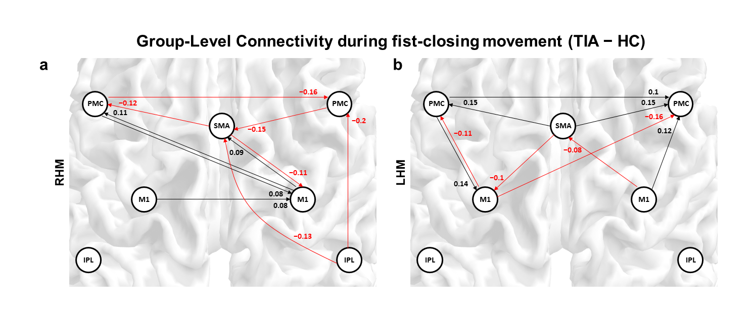
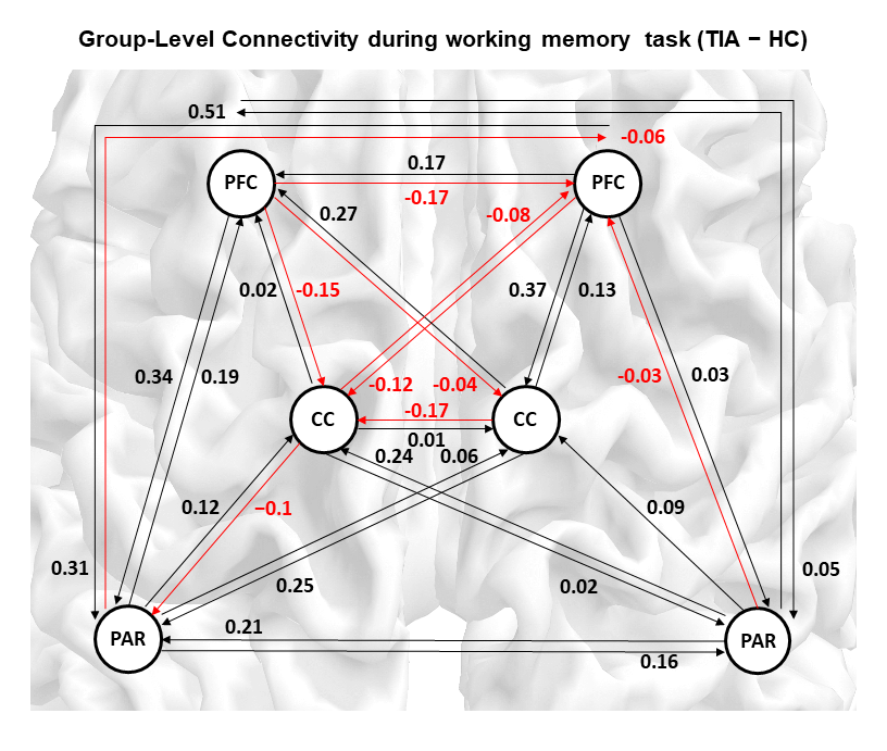
Compared to healthy controls, stronger excitatory connections from the left PMC to the ipsilateral M1 were observed in the patients with TIA, during both right-hand movement (from the left PMC to the right M1: 0.08) and left-hand movement (from the left PMC to the left M1: 0.14). Moreover, during right-hand movement, patients with TIA exhibited a stronger excitatory influence from the contralateral M1 to the ipsilateral M1 (0.08), compared to the HC. Regarding the SMA region, the effective connectivity to the SMA was decreased in the patients with TIA, compared with the HC during both right-hand movement (from the right PMC to the SMA: -0.15, the right IPL to the SMA: -0.13) and left-hand movement (the right M1 to the SMA: -0.08). Regarding the PMC regions, the patients with TIA had higher suppressive influence to the PMC during right hand movement than healthy controls. These results were shown in Figure 1. During the n-back task, patients with TIA exhibited higher connectivity strength compared to the HC group, as shown in Figure 2. Specifically, patients with TIA consistently showed increased effective connectivity among the ROIs of the PAR and CC in both hemispheres, in contrast to the HC group.Discussion and Conclusion
This study showed that while the patients with TIA performed fist-closing movements according to the visual stimuli of flickering circles (1 Hz), the patients with TIA had greater connections to the ipsilateral M1 and lower connections to the SMA and PMC than the healthy controls [3]. These experimental results may reflect the potential disruptions in the information processing (visual cue to action) and controls of voluntary movement, and decreased efficiency of primary motor function M1 in patients with TIA. We also found that patients with TIA had higher excitatory connections between the PAR and CC during working memory task, compared with HC. It has been shown that the PAR is involved in the storing and retrieving information, and the CC plays a role in performance adjustment during working memory task. Therefore, although further investigation may be required, this result suggests that the patients with TIA would have increased recruitment of the interaction between the temporary storage and manipulation of remembered information for supporting the short-term memory function. Previous TIA studies have shown changes in the brain activity as well as decreased resting-state connectivity in patients with TIA [4]. Our studies added findings to this field by revealing the disruptions in the task-residual effective connectivity of the motor and working memory network in TIA.Acknowledgements
This work was supported by a grant of National Research Foundation of Korea (NRF) grant funded by the Korean Government (MSIT) (2019R1C1C1011281, 2022R1F1A1074729), and grants from the Korea Basic Science Institute (C318300, D300500, C300300).References
1. Coull, A., Lovett, J.K. and Rothwell, P.M., 2004. Population based study of early risk of stroke after transient ischaemic attack or minor stroke: implications for public education and organisation of services. BMJ, 328 (7435), p.326.
2. Razi, A., Kahan, J., Rees, G. and Friston, K.J., 2015. Construct validation of a DCM for resting state fMRI. Neuroimage, 106, pp.1-14.
3. Chu, T., Lee, S., Jung, I.Y., Song, Y., Kim, H.A., Shin, J.W. and Tak, S., 2023. Task-residual effective connectivity of motor network in transient ischemic attack. Communications Biology, 6(1), p.843.
4. Li, R., Wang, S., Zhu, L., Guo, J., Zeng, L., Gong, Q., He, L. and Chen, H., 2013. Aberrant functional connectivity of resting state networks in transient ischemic attack. PloS one, 8(8), p.e71009.
Figures