4414
Comparative analysis of brain network connectivity changes in Alzheimer's and Parkinson's disease with mild cognitive impairment: an ICA study1The first hospital of China Medical University, Shenyang, China, 2MR Research Collaboration,Siemens Healthineers,Beijing China, Beijing, China
Synopsis
Keywords: fMRI Analysis, Alzheimer's Disease, Parkinson's Disease fMRI
Motivation: The pathogenesis of cognitive dysfunction may be different, particularly in terms of macroscopic manifestations within neural transmission pathways, which still require further exploration.
Goal(s): The aim of this study was to quantify changes in fMRI intra and inter-network connectivity in AD-MCI and PD-MCI using independent component analysis (ICA).
Approach: Nine RSNs were identified in 33 AD-MCI, 55 PD-MCI and 34 HCs using ICA method, and further their intra-and inter-network FC were compared in three groups.
Results: The results showed that the brain network changes in AD-MCI patients and PD-MCI patients were mainly concentrated in different brain network.
Impact: By investigating the pathways and compensatory mechanisms underlying cognitive dysfunction arising from different etiologies, we aim to explore potential imaging-based diagnostic approaches and therapeutic strategies for clinical treatment strategy, which is enhancing the prognosis and quality of life for patients.
Introduction
Cognitive dysfunction is a common observed in various diseases, with Alzheimer's disease (AD) being characterized by cognitive impairment as the primary symptom. While in diseases such as Parkinson's disease (PD), cognitive dysfunction is a concomitant symptom. Mild cognitive impairment (MCI) serves as an intermediary phase between normal aging and the onset of dementia, presenting a pivotal opportunity for clinical intervention. By exploring the distinctive changes in AD and PD during the early phases of cognitive decline, this study explores to the specific network-level modifications associated with cognitive dysfunction in both diseases.Methods
33 AD-MCIs, 55 PD-MCIs and 34 health controls (HC) were enrolled in this study. All participants underwent a comprehensive assessment, including Mini-Mental State Examination (MMSE) and Montreal Cognitive Assessment (MoCA) for global cognitive function. 33 AD-MCI patients, 55 PD-MCI patients were observed by experienced neurologists during hospital visits, self-reported by patients, or described by their caregivers. All participants underwent MRI scanning on a 3T system (MAGNETOM Verio, Siemens Healthineers, Erlangen, Germany). The MRI examinations included a 3D T1WI magnetization-prepared rapid gradient-echo (MPRAGE) sequence and echo planner imaging (EPI) functional images. The acquired T1WI MPRAGE sequence and functional data were preprocessed using the Gretna toolbox based on Statistical Parametric Mapping (SPM, version12, http://www.fil.ion.ucl.ac.uk/spm) in MATLAB software (version2018a, MathWorks). Then spatial group independent component analysis was conducted for all 122 participants using the GIFT4.0 (Group ICA of fMRI Toolbox, GIFT). Next, to explore changes in functional connectivity at the network level associated with cognitive dysfunction, differences in in-network and inter-network functional connectivity among the three groups were compared using one-way analysis of variance (ANOVA).Results
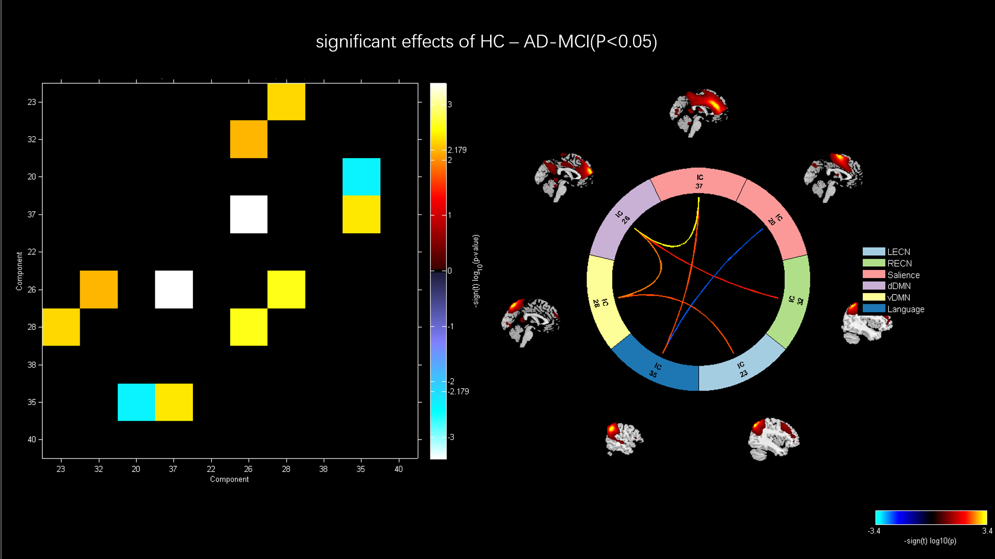
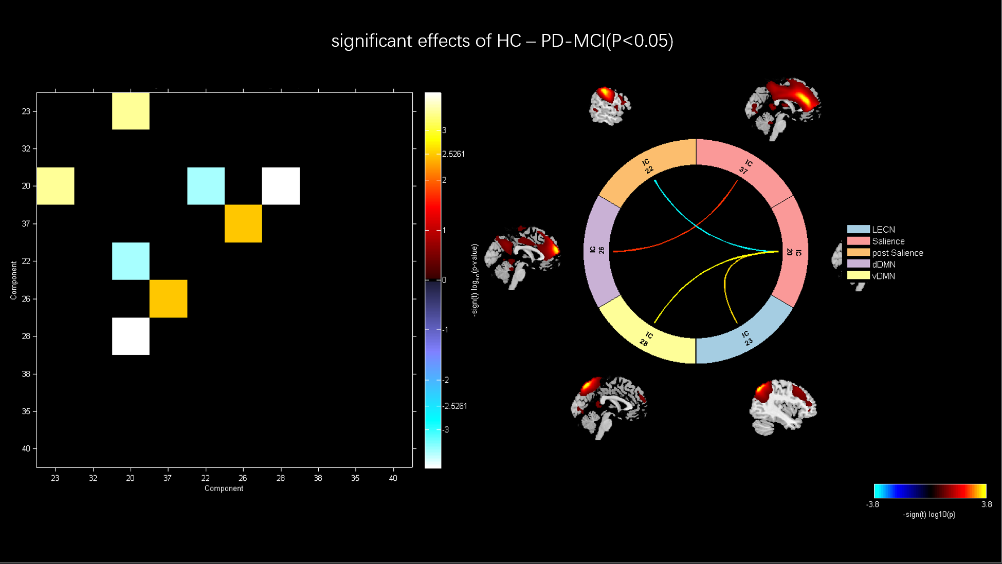
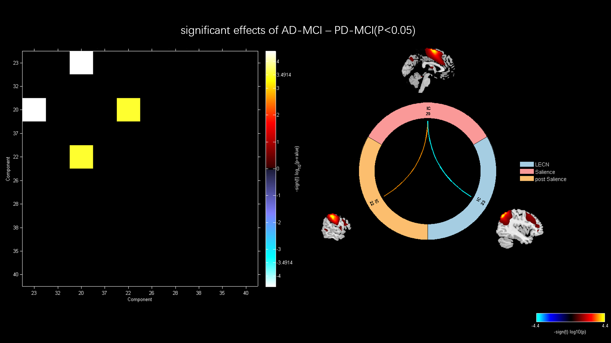
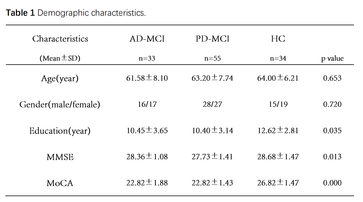
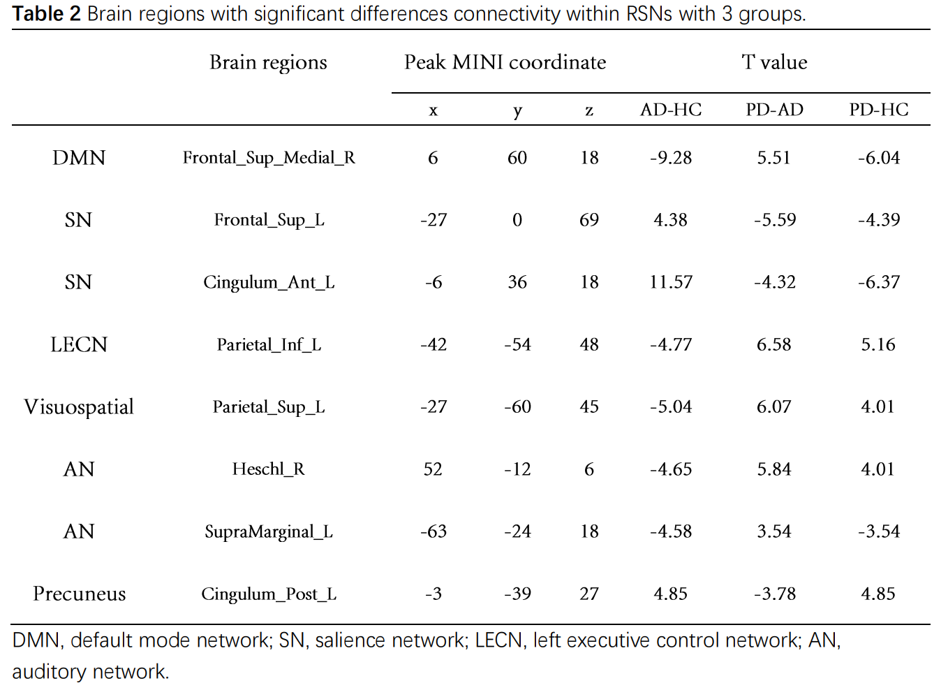
Demographic and clinical characteristics No significant differences were detected in age and gender (P = 0.653, P = 0.720) with the AD-MCI, PD-MCI and HC groups. Compared with HCs, AD-MCI and PD-MCI had lower educational attainment and showed lower MoCA scores, indicating more severe cognitive disability in the patients. The demographic and neuropsychological data from this study are shown in Table 1. Internetwork functional connectivity changes through three groups In order to further obtain the differences between groups, the network comparisons were carried out. Compared with HCs, AD-MCI group shows increased connection between salience network (SN) and language network, decreased connections between dorsal default mode network (DMN) with RECN, dDMN with SN, dDMN with vDMN, vDMN with LECN and SN with Language. Compared with HCs, PD-MCI group shows increased connection between SN and pSN, decreased connections between SN with dDMN, SN with vDMN and SN with LECN. Compared with AD-MCI group, PD-MCI group shows increased connection between SN with pSN, decreased connections between SN with LECN (p < .05, FDR corrected). Intra-network functional connectivity changes through three groups. Compared HC with AD-MCI Compared to HC, AD-MCI shows decreased FC in right frontal superior medial gyrus and right precuneus within dDMN, left parietal inferior gyrus within LECN, left parietal superior gyrus within visuospatial network, increased FC in left frontal superior and left cingulum anterior in SN, left cingulum post in Precuneus (p < .05, FDR corrected). Compared HC with PD-MCI Compared to HC, PD-MC shows decreased FC in left frontal superior and left cingulum anterior in SN, right frontal superior medial gyrus and right precuneus within dDMN, increased FC in left parietal inferior gyrus within LECN, left cingulum post in Precuneus (p < .05, FDR corrected). Compared PD-MCI with AD-MCI Compared to AD-MCI, PD-MCI shows decreased FC in left frontal superior and left cingulum anterior in SN, left cingulum post in Precuneus, increased FC in left parietal inferior gyrus within LECN, right frontal superior medial gyrus within dDMN, left parietal superior gyrus within visuospatial network (p < .05, FDR corrected).Discussion
The results showed that the brain network changes in AD-MCI were mainly concentrated in the DMN, while the changes in PD-MCI were mainly concentrated in the SN. Compared with AD-MCI, the inter-network connections in PD-MCI are lower, and the intra-network functional connections are higher,so cognitive dysfunction in PD patients may begin with the disturbance of functional connections between networks. On the other hand, increased intra-network functional connectivity may also be a compensatory mechanism for decreased inter-network functional connectivity in both diseases.Conclusion
This study explains the different or similar changes of the two diseases in the process of cognitive dysfunction from the perspective of brain networks. It provides an important neural network basis for a deeper understanding of the pathophysiological mechanisms of these two diseases in the early stage of cognitive dysfunction. The findings are expected to provide additional insights into future diagnostic and treatment strategies.Acknowledgements
No acknowledgement found.References
No reference found.Figures