4408
Gray matter morphological abnormities are constrained by normal structural covariance network in OCD1the First Affiliated Hospital of Zhengzhou University, Zhengzhou, China
Synopsis
Keywords: fMRI Analysis, Psychiatric Disorders
Motivation: Whether abnormal gray matter morphology is constrained by normal brain network architecture in in obsessive-compulsive disorder (OCD) remains unknown.
Goal(s): We investigated the association between gray matter morphological abnormities and normal structural covariance network architecture.
Approach: Ninety-eight first-episode and drag-naive patients with OCD and matched healthy controls (HCs) were included in this study.
Results: Gray matter abnormities are constrained by structural connectome and provide new insights into the possible pathological progression in OCD.
Impact: We investigated the association between gray matter morphological abnormities and normal structural covariance network architecture in OCD. We found that the structural abnormalities are constrained by structural covariance connectome of multiple disease epicenters in OCD.
Background and purpose
Brain regions are not isolated but form a highly interconnected network or ‘connectome’, that enables efficient communication among brain regions to support diverse behavioral and cognitive functions[1]. Recent studies have demonstrated that pathological propagation of brain disorders is also constrained by connectome architecture[2,3]. Accordingly, modern neuroimaging studies reveal that pathological perturbations begin with focal “epicenters” whose connectome architecture resemble the patterns of brain tissue volume loss and then propagate to connected unaffected brain regions[2-5]. Recently, these findings are found to held true in psychiatric disorders. Further evidence points that structural and functional connectome profiles of the anterior cingulate cortex account for 25% to 35% variance of regional deformation, establishing that gray matter volume atrophy is conditioned by connectome topology[6]. These findings provide clues about how pathological perturbations propagate from the initial disease epicenters to other affected brain regions, deepening our understanding of widespread pathophysiological effects and facilitating targeted treatment in these diseases. However, whether structural brain abnormalities are also associated with normal connectome topology remains unknown in OCD. In this study, we aimed to investigate the association between normal connectome architecture and gray matter volume abnormalities in OCD.Methods
Ninety-eight first-episode and drag-naive patients with OCD and matched healthy controls (HCs) were included in this study. First, gray matte volume (GMV) was measured with voxel based morphometry analysis (VBM)[7] and compared between patients with OCD and HCs. Then, we identified the putative disease epicenter(s) using a backfoward stepwise regression analysis. According to previous studies, we had two main hypotheses: First, the connectome profiles of the putative disease epicenter(s) could significantly explain observed gray matter volume differences in OCD. Second, brain regions with stronger connections with the identified disease epicenter(s) would show greater vulnerability defined by atrophy severity to disease.Results
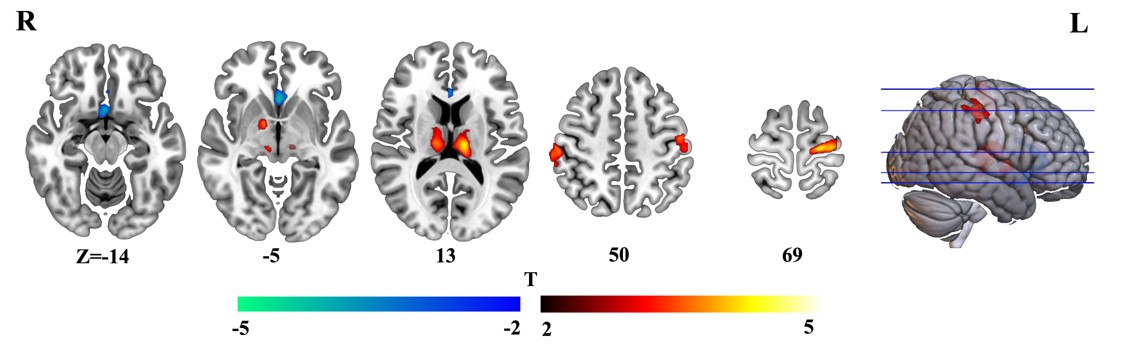
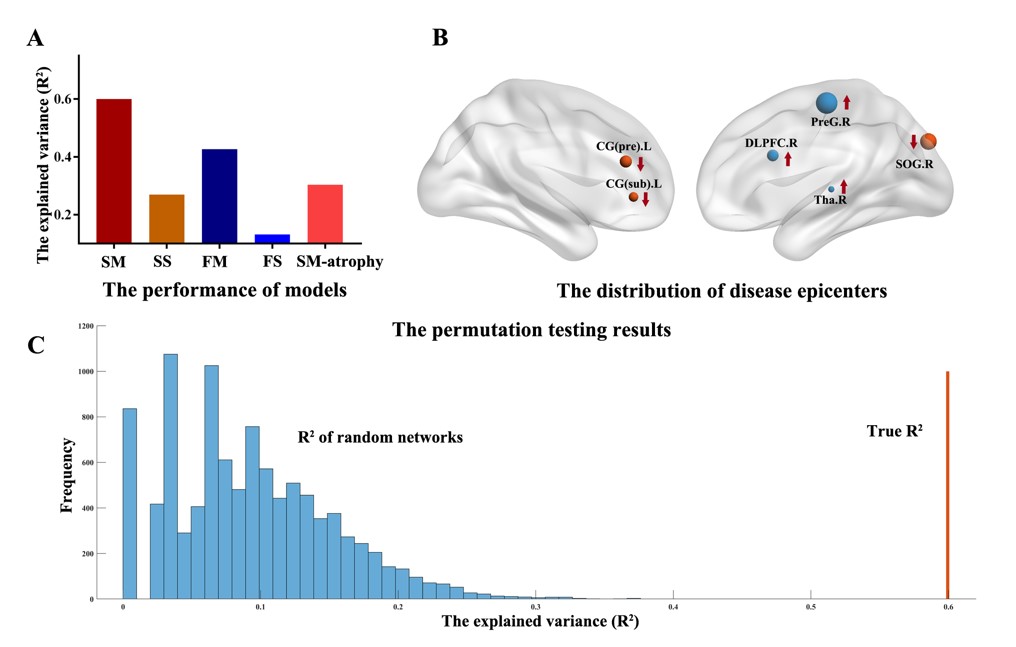
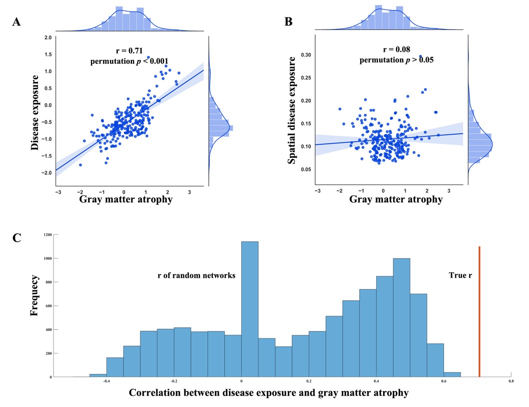
Patients with OCD exhibited significant gray matter atrophy in bilateral anterior cingulate while exhibited increased gray matter volume in the bilateral thalamus, striatum and precentral and postcentral gyrus. The details were included in Figure 1 and Table 1. To investigate whether gray matter atrophy pattern was recapitulated by normal connectome architecture, we built the single-epicenter and multiple-epicenter model based on structural and functional network respectively (in all 4 model). The structural single-epicenter model explained 26.92%, the structural multi-epicenter model explained 59.94%, the functional multi-epicenter model explained 42.61% and the functional single-epicenter model explained 13.09% of the observed gray matter abnormalities (Figure 2A). The structural multiple-epicenter model performed best and significantly explained gray matter atrophy pattern in OCD than chance (Figure 2C). Using structural multiple-epicenter model, we identified a set of disease epicenters, such as the left anterior cingulate gyrus, right dorsal lateral prefrontal cortex (DLPFC) and right thalamus with their corresponding regression coefficients in the model were significantly not zero (Figure 2B). Structural multi-epicenter model based on the identified disease epicenters performed better than that based on most atrophied brain regions (Figure 2A).The disease exposure significantly correlated with gray matter atrophy pattern in OCD (Figure 3A and Figure 3C). To further rule out the effect of spatial proximity, we also defined the ‘spatial disease exposure’ and calculated its correlation with gray matter atrophy pattern. There was no significant correlation between ‘spatial disease exposure’ values and regional gray matter atrophy in OCD (Figure 3B).Discussion
In this study, we investigated the association between the normal connectome architecture between gray matter volume abnormalities in OCD for the first time. In previous studies, the authors just hypothesize that these is only one disease epicenter. However, if this is the truth in OCD is not unclear. At the same time, both structural covariance network and functional network are found to underlay the structural abnormalities in schizophrenia, which one better recapitulates structural abnormalities in OCD is also unclear. In this study, we found that the structural abnormalities could be better explained by the structural multi-epicenter model, suggesting structural abnormalities are constrained by structural covariance connectome of multiple disease epicenters in OCD.Conclusion
In summary, first, the gray matter atrophy could be significantly explained by structural covariance network of the putative disease epicenters. Second, brain regions with stronger structural covariance connections with the identified disease epicenters presented greater vulnerability to OCD.Acknowledgements
This research study was supported by the Natural Science Foundation of China (81601467, 81871327, 62106229) and Medical science and technology research project of Henan province (201701011, SBGJ202102103, SBGJ202101013).References
1. Bullmore, E. and O. Sporns, Complex brain networks: graph theoretical analysis of structural and functional systems. Nat Rev Neurosci, 2009. 10(3): p. 186-98.2. Zhou, J., et al., Predicting regional neurodegeneration from the healthy brain functional connectome. Neuron, 2012. 73(6): p. 1216-27.3. Yau, Y., et al., Network connectivity determines cortical thinning in early Parkinson's disease progression. 2018. 9(1): p. 12.4. Zeighami, Y., et al., Network structure of brain atrophy in de novo Parkinson's disease. 2015. 4.5. Brown, J.A., et al., Patient-Tailored, Connectivity-Based Forecasts of Spreading Brain Atrophy. Neuron, 2019. 104(5): p. 856-868.e5.6. Shafiei, G., et al., Spatial Patterning of Tissue Volume Loss in Schizophrenia Reflects Brain Network Architecture. Biol Psychiatry, 2020. 87(8): p. 727-735.7. Ashburner, J. and K.J. Friston, Voxel-based morphometry--the methods. Neuroimage, 2000. 11(6 Pt 1): p. 805-21.Figures