4407
Selective Vulnerability of the Brain Regions in Neurodegenerative Disorders1Biomedical Engineering, The University Of Arizona, Tucson, AZ, United States, 2Biomedical Engineering, The university of Arizona, Tucson, AZ, United States
Synopsis
Keywords: Functional Connectivity, Brain Connectivity, Virtual lesion
Motivation: Understanding neurodegenerative diseases requires considering selective brain region vulnerability and progressive atrophy.
Goal(s): We seek to develop a computational method to identify dynamic, selectively vulnerable brain regions affected by specific diseases.
Approach: This study introduces a novel graph theory-based technique focusing on functional interactions in atrophy progression.
Results: In Parkinson's disease, our approach demonstrated widespread chronological atrophy across motor and non-motor brain regions, showcasing its effectiveness.
Impact: This pioneering study reveals selective vulnerability in functional brain networks, by employing graph theory and computational methods, advancing neurodegenerative disease understanding. These findings hold promise for tracking disease-specific atrophy patterns, enabling longitudinal assessment and the development of effective therapeutic strategies.
INTRODUCTION
Neurodegenerative diseases are characterized by two key features: selective regional vulnerability and the progressive spread of brain atrophy1,2. These characteristics are crucial for understanding the impact of these diseases. We aimed to efficiently characterize the underlying pathological processes of specific neurodegenerative diseases, focusing on brain vulnerability3-5. In this paper, we introduce a novel graph theory-based approach for locating potential atrophic brain regions associated with specific neurodegenerative diseases. This approach identifies brain atrophy patterns related to these diseases by simulating the dynamic failure process within functional brain connectivity networks. The strength of this novel technique is the inclusion of the experimental group that allows for the detection of central regions with selective vulnerability based on the functional impact of the disease on brain regions. To our knowledge, this is the first study to computationally investigate selective vulnerability and progressive atrophy of the brain given a specific neurodegenerative disease.METHODS
Our method leverages the fundamental attributes of neurodegenerative diseases, namely selective regional vulnerability and the progressive spread of brain atrophy. We illustrate the proposed approach in Figure 1. The methodology involves an experimental group (EG) to identify brain regions prone to atrophy due to a particular disease. Pre-processed fMRI data is used to construct functional connectivity networks. To assess the extent of functional damage to brain regions under a specific disease, we introduce a new risk assessment method that employs an attack graph and local efficiency. Local efficiency measures a region's ability to efficiently distribute information through its neighbors. We utilize local efficiency-based graph signals to generate attack paths in a recursive dynamic manner. The selective vulnerability maximization unit employs a dynamic fault model to assess the consequences of a successful attack on specific brain regions. Cascading failures propagate through the functional networks of a control group (CG) to find optimal attack paths that represent the disease impact on the EG. A functional similarity metric is used for graph comparison, measuring the similarity between virtually attacked graphs and the graphs of the EG. Notably, the model uses the local efficiency measure to assess the risk of functional disruption following a local attack, thus focusing on the functional contribution of a region to the disease pathology.RESULTS
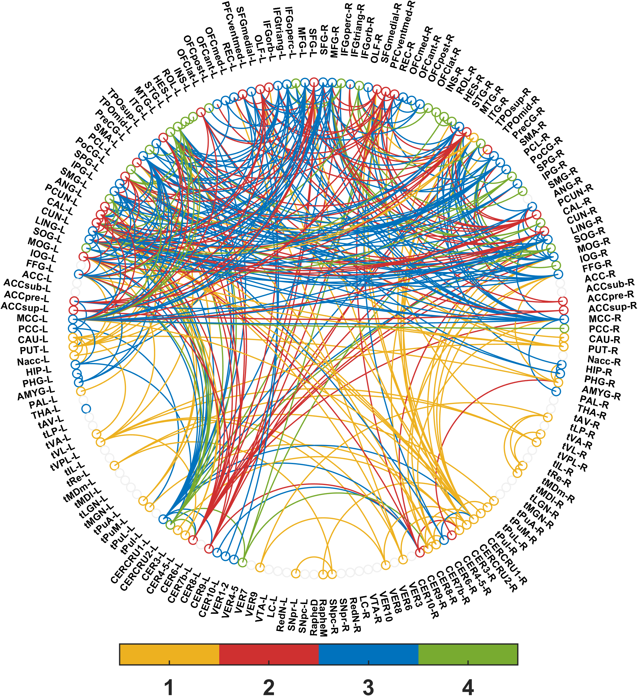
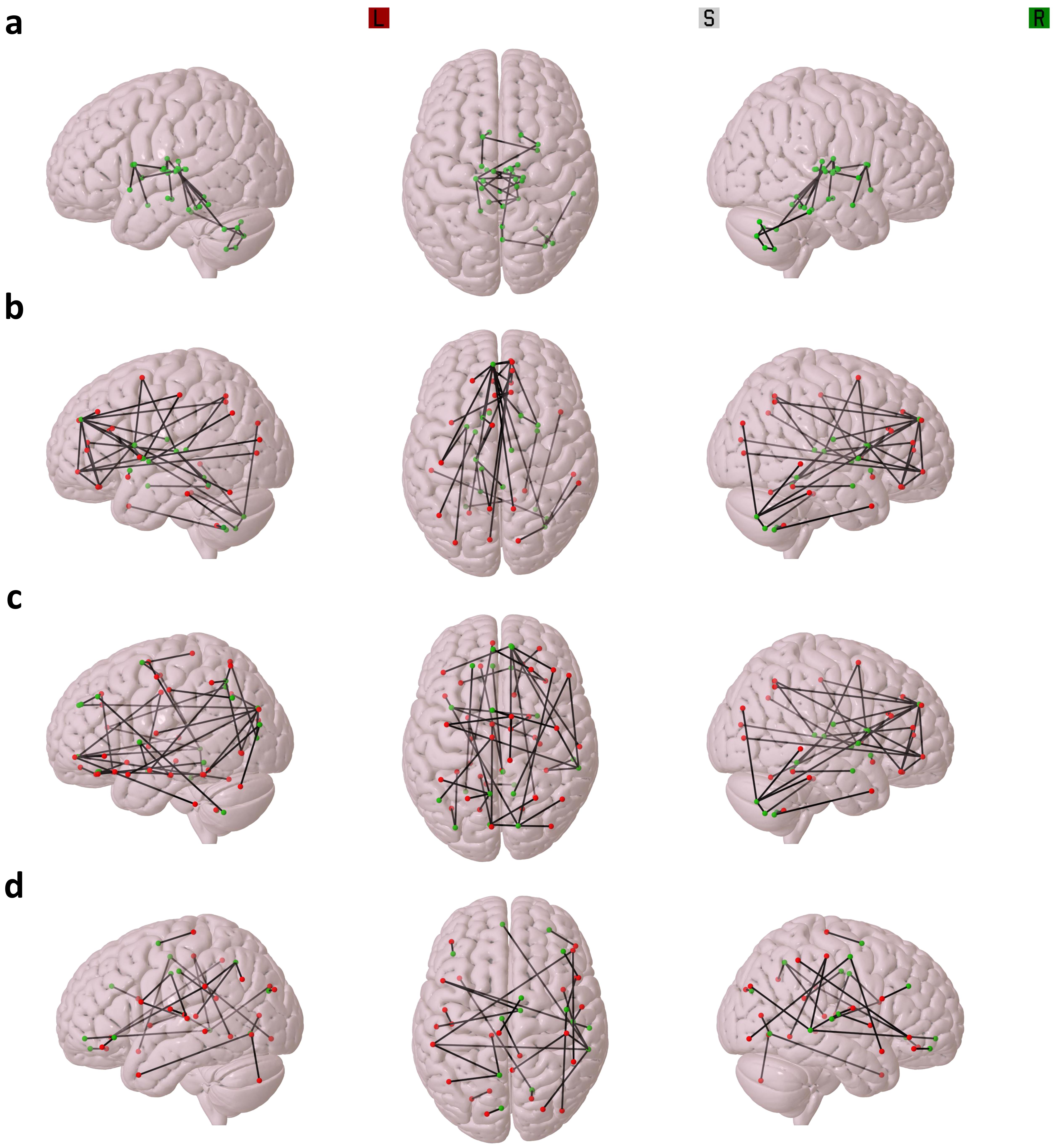
The proposed approach was applied to study the selective vulnerability of brain regions in Parkinson's disease (PD) using data from 28 PD patients and 30 healthy controls. For each participant, we generated a 170×170 undirected and weighted functional connectivity matrix, based on the third version of the Automated Anatomical Labeling (AAL3) atlas6. For each subject in the control group, 170 cascading chains were triggered following an initial attack on each node. This process generated a weighted and directed graph that characterized the selective progressive brain atrophy in PD across all subjects, revealing potential attack paths. These attack paths were weighted based on the participation frequency and the strength of fault transmission between functionally adjacent nodes. To reduce the influence of the initial node attack and intra-group variability, we extracted a fault propagation graph over all subjects, focusing on highly probable edges. Figure 2 illustrates the results for the fault propagation network, presenting a directed graph that organizes nodes and edges hierarchically to represent the progression of PD across brain regions over different time steps. To simplify this figure, the causal relationship between adjacent nodes is determined by a color order system defined in the color bar. The propagation graph was categorized into four different patterns of atrophy propagation, as shown in Figure 3. These patterns describe the hierarchical propagation of the atrophic process and may vary depending on the presence of specific factors. The results indicated that PD led to progressive brain atrophy affecting both motor and non-motor functions. The progression appears to initiate from the base of the brainstem, extending to the upper parts of the brainstem, basal ganglia (BG), motor cortex, and cerebellum, resulting in motor impairments. Subsequently, the atrophy progresses to subcortical nuclei, leading to cognitive impairments and mood disorders7.CONCLUSION
Selective regional vulnerability and progressive atrophic changes are essential aspects of understanding neurodegenerative diseases. This study introduced a novel graph theory-based framework to identify dynamic patterns of brain regions vulnerable to specific diseases. The approach utilizes a fault model to assess the extent of functional damage caused by a disease's dynamic progression through the brain network. The method was successful in detecting atrophied regions and their interactions in Parkinson's disease. The results demonstrated that this approach holds promise for detecting complex patterns of selective atrophy, which can have implications for diagnostics and therapeutics.Acknowledgements
The study was supported by NIH grant R01 NS102220.References
[1] H. Fu, J. Hardy, and K. E. Duff, “Selective vulnerability in neurodegenerative diseases,” Natureneuroscience, vol. 21, no. 10, pp. 1350–1358, 2018.
[2] A. Fornito and E. T. Bullmore, “Connectomics: A new paradigm for understanding brain disease,” European Neuropsychopharmacology, vol. 25, no. 5, pp. 733–748, 2015.
[3] S. Achard, R. Salvador, B. Whitcher, J. Suck-ling, and E. Bullmore, “A resilient, low-frequency,small-world human brain functional network with highly connected association cortical hubs,” Journal of Neuroscience, vol. 26, no. 1, pp. 63–72, 2006.
[4] M. Kaiser, R. Martin, P. Andras, and M. P. Young, “Simulation of robustness against lesions of cortical networks,” European Journal of Neuroscience, vol. 25, no. 10, pp. 3185–3192, 2007.
[5] O. Sporns, C. J. Honey, and R. Kötter, “Identification and classification of hubs in brain networks,” PloS one, vol. 2, no. 10, e1049, 2007.
[6] E. T. Rolls, C.-C. Huang, C.-P. Lin, J. Feng, and M. Joliot, “Automated anatomical labelling atlas3,” Neuroimage, vol. 206, p. 116 189, 2020.
[7] K. Singh et al., “Functional connectome of arousal and motor brainstem nuclei in living humans by 7tesla resting-state fMRI,” NeuroImage, p. 118 865,2022
Figures
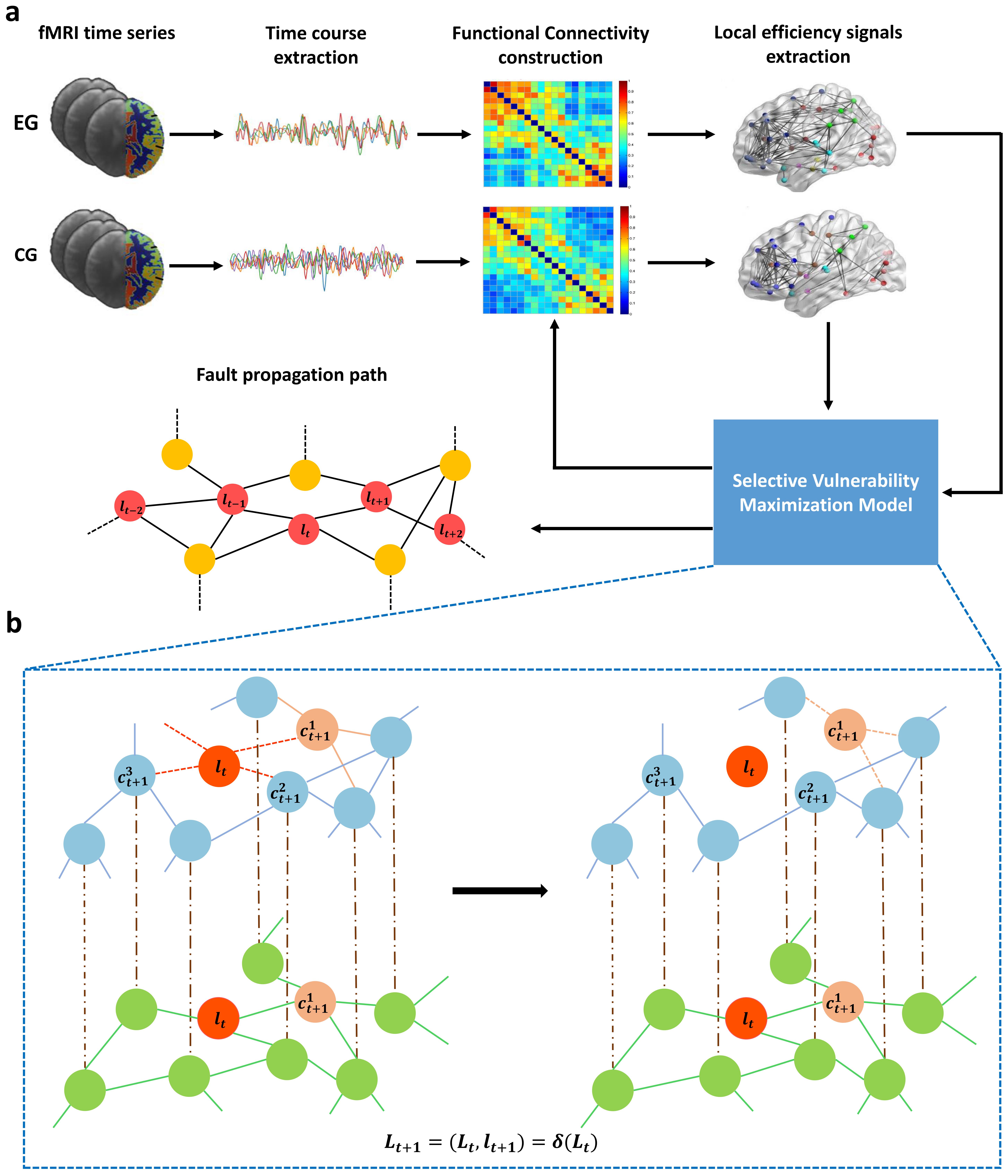
Fig. 1: Schematic of the proposed approach: (a) Utilizing fMRI data from EG and CG subjects, the model identifies fault paths related to brain atrophy progression. It constructs functional connectivity networks, extracts local efficiency signals, and dynamically updates CG signals post-attack. (b) Iteratively refines the CG local efficiency signals (shown in blue) while revealing the EG local efficiency signals (in green) to determine globally optimal paths.