4406
Pattern separation engages regions beyond the hippocampus among nondemented elderly subjects: a 7T task fMRI study1Cleveland Clinic Lou Ruvo Center for Brain Health, Las Vegas, NV, United States, 2Cleveland Clinic, Cleveland, OH, United States, 3University of Colorado, Boulder, CO, United States
Synopsis
Keywords: Task/Intervention Based fMRI, fMRI (task based), pattern separation, mnemonic similarity task
Motivation: Investigating the ability to differentiate similar representations is extensively focused on hippocampus. The roles of cortical regions and their interaction with the hippocampus largely remain unclear.
Goal(s): We aim to address this issue with whole-brain high-resolution fMRI data collected during a mnemonic similarity task.
Approach: Whole-brain and hippocampal ROI analysis were conducted to examine brain activity during the task.
Results: The right frontoparietal network showed increased activity when a lure or the same object was presented compared to a new object. The left anterior CA3/DG and left frontoparietal network had higher activity when a lure was correctly identified as “similar” instead of “same”.
Impact: Besides the CA3/DG, left frontoparietal regions were also involved in discriminating similar objects while right frontoparietal regions were mainly involved in successful retrieval, suggesting the functional lateralization of frontoparietal network.
INTRODUCTION
Pattern separation is the process of forming distinct memory representations to discriminate among similar but not the same stimuli, which is a critical aspect of episodic memory [1]. The Hippocampus and its subdivisions have drawn extensive attention in understanding the biological mechanism of the pattern separation process by using task fMRI data [2,3,4]. Increasing fMRI spatial resolution to investigate hippocampal subdivisions came with the cost of limited spatial coverage, the roles of cortical regions and their interaction with the hippocampus largely remain unclear. In addition, the 3T MR scanners used in these studies had a lower signal-to-noise ratio compared to ultra-high field MR scanners. In this study, we aim to address these issues with whole-brain high-resolution fMRI data collected at a 7T MR scanner.METHODS
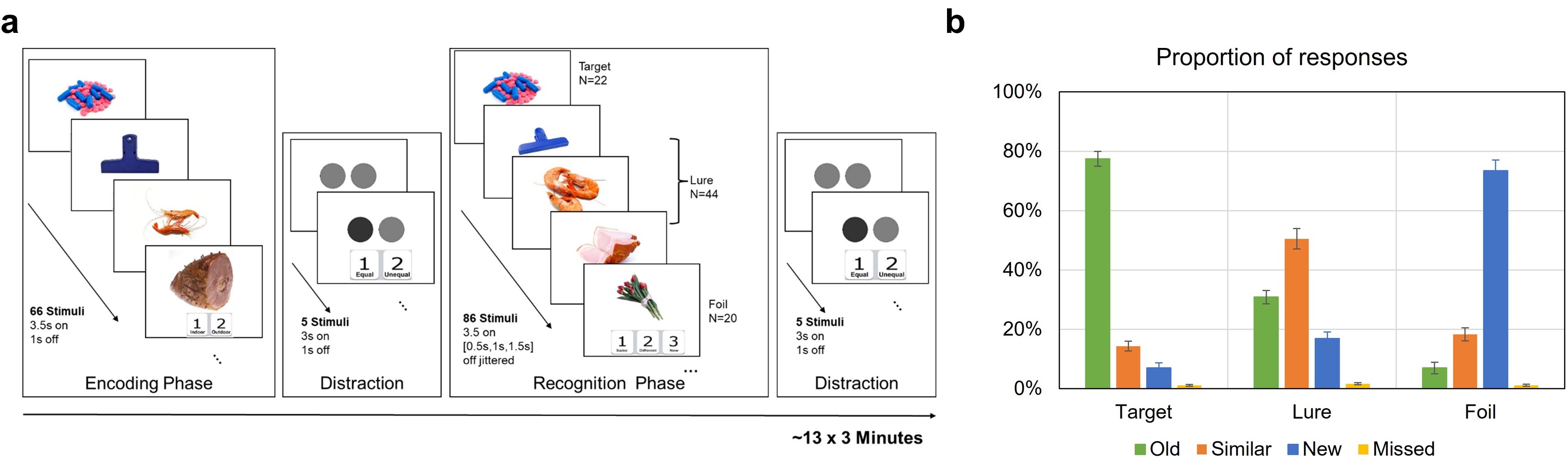
MRI data were acquired from 30 non-demented elderly subjects (71.43±5.18 years) at a 7T Siemens MAGNETOM Terra MR scanner, including whole brain T1-weighted structural image (0.8mm isotropic), high-resolution T2-weighted image acquired perpendicular to the long axis of the hippocampus (in-plane resolution 0.44 x 0.44 mm, slice thickness 1mm with a 0.1mm gap), and three 13-minute whole-brain mnemonic similarity task (MST) [5] fMRI runs (1.5mm isotropic). In MST (see Figure 1a), 66 everyday objects were first presented in the encoding phase. The same objects (targets), similar objects (lures) and new objects were later presented in the recognition phase, during which participants were asked to respond whether the objects were old, similar, or new. The segmentation of hippocampal ROIs was carried out with FreeSurfer software suite (v7.2, https://surfer.nmr.mgh.harvard.edu/). The T1-weighted image was first analyzed with the main FreeSurfer command “recon-all” with default settings and then together with the T2-weighted image to segment hippocampal subdivisions with the command “segmentHA_T2.sh”. Six hippocampal ROIs were extracted in the study, including anterior CA1, anterior subiculum, anterior CA3+DG, posterior CA1, posterior subiculum, and posterior CA3+DG. The affine transformation in Advanced Normalization Tools (ANTs, http://stnava.github.io/ANTs/) was used for co-registering fMRI data and T1-weighted image with dilated gray matter and white matter mask, using this mask was found to substantially reduce mis-alignment for 7T fMRI data. The labelling of hippocampal ROIs was transformed into individual fMRI space and the mean time series were computed for the hippocampal ROI analysis. For the whole-brain voxel-wise analysis, fMRI data was first co-registered to T1 space and then spatially normalized to standard MNI space by using the symmetric diffeomorphic transformation. General linear model was applied on data collected during recognition phase with five conditions, including new objects correctly identified as new (foil), targets correctly identified as same (hits), lures correctly identified as similar (LureCR) or falsely identified as same (LureFA), and all other trials. Hippocampal ROI-specific analysis and whole-brain voxel-wise analysis were conducted to examine the differential activities for lure discrimination (“similar” versus “same” response to lure; namely LureCR versus LureFA). Cluster-wise correction was used to detect significant clusters in the whole-brain analysis. The correlation between the activity difference and participants’ age or task performance was then tested. Functional connectivity between hippocampal ROIs and significant clusters was examined.RESULTS
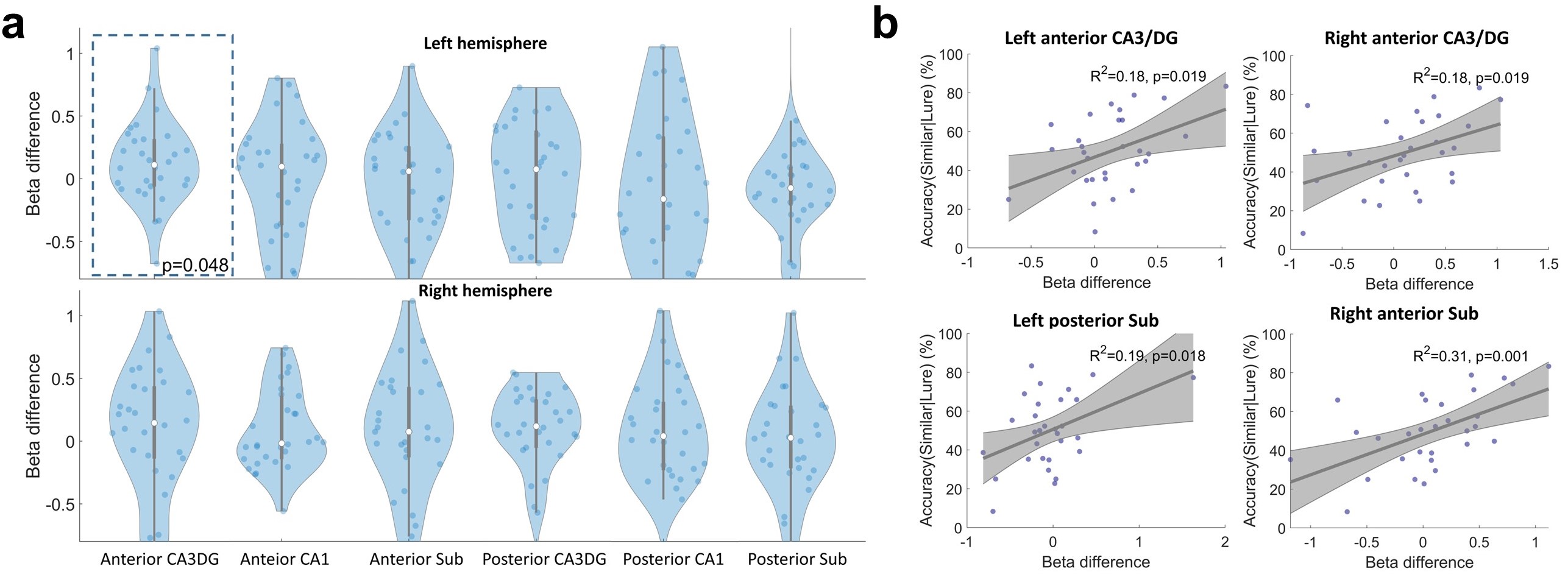
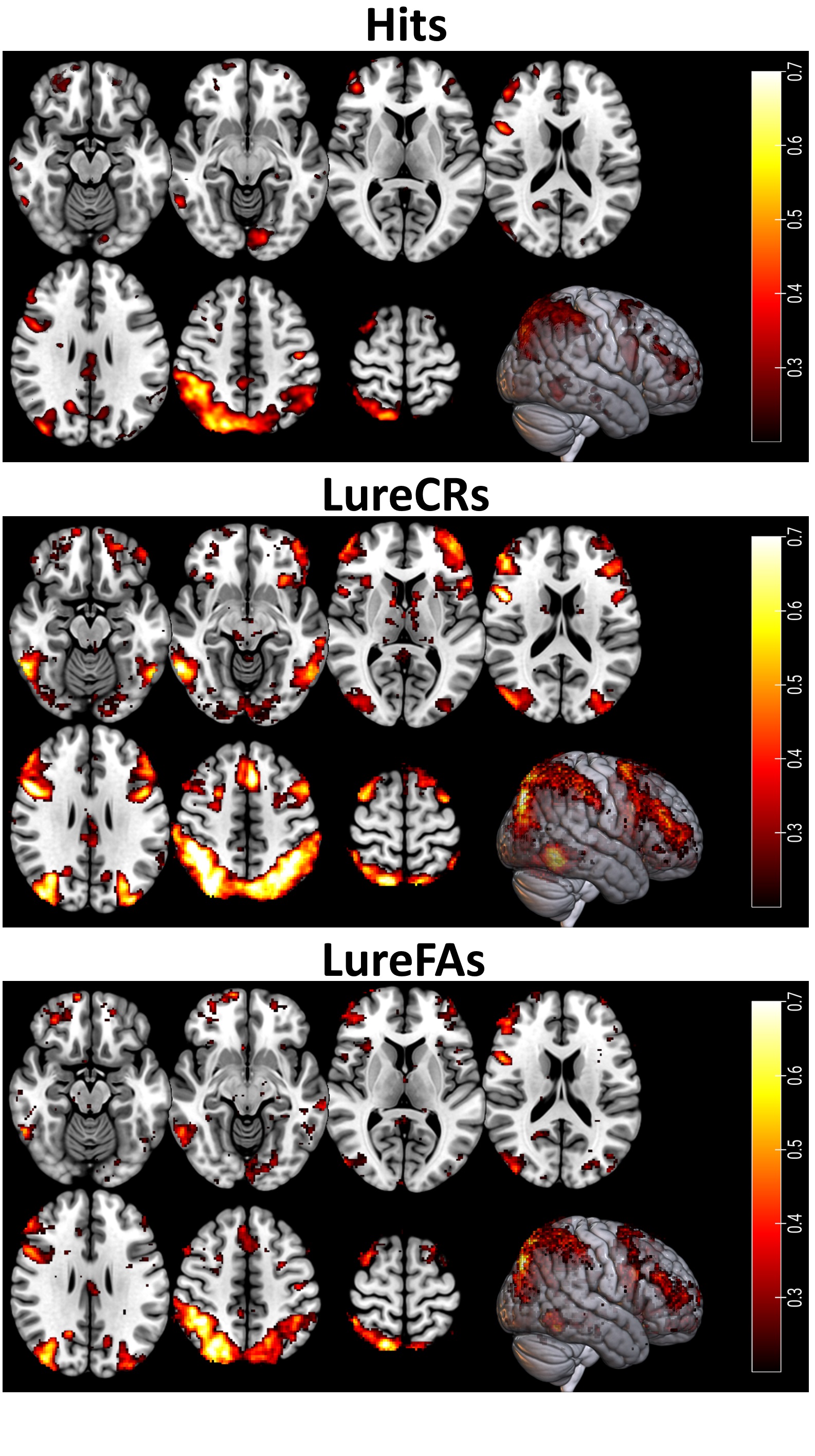
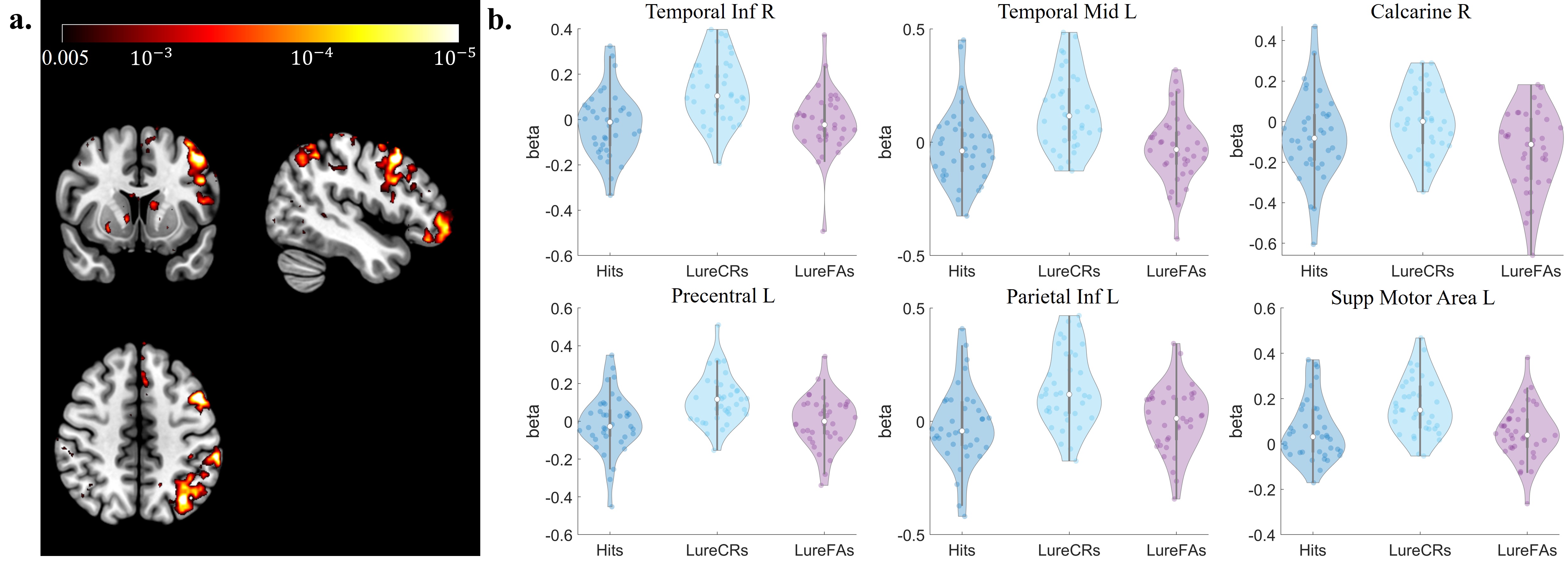
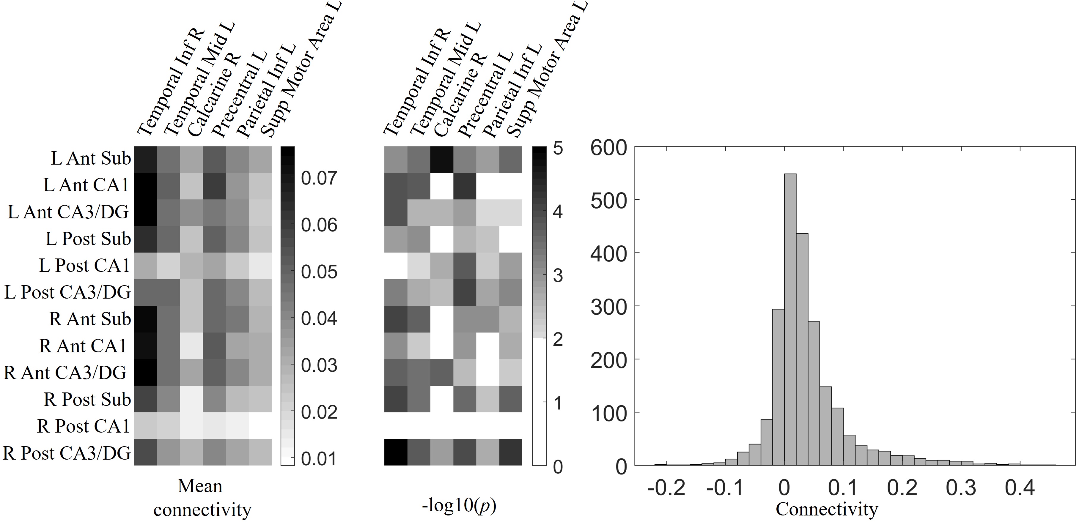
The average proportions of same, similar, new, and missed responses for target, lure and foil stimuli were shown in Figure 1b. The participants correctly identified the target as same and identified the foil as new with accuracy above 70%. 50.51% of lure stimuli were correctly identified as similar, and the lure stimuli were more frequently incorrectly identified as same (30.88%) than new (16.94%). In the hippocampal ROI analysis, left anterior CA3/DG showed higher activity in LureCR condition compared to LureFA condition (Figure 2). All the other hippocampal ROIs did not show discriminative activity. The activity difference between LureCR and LureFA was positively associated with higher accuracy in responding to lures as “similar” at bilateral anterior CA3/DG, left posterior subiculum and right anterior subiculum. In the whole-brain analyses, compared to Foil condition, the right frontoparietal network consistently showed increased activity in the Hit, LureCR and LureFA conditions (Figure 3). When we compared LureCR and LureFA to detect activity of “pattern separation” signature, six clusters were found to have significantly greater activity in LureCR than LureFA, with most voxels located within left frontoparietal network (Figure 4). These regions were significantly correlated with hippocampal ROIs (Figure 5).DISCUSSION
By using ultra-high field MR scanner and advanced fMRI protocol, our study found that left frontoparietal network, besides CA3/DG, could be involved in discriminating similar visual objects, and right frontoparietal network could be involved in memory retrieval in MST test. These findings suggest the functional lateralization of frontoparietal network in MST.Acknowledgements
This research project was supported by the NIA Grant 1RF1AG071566, Cleveland Clinic Keep Memory Alive Young Investigator Award, a private grant from Stacie and Chuck Matthewson, a private grant from Peter and Angela Dal Pezzo, and a private grant from Lynn and William Weidner.References
1. Sahay, A., D.A. Wilson, and R. Hen, Pattern separation: a common function for new neurons in hippocampus and olfactory bulb. Neuron, 2011. 70(4): p. 582-588.
2. Rolls, E.T., The mechanisms for pattern completion and pattern separation in the hippocampus. Frontiers in systems neuroscience, 2013. 7: p. 74.
3. Leutgeb, J.K., et al., Pattern separation in the dentate gyrus and CA3 of the hippocampus. science, 2007. 315(5814): p. 961-966.
4. Neunuebel, J.P. and J.J. Knierim, CA3 retrieves coherent representations from degraded input: direct evidence for CA3 pattern completion and dentate gyrus pattern separation. Neuron, 2014. 81(2): p. 416-427.
5. Kirwan, C.B. and C.E. Stark, Overcoming interference: An fMRI investigation of pattern separation in the medial temporal lobe. Learning & Memory, 2007. 14(9): p. 625.
Figures